Note: Descriptions are shown in the official language in which they were submitted.
CA 02726511 2010-11-30
WO 2009/147201 PCT/EP2009/056862
1
ANTI-INFLAMMATORY AGENTS
Field of the Invention
This invention is in the field of treating or preventing inflammation in
humans and animals and relates to pharmaceutical compositions and methods for
treating
or preventing various inflammatory conditions. In particular, the invention
relates to
compositions and methods for preventing or treating inflammatory conditions
such as
citrulline related diseases, preferably inflammatory diseases. The invention
provides
specific binding molecules directed against citrulline-containing epitopes for
use in the
therapy and prevention of inflammatory conditions.
Background of the invention
Inflammatory conditions, whether of a chronic or acute nature, represent
a substantial problem in the healthcare industry. Briefly, chronic
inflammation is
considered to be inflammation of a prolonged duration (weeks or months) in
which active
inflammation, tissue destruction and attempts at healing are proceeding
simultaneously
(Robbins Pathological Basis of Disease by R. S. Cotran, V. Kumar, and S. L.
Robbins, W.
B. Saunders Co., p. 75, 1989). Although chronic inflammation can follow an
acute
inflammatory episode, it can also begin as an insidious process that
progresses with time,
for example, as a result of a persistent infection (e.g., tuberculosis,
syphilis, fungal
infection) that causes a delayed hypersensitivity reaction, prolonged exposure
to
endogenous (e.g., elevated plasma lipids) or exogenous (e.g., silica,
asbestos, cigarette
tar, surgical sutures) toxins, or autoimmune reactions against the body's own
tissues (e.g.,
rheumatoid arthritis, systemic lupus erythematosus, multiple sclerosis,
psoriasis).
Inflammatory arthritis is a serious health problem in developed
countries, particularly given the increasing number of aged individuals. For
example, one
form of inflammatory arthritis, rheumatoid arthritis (RA) is a multisystem
chronic, relapsing,
inflammatory disease affecting 1 to 2% of the world's population.
Although many organs can be affected, RA is basically a severe form of
chronic synovitis that sometimes leads to destruction and ankylosis of
affected joints
(Robbins Pathological Basis of Disease, by R. S. Cotran, V. Kumar, and S. L.
Robbins,
W.B. Saunders Co., 1989). Pathologically the disease is characterized by a
marked
thickening of the synovial membrane which forms villous projections that
extend into the
joint space, multilayering of the synoviocyte lining (synoviocyte
proliferation), infiltration of
the synovial membrane with white blood cells (macrophages, lymphocytes, plasma
cells,
and lymphoid follicles; called an "inflammatory synovitis"), and deposition of
fibrin with
CA 02726511 2010-11-30
WO 2009/147201 PCT/EP2009/056862
2
cellular necrosis within the synovium. The tissue formed as a result of this
process is
called pannus and eventually the pannus grows to fill the joint space. The
pannus
develops an extensive network of new blood vessels through the process of
angiogenesis,
which is essential to the evolution of the synovitis. Release of digestive
enzymes (matrix
metalloproteinases (e.g., collagenase, stromelysin)), and other mediators of
the
inflammatory process (e.g., hydrogen peroxide, superoxides, lysosomal enzymes,
and
products of arachadonic acid metabolism), from the cells of the pannus tissue
leads to the
progressive destruction of the cartilage tissue. The pannus invades the
articular cartilage
leading to erosions and fragmentation of the cartilage tissue. Eventually
there is erosion of
the subchondral bone with fibrous ankylosis, and ultimately bony ankylosis, of
the involved
joint.
It is generally believed that RA is an autoimmune disease and that many
different arthrogenic stimuli activate the immune response in an
immunogenetically
susceptible host. Both exogenous infectious agents (Epstein-Barr virus,
rubella virus,
cytomegalovirus, herpes virus, human T-cell lymphotropic virus, Mycoplasma,
and others)
and endogenous proteins such as collagen, proteoglycans, altered
immunoglobulins and
post-translationally modified proteins like citrullinated proteins have been
implicated as a
causative agent that triggers an inappropriate host immune response.
Regardless of the
inciting agent, autoimmunity plays a role in the progression of the disease.
In particular,
the relevant antigen is ingested by antigen-presenting cells (macrophages or
dendritic
cells in the synovial membrane), processed, and presented to T lymphocytes.
The T cells
initiate a cellular immune response and stimulate the proliferation and
differentiation of B
lymphocytes into plasma cells. The end result is the production of an
excessive
inappropriate immune response directed against the host tissues (e.g.,
antibodies directed
against type II collagen, antibodies directed against the Fc portion of
autologous IgG
(called "Rheumatoid Factor")), and antibodies directed against different
citrullinated
epitopes (anti-CCP). This further amplifies the immune response and hastens
the
destruction of the cartilage tissue. Once this cascade is initiated numerous
mediators of
cartilage destruction are responsible for the progression of rheumatoid
arthritis.
The above mentioned anti-CCP antibodies have been demonstrated to
be highly specific for RA. Recent evidence shows that each individual that is
seropositive
for these antibodies either already has RA or will develop this disease in the
future. The
presence of anti-CCP antibodies (especially when high titers are present) is
predictive of
erosive disease outcome (Nijenhuis et al., Clin. Chim. Acta, vol 350, 17-34,
2004).
Furthermore, it has been demonstrated that anti-CCP antibodies are produced
locally at
the site of inflammation. The proportion of anti-CCP antibodies with respect
to total IgG
CA 02726511 2010-11-30
WO 2009/147201 PCT/EP2009/056862
3
found in synovial material from IRA patients appeared to be significantly
higher than that in
serum of the same patients (Masson-Bessiere et al, Clin Exp Immunol, vol 119,
544-552,
2000) (Reparon-Schuijt et al, Arthritis Rheum, vol 44, 41-47, 2001).
The presence of anti-CCP producing plasma cells in the synovium is
indicative of an antigen-driven maturation of CCP-specific B cells at the site
of
inflammation. Once anti-CCP antibodies are produced, the formation of immune
complexes with citrullinated proteins in the synovia may trigger the
progression of the
inflammatory process. These and other data supported the hypothesis that anti-
CCP
antibodies actually caused at least part of the disease symptoms of RA. A role
for the anti-
CCP antibodies in the pathogenesis of RA is supported by the results of B
lymphocyte
depletion experiments in patients with RA (Cambridge et al., Arthritis Rheum,
vo148, 2146-
2154, 2003).
People with advanced rheumatoid arthritis have a mortality rate greater
than some forms of cancer and because of this, treatment regimes have shifted
towards
aggressive early drug therapy designed to reduce the probability of
irreversible joint
damage. Recent recommendations of the American College of Rheumatology
(Arthritis
and Rheumatism 39(5):713-722, 1996) include early initiation of disease-
modifying anti-
rheumatic drug (DMARD) therapy for any patient with an established diagnosis
and
ongoing symptoms. Anticancer drugs have become the first line therapy for the
vast
majority of patients, with the chemotherapeutic drug methotrexate being the
drug of
choice for 60 to 70% of rheumatologists. The severity of the disease often
warrants
indefinite weekly treatment with this drug, and in those patients whose
disease progresses
despite methotrexate therapy (over 50% of patients), second line
chemotherapeutic drugs
such as cyclosporin and azathioprine (alone or in combination) are frequently
employed.
There remains a need for compounds for the treatment or prevention of
inflammatory diseases that are capable of inhibiting the pathogenesis of
inflammatory
diseases, in particular diseases wherein the synovium is involved and
citrulline related
inflammatory diseases.
Summary of the invention
The invention provides a binding molecule specifically reactive with a
citrullinated epitope on p15 andior p17 for use in the treatment or prevention
of
inflammatory diseases.
The invention also provides a method for treating or preventing an
inflammatory disease, comprising the step of administering to a patient in
need thereof a
therapeutically effective amount of an anti-inflammatory composition
comprising a binding
CA 02726511 2016-12-05
54013-15
4
molecule specifically reactive with a citrulline epitope on p15 and/or p17.
The compositions and methods of the present invention include
pharmaceutically acceptable formulations of specific binding molecules
reactive with
citrulline residues. In particular, the binding molecules are specifically
reactive with
citrullinated epitopes on two polypeptides as identified herein, termed p15
and p17.
More specifically, in an embodiment, the invention relates to the use of
an antibody specifically reactive with a citrullinated epitope on a peptide
comprising
the amino acid sequence of SEQ ID NO: 21 for the prevention or treatment of
rheumatoid arthritis or joint damage.
In another embodiment, the invention relates to an antibody specifically
reactive with a citrullinated epitope on a peptide comprising the amino acid
sequence
of SEQ ID NO: 21 that competes with a) a recombinant human monoclonal antibody
comprising a heavy chain variable region according to SEQ ID NO: 13 and a
light
chain variable region according to SEQ ID NO: 15, b) a recombinant human
monoclonal antibody comprising a light chain variable region according to SEQ
ID
NO: 17 and a heavy chain variable region according to SEQ ID NO: 19, c) a
recombinant human monoclonal antibody comprising a heavy chain as deposited at
the EMBL database with accession number: AJ430749 and a light chain as
deposited
at the EMBL database with accession number: AJ430773, d) a recombinant mouse
monoclonal antibody comprising a heavy chain as deposited at the EMBL database
with accession number: AJ430749 and a light chain as deposited at the EMBL
database with accession number: AJ430773, e) a recombinant human monoclonal
antibody comprising a heavy chain as deposited at the EMBL database with
accession number: AJ430732 and a light chain as deposited at the EMBL database
with accession number: AJ430753, f) a recombinant mouse monoclonal antibody
comprising a heavy chain as deposited at the EMBL database with accession
number: AJ430732 and a light chain as deposited at the EMBL database with
CA 02726511 2016-12-05
,
54013-15
4a
accession number: AJ430753, g) a recombinant monoclonal antibody comprising a
heavy chain variable region according to SEQ ID NO: 39 and a light chain
variable
region according to SEQ ID NO: 40, or h) a recombinant human monoclonal
antibody
comprising a heavy chain variable region according to SEQ ID NO: 41 and a
light
chain variable region according to SEQ ID NO: 42, for binding to the peptide
comprising the amino acid sequence of SEQ ID NO: 21.
In another embodiment, the invention relates to a polypeptide
comprising a heavy chain variable region or light chain variable region
according to a
sequence selected from the group consisting of SEQ ID NO: 13, SEQ ID NO: 15,
SEQ ID NO: 17, SEQ ID NO: 19, SEQ ID NO: 39, SEQ ID NO: 40, SEQ ID NO: 41
and SEQ ID NO: 42.
In another embodiment, the invention relates to a nucleic acid encoding
the polypeptide as described herein.
These and other aspects of the present invention will become evident
upon reference to the following detailed description, figures and examples. In
addition, various references are set forth herein which describe in more
detail certain
procedures, devices, or compositions.
Detailed description of the invention.
The invention provides a binding molecule specifically reactive with a
citrullinated epitope on p15 and/or p17 for use in the treatment or prevention
of
inflammatory diseases.
The term "specific binding molecule" is used herein to indicate a
molecule, preferably a small molecule, capable of specific binding. Specific
binding in
this respect is intended to mean that the molecule is capable of binding to a
selected
target molecule whereas it will not bind to another non-related target
molecule under
the same conditions. For instance, a binding molecule is said to specifically
bind to
CA 02726511 2016-12-05
,
54013-15
4b
serum albumin when it binds to serum albumin and less or not at all to another
or
preferably any other protein found in serum.
The term: "specifically reacts with citrulline" or "reactive with a
citrullinated epitope" or "reactive with a citrulline epitope" in this context
means that
the antibody reacts with a structure such as a peptide or peptide-like
molecule
containing a citrulline residue whereas the antibody reacts less or preferably
not at all
with the same structure containing an arginine residue instead of the
citrulline
residue. The term peptide or peptide-like molecule should be interpreted as
structures that are capable of presenting the citrulline residue in the
correct context
for immunoreactivity with the specific binding molecules as described herein,
preferably in the same context as it appears in the human or animal body,
preferably
in the context of a native polypeptide.
The "specific binding molecule" may be a molecule, preferably a small
molecule composed of DNA, RNA, peptide, protein domain, whole proteins, or
combinations thereof or parts thereof, that are capable of specifically
binding to a
target compound. Preferred examples of specific binding molecules are peptides
or
antibodies or parts thereof, such as Single Chain Variable Fragments (scFvs),
Fragment antigen binding regions (Fabs), single domains antibodies (sdabs),
also
known as VHH
CA 02726511 2010-11-30
WO 2009/147201 PCT/EP2009/056862
antibodies, nanobodies (Camelids derived single domain antibodies), or shark
IgNAR
derived single domain antibody fragments called VNAR, or other active
components
thereof, Anticalins, or aptamers (DNA or RNA). In a preferred embodiment, a
specific
binding molecule is a fusion protein comprising the antigen-binding domain of
an antibody
5 or an aptamer, such as an aptamer in the form of DNA or RNA. In an even
more preferred
embodiment, the specific binding molecule comprises antibodies, or derivatives
thereof,
such as antibody fragments, nanobodies, single domain antibodies, or active
parts
thereof. The invention therefore in particular relates to specific binding
molecules as
described above which are peptides or antibodies.
The term "Antibodies" or "antibody" refers to a protein or polypeptide
capable of specific binding to a target molecule often referred to as
"antigen". Antibodies
(also known as immunoglobulins) are gamma globulin proteins that are found in
blood or
other bodily fluids of vertebrates, and are used by the immune system to
identify and
neutralize foreign objects, such as bacteria and viruses.
Antibodies are typically made of basic structural units - each with two
large heavy chains and two small light chains - to form, for example, monomers
with one
unit, dimers with two units or pentamers with five units. Antibodies are
produced by a kind
of white blood cell called a B cell. There are several different types of
antibody heavy
chain, and several different kinds of antibodies, which are grouped into
different isotypes
based on which heavy chain they possess. Five different antibody isotypes are
known in
mammals which perform different roles, and help direct the appropriate immune
response
for each different type of foreign object they encounter. Some animal species
such as
Camelids (e.g. llamas) and sharks may have aberrant antibody structures.
Although the general structure of all antibodies is very similar a small
region at the tip of the protein is extremely variable, allowing millions of
antibodies with
slightly different tip structures to exist. This region is known as the
hypervariable region.
Each of these variants can bind to a different target, known as an antigen.
This huge
diversity of antibodies allows the immune system to recognize an equally wide
diversity of
antigens. The unique part of the antigen recognized by an antibody is called
an epitope.
These epitopes bind with their antibody in a highly specific interaction that
allows
antibodies to identify and bind only their unique antigen in the midst of the
millions of
different molecules that make up an organism. Recognition of an antigen by an
antibody
tags it for attack by other parts of the immune system. Antibodies can also
neutralize
targets directly, for example, by binding to a part of a pathogen that it
needs to cause an
infection.
The large and diverse population of antibodies is generated by random
CA 02726511 2010-11-30
WO 2009/147201 PCT/EP2009/056862
6
combinations of a set of gene segments that encode different antigen binding
sites (or
paratopes), followed by random mutations in this area of the antibody gene,
which create
further diversity. Antibody genes also re-organize in a process called class
switching that
changes the base of the heavy chain to another, creating a different isotype
of the
antibody that retains the antigen specific variable region. This allows a
single antibody to
be used in several different isotypes by several different parts of the immune
system.
The term "Antibody" as used herein includes single chain antibodies,
fragment antigen binding regions, recombinantly produced antibodies,
monoclonal
antibodies, single domain antibodies, and the like.
The term "or part thereof" in the context of an antibody or other specific
binding molecule is meant to refer to the part of the antibody or specific
binding molecule
that makes up the specific binding site of the antibody or specific binding
molecule and
may be interpreted as the part of an antibody or specific binding molecule
that is still
capable to react with the same epitope as the entire antibody or specific
binding molecule.
All kind of specific binding molecules, and derivatives thereof such as
antibodies, fusion proteins comprising a specific binding domain of an
antibody, aptamers,
antibody fragments, single domain antibody fragments, other proteinacous
binding
domains such as anticalins, and small molecules that specifically bind
citrullinated
epitopes can be used in the invention. However, human antibodies or fragments
thereof
are a preferred embodiment of the invention. Preferably IgG1 (e.g., lgG1.)
antibodies
having an IgG1 heavy chain and a lambda light chain are used. However, other
human
antibody isotypes are also encompassed by the invention, including IgG2, IgG3,
IgG4,
IgM, IgA1, IgA2, IgAsec, IgD and IgE in combination with a kappa or lambda
light chain.
Also all animal-derived antibodies of various isotypes can be used in the
invention. The
antibodies can be full-size antibodies or antigen-binding fragments of
antibodies, including
Fab, F(ab')2, single chain Fv fragments, or single domain VHH, VH or VL single
domains.
"Specific binding molecules reactive with a citrullinated epitope" are to
be interpreted as specific binding molecules that specifically react with a
citrulline residue
in the context of a larger structure such as a peptide or a peptide nucleic
acid or an
aptamer or a peptide mimicking structure.
Citrulline is an amino acid that is not incorporated into proteins during
translation, however, it can be generated by post-translational modification
of an arginine
residue by peptidylarginine deiminase (PAD).
Citrullination is the posttranslational conversion of arginine residues to
citrulline residues, which is catalyzed by peptidylarginine deiminase (PAD).
Peptidylarginine deiminase (PAD; EC 3.5.3.15) enzymes catalyse the conversion
of
CA 02726511 2010-11-30
WO 2009/147201 PCT/EP2009/056862
7
arginine residues to citrulline residues in proteins. No tRNA exists for
citrulline, the
presence of citrulline residues in proteins is exclusively the result of post-
translational
modification. In mammals (humans, mice and rats) five PAD isotypes (PAD1 ¨
PAD6;
'PAD4' and 'PAD5' are used for the same isotype), each encoded by a distinct
gene, have
been identified (Vossenaar et al, Bioessays 25, 1106-1118, 2003). All these
enzymes rely
strongly on the presence of Ca2+ for activity and are unable to convert free L-
arginine into
free L-citrulline. Free L-arginine can be converted to free L-citrulline by
nitric oxide
synthase (EC 1.14.13.39) in eukaryotes or by arginine deiminase (EC 3.5.3.6)
in bacteria.
These enzymes are not Ca2+ dependent.
The most pronounced difference between the highly homologous PAD
enzymes is their tissue-specific expression. In epidermis PAD1 (synonyms: PAD
I, PAD
type l) is involved in the citrullination of keratin filaments during the
final stages of
keratinocyte differentiation, which is important for the reorganization of the
cornified
envelope. Another site of citrullination in the epidermis is the hair
follicle, which contains
PAD3 (synonyms PAD III, PAD type III) and its natural substrate trichohyalin
(THH). THH
is a major structural protein of the inner root sheath cells and the medulla
layer of the hair
follicle and, to a lesser extent, of other specialized epithelia. The most
recently identified
PAD isotype, PAD6 (synonym: ePAD), was found in cytoplasmic sheets of mouse
oocytes, which play an important role in early embryogenesis. The expression
of its
human orthologue was found to be restricted to ovary, testis and peripheral
blood
leukocytes (Chavanas et al., Gene vol 330; 19-27, 2004). Originally, this PAD
isotype was
designated ePAD, but based upon the systematic numbering of other PADs, this
isotype
was renamed PAD6 (Vossenaar et al., Bioessays vol 25 1106-1118, 2003). The
most
widely expressed isotype, PAD2 (synonyms PAD II, PAD type II, PAD-H19), is
present in
many different tissues, like skeletal muscle, brain, spleen, secretory glands
and
macrophages. Despite this broad expression pattern, only myelin basic protein
(MBP) and
vimentin have been identified as natural substrates. In multiple sclerosis
(MS) patients
develop an autoimmune response against MBP. MBP is an abundant protein of the
myelin
sheath, and its citrullination occurs during development of the central
nervous system.
Citrullination of vimentin was observed during calcium-ionophore induced
apoptosis of
human and mouse macrophages and, as described above, citrullinated vimentin
was
shown to be the target of the RA-specific anti-Sa autoantibodies. In contrast
to the PADs
discussed above, which are all mainly localized in the cytoplasm of cells, the
PAD4
isotype (synonyms: PAD IV, PAD type IV, HL-60 PAD, PAD V, PAD type V, PADI4)
is
localized in the nucleus. The nuclear localization signal of PAD4 was found in
the N-
terminal region of the protein. PAD4 is mainly expressed in peripheral blood
granulocytes
CA 02726511 2010-11-30
WO 2009/147201 PCT/EP2009/056862
8
and monocytes. Substrates of PAD4 in the nucleus are histone core proteins
(H2A, H3
and H4) and nucleophosmin/B23, a nucleolar protein that functions in ribosome
assembly,
nucleocytoplasnnic transport and centrosome duplication.
Specific binding molecules according to the invention are directed
against a citrullinated epitope on p15 and/or p17, two polypeptides
characterized by their
molecular weights of 15 kDa and 17 kDa, respectively.
Such specific binding molecules were found to be particularly suited for
the treatment or prevention of inflammatory diseases.
"Inflammatory Conditions" or Inflammatory diseases" as used herein
refers to any of a number of conditions or diseases which are characterized by
vascular
changes: edema and infiltration of neutrophils (e.g., acute inflammatory
reactions);
infiltration of tissues by mononuclear cells; tissue destruction by
inflammatory cells,
connective tissue cells and their cellular products; and attempts at repair by
connective
tissue replacement (e.g., chronic inflammatory reactions).
Representative examples of such conditions include citrulline related
inflammatory diseases and autoimmune diseases. Citrulline related inflammatory
diseases
are herein defined as those diseases wherein citrullination plays a role in
the
pathogenesis of the disease. Whether or not citrullination plays a role in the
pathogenesis
of the disease, may be easily determined by a skilled person using routine
tests available
in the art. For example, these diseases may be characterized by the presence
of an
abnormal level of citrullinated proteins in affected or disease related
tissue. Such may be
accomplished by an immunological test such as a western blot or an ELISA
wherein the
affected tissue is used as an antigen and citrullination of that antigen may
be detected
with the aid of an anti-citrullin antibody as described herein.
Alternatively, a person skilled in the art can use Proteomics applications
such as mass spec. analysis to compare the level and type of citrullinaton in
a diseased
versus healthy tissue from affected patients.
The disease may also be characterized by the presence of an immune
response against citrulline containing peptides or proteins. This may be a
humoral or a
cellular immune response, such as a response mediated by T-cells or B-cells.
Tests for
detecting anti-citrulline antibodies have been described in the art and are
commercially
available.
The invention therefore relates to a specific binding molecule for use in
treating or preventing citrulline related inflammatory diseases.
Such diseases are for instance inflammatory arthritis, including
rheumatoid arthritis and osteoarthritis, multiple sclerosis, psoriatic
arthritis, psoriasis,
CA 02726511 2010-11-30
WO 2009/147201 PCT/EP2009/056862
9
Alzheimer's disease, autoimmune hepatitis, juvenile idiopathic arthritis,
spondylo-
arthropathy, Down's syndrome, multiple system atrophy, Parkinson's disease and
Lewy
body dementia. The invention therefore relates to a specific binding molecule
for use in
treating or preventing diseases selected from the group consisting of
arthritis, rheumatoid
arthritis, osteoarthritis, multiple sclerosis, psoriatic arthritis, psoriasis,
Alzheimer's disease,
autoimmune hepatitis, juvenile idiopathic arthritis, spondyloarthropathy,
Down's syndrome,
multiple system atrophy, Parkinson's disease and Lewy body dementia.
The invention in particular relates to specific binding molecules for the
treatment or prevention of autoimmune diseases, more in particular rheumatoid
arthritis or
osteoarthritis
Multiple sclerosis or MS is a chronic inflammatory disorder of the CNS,
characterized by autoimmunity mediated destruction of the myelin sheath. The
cells of the
myelin sheath form a multibilayer structure around the axons consisting of
lipid-protein
complexes in a ratio of about 3: 1. Two major proteins, MBP and proteolipid
protein,
account for 85% of the protein fraction. MBP is a highly cationic protein,
capable of
forming strong interactions with negatively charged phospholipids such as
phosphatidylserine. In approximately 18% of the MBP molecules of healthy adult
humans
6 (out of 19) arginines are citrullinated (Wood et al., J Bio I Chem, vo1264,
5121-5127,
1989, Wood et al., Ann Neurol, vol40, 18-24, 1996). The remaining MBP
molecules do not
contain citrulline. In MS patients the proportion of MBP-cit6 is increased to
45% of total
MBP. The decreased net positive charge of MBP-cit6 causes partial unfolding of
MBP
molecules and weakens their interaction with the phospholipids (Boggs et al.,
J Neurosci
Res, vo157, 529-535, 1999, Pritzker et al., Biochemistry, vo139, 5374-5381,
2000).
Although MBP-cit6 is capable of forming lipid complexes more rapidly than non-
citrullinated MBP, the complexes that are formed are not as densely packed as
those
formed with non-citrullinated MBP (Boggs et al, J Neurosci Res, vol57, 529-
535, 1999,
Beniac et al, J Struct Biol, vol129, 80-95, 2000). MBP-cit6 is degraded 4
times more
rapidly by cathepsin D than non-citrullinated MBP (Cao et al., Biochemistry,
vo138, 6157-
6163, 1999). In a rare case of acute fulminating MS (Marburg type), 80% of the
MBP
molecules are heavily citrullinated (MBPcit18) (Wood et al., Ann Neurol,
vo140, 18-24,
1996). The severely unfolded MBP-cit18 is degraded 45 times more rapidly by
cathepsin
D than normal MBP (Cao et al., Biochemistry, vo138, 6157-6163, 1999). Clinical
trials with
paclitaxel, the active component of the anti-cancer drug taxol, are in
progress (O'Connor
et al., Ann Neurol, vo146, 470, 1999). Low doses of paclitaxel can inhibit
citrullination of
MBP by PAD2 in vitro (Pritzker et al., Biochim Biophys Acta, vol1388, 154-160,
1998).
Treatment with paclitaxel attenuates clinical symptoms and induces
remyelination of
CA 02726511 2010-11-30
WO 2009/147201 PCT/EP2009/056862
damaged sheaths (Moscarello et al., Mult Soler, voI8, 130138, 2002),
underlining the
possible importance of PAD as a candidate factor in demyelinating disease
(Moscarello et
al., J Neurochem, vol81, 335-343, 2002).
In psoriasis, keratinocytes proliferate very rapidly and travel from the
5 basal layer to the surface in only about four days. The skin can not shed
these cells
quickly enough so they accumulate in thick, dry patches, or plaques. In normal
keratinocytes, keratin K1 is citrullinated by PAD1 during terminal
differentiation. This
process causes the keratin filaments to become more compact, which is
essential for the
normal cornification process of the epidermis. The keratinocytes in the
psoriatic
10 hyperproliferative plaques do not contain citrullinated keratin K1
(Ishida-Yamamoto et al.,
J Invest Dermatol, vol114, 701-705, 2000). It is not clear whether the
increased cell
proliferation prevents adequate citrullination by PAD or that inactivity of
PAD allows
hyperproliferation and accumulation of keratinocytes. Although the mechanism
is
unknown, aberrant citrullination in psoriatic epidermis obviously is related
to PAD1.
In a preferred embodiment, the composition according to the invention is
in a form selected from the group consisting of an aqueous solution, a gel, a
hydrogel, a
film, a paste, a cream, a spray, an ointment, or a wrap. In further
embodiments, the above
methods are used to administer the compositions described herein by a route
selected
from intra-articular, intraperitoneal, topical, rectal, intravenous, oral,
ocular, or to the
resection margin of tumors.
In certain embodiments, a pharmaceutically acceptable carrier
comprises at least one carrier selected from the group consisting of a co-
solvent solution,
liposomes, micelles, liquid crystals, nanocrystals, nanoparticles, emulsions,
microparticles, microspheres, nanospheres, nanocapsules, polymers or polymeric
carriers, surfactants, suspending agents, complexing agents such as
cyclodextrins or
adsorbing molecules such as albumin, surface active particles, and chelating
agents. In
further embodiments, a polysaccharide comprises hyaluronic acid and
derivatives thereof,
dextran and derivatives thereof, cellulose and derivatives thereof (e.g.,
methylcellulose,
hydroxy-propylcellulose, hydroxy-propylmethylcellulose,
carboxymethylcellulose, cellulose
acetate phthalate, cellulose acetate succinate, cellulose acetate butyrate,
hydroxypropylmethyl-cellulose phthalate), chitosan and derivative thereof,
[beta]-glucan,
arabinoxylans, carrageenans, pectin, glycogen, fucoidan, chondrotin, dermatan,
heparan,
heparin, pentosan, keratan, alginate, cyclodextrins, and salts and
derivatives, including
esters and sulfates, thereof.
In a further aspect, the method according to the invention comprises
delivering a composition according to the invention to a target site, most
notably a
CA 02726511 2010-11-30
WO 2009/147201 PCT/EP2009/056862
11
synovial joint.
In one specific embodiment of the present invention, the specific binding
molecule competes with monoclonal antibodies RhmAb2.102, RmmAb1.102,
RhmAb2.103, RmmAb1.103, RhmAb2.104, RmmAb1.104, RhmAb2.105 and
RhmAb2.107 for binding to p15 and/or p17.
The primary mRNA sequences of the variable regions of monoclonal
antibodies RhmAb2.101, RhmAb2.103, and RhmAb2.104, RmmAb1.101, RmmAb1.103
and RmmAb1.104 have been published and were deposited in the EMBL database
under
accession numbers as shown in table 1. The primary sequence of the variable
regions of
monoclonal antibodies RhmAb2.102, RmmAb1.102, RhmAb2.105 and RhmAb2.107 are
disclosed herein in SEQ ID NO: 13, SEQ ID NO: 15, SEQ ID NO: 17, SEQ ID NO:
19,
SEQ ID NO: 39, SEQ ID NO: 40, SEQ ID NO: 41 and SEQ ID NO: 42.
The invention therefore relates to a polypeptide comprising a variable
heavy or light chain according to SEQ ID NO: 13, SEQ ID NO: 15, SEQ ID NO: 17,
SEQ
ID NO: 19, SEQ ID NO: 39, SEQ ID NO: 40, SEQ ID NO: 41 and SEQ ID NO: 42. The
invention also relates to a nucleic acid encoding a polypeptide according to
SEQ ID NO:
13, SEQ ID NO: 15, SEQ ID NO: 17, SEQ ID NO: 19, SEQ ID NO: 39, SEQ ID NO: 40,
SEQ ID NO: 41 and SEQ ID NO: 42.
In another preferred embodiment, the specific binding molecule is an
antibody selected from the group consisting of monoclonal antibodies
RhmAb2.102,
RmmAb1.102, RhmAb2.103, RmmAb1.103, RhmAb2.104, RmmAb1.104, RhmAb2.105
and RhmAb2.107.
In another preferred embodiment, the specific binding molecule
comprises VH and /or VL domains derived from an antibody selected from the
group
consisting of monoclonal antibodies RhmAb2.102, RmmAb1.102, RhmAb2.103,
RmmAb1.103, RhmAb2.104, RmmAb1.104, RhmAb2.105 and RhmAb2.107.
Specific binding molecules according to the invention may be generated
essentially in two ways. First, they may be derived from the antibodies and
its sequences
as presented herein. Reactivity of the antibodies may even be improved by side-
directed
mutagenesis, chain shuffling, sexual PCR, or by other means for antibody
derivation and
optimisation known to the person skilled in the art. Alternatively, specific
binding
molecules, in particular antibodies may be obtained by panning with any of the
specifically
reactive epitopes as described herein, in particular PAD4 treated Histon 2A,
peptide 1
(SEQ ID NO: 21) and other particularly reactive peptides.
The term "derived" in this respect means that the essential residues
responsible for the specific binding properties of the VH and /or VL domains
in a particular
CA 02726511 2010-11-30
WO 2009/147201 PCT/EP2009/056862
12
antibody are identified and that these essential residues are then transferred
into the
context of another peptide.
A person skilled in the art may use the sequences described herein to
clone or generate cDNA or genomic sequences for instance such as described in
the
below examples. Cloning of these sequences in an appropriate eukaryotic
expression
vector, like pcDNA3 (In Vitrogen), or derivates thereof, and subsequent double
transfection of mammalian cells (like CHO cells) with combinations of the
appropriate light
chain and heavy chain containing vectors will result in the expression and
secretion of the
listed antibodies RhmAb2.101, 2.102, 2.103, 2.104, 2.105 and/or 2.107, and
RmmAb1.101, 1.102, 1.103, 1.104.
He may also make analogues of the specific binding molecules as
described herein by using the specific binding domains of the antibody
sequences and
express them in a different context such as a polypeptide such as a fusion
protein. This is
well known in the art.
Recombinant Human and Mouse monoclonal anti-citrulline antibodies
were obtained as described in Examples 1 and 15. Monoclonal antibodies were
obtained
with a human IgG1 Fc region (RhmAb2.101, RhmAb2.102, RhmAb2.103, RhmAb2.104,
RhmAb2.105 and RhmAb2.107) and a mouse IgG2a Fc region (RmmAb1.101,
RmmAb1.102, RmmAb1.103 and RmmAb1.104). The human and mouse recombinant
antibody pairs (RhmAb2.101 and RmmAb1.102, RhmAb2.102 and RmmAb1.102,
RhmAb2.103 and RmmAb1.103, and RhmAb2.104 and RmmAb1.104) contain identical
VH and VL domains but contain human IgG1 (SEQ ID NO: 14) or mouse IgG2a Fc
domains (SEQ ID NO: 20) respectively. The three mouse and human monoclonal
antibody pairs were analysed on western blots and each pair was found to have
the same
specificity for their respective antigens.
Mouse monoclonal anti-citrullin-peptide antibodies RmmAb13.101,
RmmAb13.102 and RrrimAb13.103 were obtained from a commercial source
(ModiQuest
Research BV Nijmegen, The Netherlands; Cat no, MQ13.101, MQ13.102and
MQ13.103).
Anti-citrullin antibodies were tested in an experimental model wherein
inflammation is induced by injecting anti-collagen antibodies into a mouse.
This model is
known as collagen antibody induced arthritis (CAIA) (Nandakumar and Holmdahl,
J
Immunol Methods, vo1304, 126-136, 2005). Anti collagen antibodies were
obtained from a
commercial source (ModiQuest Research BV Nijmegen, The Netherlands; Cat no,
MQ18.101).
Mouse monoclonal anti-citrulline antibodies RmmAb13.101,
RmmAb13.102 and RmmAb13.103 were confirmed to enhance the severity of the
CA 02726511 2010-11-30
WO 2009/147201 PCT/EP2009/056862
13
collagen antibody induced arthritis, as has been described also by Kuhn et al.
(J. Clin.
Invest, vol116, 961-871, 2006); and Hill et al. (J Exp Med, vol 205, 967-979,
2008). This is
shown in figures la and lb.
Furthermore, several studies in human patients indicate that antibodies
against citrullinated epitopes add to the pathogenesis of RA (Masson-Bessiere
et al, J.
lmmunol, vo1166, 4177-4184, 2001; Vossenaar and van Venrooij, Arthritis Res
Ther, voI6,
107-111, 2004). This is shown in Figure la and b, which shows the "mean
arthritis score"
and "arthritis incidence" respectively of the same experiment.
Surprisingly, however, human monoclonal antibodies RhmAb2.104 and
RhmAb2.105 reduced the clinical signs of arthritis in the experimental CAIA
model,
whereas RhmAb2.103, RhmAb2.102 and RhmAb2.107 even abolished the clinical
signs
of arthritis in the experimental CAIA model.
RhmAb2.103 and RhmAb2.102 performed identical, only the results
obtained with RhmAb2.102 are shown in Figures lc and 1d. Results obtained with
RhmAb2.105 and RhmAb2.107 are shown in Figure 10.
The human monoclonal antibody RhmAb2.101 had no effect at all on
the clinical signs of arthritis at the dose applied. The commercially
available antibody
RhmAb2.201 is used as an irrelevant antibody control in this experiment
(ModiQuest
Research B.V., cat no: MQR2.201). This antibody does not recognize
citrulinated
epitopes.
The same experiments were also performed with the equivalent mouse
Fc IgG2a monoclonal antibodies RmmAb1.101, RmmAb1.102, RmmAb1.103 and
RmmAb1.104 which contain identical VH and VL domains compared to their human
counterparts and also recognize the same epitopes as their human counterparts.
Identical
results were obtained as with their human counterparts. RmmAb1.102, RmmAb1.103
and
RmmAb1.104 abolished (RmmAb1.102, RmmAb1.103) or reduced (RmmAb1.104) the
clinical signs of arthritis whereas RmmAb1.101 had no effect at all.
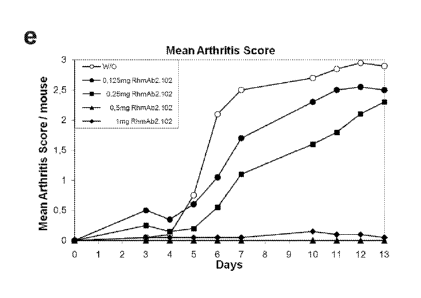
Figure le and 1f show an independent CAIA experiment in which the
clinical dose for RhmAb2.102 has been evaluated. The lowest dose that gave
maximum
inhibition was 0,5 mg Ab/mouse which corresponds to 28 mg/kg at IP injection.
From these experiments it is concluded that the specific epitopes
recognized by monoclonal antibodies selected from the group consisting of
RhmAb2.102,
RhmAb2.103, RhmAb2.104, RmmAb1.102, RmmAb1.103, RmmAb1.104, RhmAb2.105
and RhmAb2.107 play an important role in the treatment or prevention of
inflammatory
diseases.
In order to further analyze the antigen or antigens recognized by these
CA 02726511 2010-11-30
WO 2009/147201 PCT/EP2009/056862
14
monoclonal antibodies, they were tested for their reactivity towards cell
extracts that were
deiminated using Peptidylarginine deiminase (PAD enzyme) as described in
Example 3.
Western blots containing hPAD2 or hPAD4 transfected COS-1 lysates that were
post-
lytically deiminated were incubated with the monoclonal antibodies RhmAb2.101,
RhmAb2.102, RhmAb2.103 and RhmAb2.104. It was observed, that only strips
incubated
with RhmAb2.102, RhmAb2.103 and RhmAb2.104 showed reactivity with a doublet of
proteins with a molecular weight of approximately 15 and 17 kiloDalton.
WO 2004/078098 discloses antibodies specific for citrullinated
peptide/MHC class II complexes to inhibit T cell activation. These antibodies
do not bind
to the separate peptide or MHC class II molecule but only to the complex of
the peptide
and the MHC class II molecule. The antibodies disclosed herein are different
from the
antibodies disclosed in WO 2004/078098 since they recognize the individual
peptides and
proteins as disclosed herein. Moreover, the antibodies recognize a polypeptide
in a
western blot that could not be a complex between a peptide and an MHC class II
molecule, since the complex between an MHC molecule and a citrullinated
peptide would
never survive the reducing conditions of an SDS gel used in the immunoblot
procedure.
The epitopes recognized by the binding molecules as disclosed herein are
therefore
different from the antibodies disclosed in WO 2004/078098. Moreover, the
antibodies as
disclosed herein are not specifically reactive with a complex of a peptide and
an MHC
class II molecule.
The above described experiments and considerations led us to conclude
that there is a clear correlation between the ability to prevent clinical
signs of inflammatory
diseases and reactivity with citrullinated epitopes on p15 and p17.
Similar data were obtained when human monoclonal antibodies
RhmAb2.101, RhmAb2.102, RhmAb2.103 and RhmAb2.104 and mouse monoclonal
antibodies RmmAb1.101, RmmAb1.102, RmmAb1.103 and RmmAb1.104 were used in
immunoprecipitation experiments as detailed in Example 5.
lmmunoprecipitations with RhmAb2.102, RmmAb1.102, RhmAb2.103
and RmmAb1.103 on both human PAD2 and PAD4 deiminated COS-1 lysates revealed
prominent p15 and p17 protein bands. These bands were somewhat less prominent
when
immuno-precipitations were performed with RhmAb2.104 and RmmAb1.104.
The intensity of recognition of p15 and p17 proteins therefore seems to
correlate well with the therapeutic properties of these antibodies (Figures la-
d).
Whether or not an antibody is reactive with p15 or pi 7 may easily be
established by performing immunoprecipitation or western blot analysis as
detailed in
Examples 4 and 5. Alternatively, competition experiments with RhmAb2.102,
CA 02726511 2010-11-30
WO 2009/147201 PCT/EP2009/056862
RhmAb2.103 or RhmAb2.104 can be performed using either Western blots
containing
deiminated COS-1 lysates as described in example 6 or purified deiminated p15
and/or
p17 proteins in Western blot or ELISA.
Proteins p15 and p17 were further characterized by Matrix-assisted
5 laser desorption/ionisation-time of flight mass spectrometry (MALDI-TOF
MS) as detailed
in example 7. Since the genome of the African Green Monkey is not completely
sequenced we screened all other mammal genome databases for homology with the
peptides found with MALDI-TOF MS. Proteins found with a high degree of
homology
turned out to be histones This is shown in Table 3 (Example 7).
10 The invention therefore also relates to a binding molecule
specifically
reactive with a citrullinated epitope on histones for use in the treatment or
prevention of
inflammatory diseases.
The citrullination of histones by enzymatic action of PAD is well
documented and therefore citrullinated histones may very well be produced in
vitro.
15 These citrullinated histones may then be used as a substrate in an
enzymatic binding
assay to screen and select for other specific binding molecules such as
peptides and
antibodies reactive with epitopes on citrullinated p15 and p17, i.e. histones.
Preferably,
specific binding molecules are selected that compete with antibodies
RhmAb2.102,
RmmAb1.102, RhmAb2.103, RmmAb1.103, RhmAb2.104, RmmAb1.104 and
RhmAb2.105 and RhmAb2.107 for binding to p15 and/or p17.
In this document and in its claims, the verb "to comprise" and its
conjugations is used in its non-limiting sense to mean that items following
the word are
included, but items not specifically mentioned are not excluded. In addition,
reference to
an element by the indefinite article "a" or "an" does not exclude the
possibility that more
than one of the element is present, unless the context clearly requires that
there be one
and only one of the elements. The indefinite article "a" or "an" thus usually
means "at least
one".
In order to further analyze which deiminated histone or histones are involved
in the therapeutic action of RhmAb2.102 and RhmAb2.104, commercial available
histones
(H1, H2A, H2B, H3 and H4) were deiminated with human peptidylarginine
deiminase
(PAD, EC 3.5.3.15) enzymes (huPAD2 or huPAD4). Deiminated as well as non-
deiminated histones were coated on 96-well ELISA plates and incubated with
serial
dilutions of RhmAb2.101, RhmAb2.102 and RhmAb2.104. The results are shown in
table
6 and Figure 2.
It is evident from the results shown in figure 2 that huPAD4 deiminated
histone 2A (H2A/p4) is best recognized by the therapeutic antibodies
RhmAb2.102 and
CA 02726511 2010-11-30
WO 2009/147201 PCT/EP2009/056862
16
RhmAb2.104, but not by RhmAb2.101 (Figure 2a, 2b and 2c). Furthermore
RhmAb2.102
has higher affinity for H2A/p4 if compared to RhmAb2.104 (Figure 2b and 2c).
These data
correlate well with the effect of these antibodies on the clinical signs of
arthritis in the
experimental CAIA model, in which RhmAb2.102 abolish, RhmAb2.104 reduce and
RhmAb2.101 has no effect on the clinical signs of arthritis (Figure lc and
1d).
We have therefore shown that a deiminated epitope on H2A/p4 or its
structural mimics play a crucial role in the RA inflammatory cascade. The same
is true for
deiminated epitopes on H3/p2, H4/p2 and H4/p4 since RhmAb2.102 shows higher
affinity
for these histones than RhmAb2.104 and RhmAb2.101 (Figure 2a, 2b and 2c).
A mimic is for instance a molecule with an acceptable level of equivalent
activity, which, in this case, would include as being recognized with higher
affinity by
RhmAb2.102 than RhmAb2.104 and RhmAb2.101.)
The invention therefore relates to a specific binding molecule as described
above, reactive with a citrullinated epitope on human PAD4 deiminated human
histone 2A
or histone 4, or on human PAD2 deiminated human histone H4 or histone H3.
To further pinpoint the exact citrullinated epitope on H2A which is recognized
by RhmAb2.102 and RhmAb2.104, biotin labeled peptides were synthesized
containing all
13 potential deimination sites of histone 2A (Table 4). These peptides were
coated on 96-
well neutravidin-ELISA plates and incubated with serial dilutions of
RhmAb2.101,
RhmAb2.102 and RhmAb2.104. The results are shown in Figure 3.
Table 6A Reactivity of deiminated histones with RhmAb2.101, shown in figure 2A
sz
2.101 H1 H1/p2 H1/p4 H2A
H2A/p2 H2A/p4 H2B H2B/p2 H2B/p4
0,141 0.151 0,126 0,14 3,141 0,522 0,105 0,216 0.114
2 0,072 0,09 0,084 0,089 1,473 0,159 0,085 0,12 0.087
0.4 0, 067 0,08 0,083 0,085 0,426 0.11 0,069 0,077 0.069
,
0,08 0,064 0.072 0,072 0,076 0,128 0,073 0,067 0,067 0.064
0,016 0,061 0.064 0,072 0,073 0,076 0,073 0,065 0,062 0.064
0,0032 0,061 0,066 0,069 0,072 0,063 0,065 0,062 0,064 0,061
0,00064 0,06 0.067 0,069 0,071 0,059 0,064 0,059 0,06 0.061
0,000128 0,064 0.063 0,071 0,066 0,058 0,063 0,058 0,065 0.062
No
H3 H3/p2 H3/p4 H4 H4/p2 H4/p4 CFC-0 CFC-1
coating
0,115 0,217 0.383 0,111 1,341 0,116 0,303 3,587 0,069
0,075 0,087 0,146 0,093 0,412 0,073 0,103 3,26 0,055
0,065 0,073 0.076 0,089 0,154 0,077 0,084 2,13 0,058
0,074 0,067 0.069 0,066 0,084 0,065 0,066 0,807 0,067
u.)
0,071 0,069 0.079 0,067 0,06 0,063 0,056 0,249 0,053
0,072 0,079 0,076 0,072 0,067 0,066 0,056 0,097 0,057
0,074 0,077 0.074 0,07 0,062 0,063 0,057 0,072 0,052
0,079 0,104 0,104 0,073 0,08 0,063 0,056 0,065 0,051
"d
Table 6B Reactivity of deiminated histones with RhmAb2.102, shown in figure 2B
sz
2.102 H1 H1/p2 H1/p4 H2A H2A/p2 H2A/p4 H2B H2B/p2 H2B/p4
10 0,9 1,214 1,045 0,428 3,411 3,425 0,247 0,31 0,229
2 0,178 0,304 0,27 0,115 3,179 3,134 0,076 0,086 0,069
0,4 0,089 0,119 0,103 0,071 3,085 2,722 0,056 0,06 0,054
0,08 0,059 0,069 0,065 0,06 1,963 1,747 0,054 0,053 0,052
0,016 0,054 0,058 0,059 0,057 0,628 0,426 0,065 0,052 0,052
0,0032 0,055 0,058 0,057 0,056 0,161 0,135 0,05 0,052 0,052
0,00064 0,102 0,058 0,058 0,057 0,077 0,075 0,052 0,052 0,055
0,000128 0,053 0,057 0,057 0,058 0,063 0,062 0,052 0,051 0,053
0
1.)
No
H3 H3/p2 H3/p4 H4 H4/p2 H4/p4 CFC-0 CFC-1
coating
0,549 2,442 1,311 0,825 2,979 1,776 0,26 3,478 0,08
oo
0,275 1,935 0,439 0,208 2,735 1,556 0,086 3,377 0,053
0
0,08 1,177 0,166 0,091 2,218 0,986 0,06 3,115 0,05
0
0,062 0,493 0,093 0,067 1,343 0,432 0,05 2,145 0,046
0,058 0,155 0,076 0,061 0,491 0,167 0,05 0,702 0,047
0.058 0,08 0,065 0,06 0,151 0,077 0,049 0,178 0,047 0
0,056 0,062 0,062 0,06 0,073 0,058 0,048 0,077 0,045
0,058 0,066 0,06 0,06 0,073 0,055 0,047 0,058 0,046
"d
Table 6C Reactivity of deiminated histones with RhmAb2.104, shown in figure 20
2.104 H1 H1 /p2 H1 /p4 H2A H2A/p2 H2A/p4 H2B
H2B/p2 H2B/p4
0,082 0 096 0,09 0,095 2,688 3.13 0,101 0,099 0,09
2 0,07 0,08 0,077 0,077 2,034 2,224 0,083 0,085 0.078
0.4 0,07 0,078 0,075 0,084 0,923 0,834 0,077 0,085 0.073
0,08 0,067 0.073 0,075 0,07 0,396 0,23 0,077 0,081 0.074
0,016 0,071 0.074 0,074 0,07 0,124 0,105 0,076 0,079 0.075
0,0032 0,069 0,08 0,074 0,071 0,086 0,082 0,075 0,086 0,077
0,00064 0,069 0.069 0,071 0,075 0,078 0,078 0,079 0,081 0.074
0,000128 0,068 0 072 0,072 0,068 0,077 0,078 0,075 0,077
0.072
No
H3 H3/p2 H3/p4 H4 H4/p2 H4/p4 CFC-0 CFC-1
coating
0,087 0,145 0,14 0,104 1,243 0,144 0,085 3,901 0,064
0,078 0,103 0,112 0,094 0,553 0,075 0,065 4,041 0,062
0,073 0,077 0,09 0,09 0,227 0,069 0,057 4,003 0,057
0,07 0,081 0.075 0,08 0,344 0,066 0,056 3,942 0,052
0,074 0,074 0,087 0,209 0,243 0,068 0,057 3,895 0,05
0,072 0,075 0,072 0,071 0,069 0,065 0,056 2,27 0,053
0,07 0,077 0,075 0,069 0,067 0,068 0,055 0,536 0,051
0,068 0,082 0,089 0,068 0,068 0,069 0,053 0,205 0,051
"d
Table 7 Reactivity of selected peptides with mAbs RhmAb2.102, RhmAb2.104 and
RhmAb2.101 as indicated 0
t,..)
=
=
s.c
,
-
2.101 peptide 1 2 3 4 5 6 7 8 9 10
11 12 CFC-0 CFC-1 No coating .r.,
-.1
ng/wel I 0,266 0,457 0,393
0,095 0,083 0.750 1,178 0,090 0,087 0,073 0,148 0,072 0,095 2,841
0,076 r..1
=
2
0,102 0,136 0,121 0,048 0,051 0.218 0,459 0,053 0,053 0,069 0,064 0,053 0,071
2,717 0,055
0,4
0,086 0,071 0,068 0,051 0,064 0.090 0,174 0,050 0,056 0,061 0,058 0,050 0,068
1,827 0,050
0,08 0,062 0,054
0,053 0,056 0,051 0 062 0,080 0,051 0,052 0,052 0,051 0,050 0,065 0,951
0,051
0,016
0,057 0,049 0,049 0,051 0,054 0.058 0,055 0,050 0,049 0,048 0,050 0,050 0,055
0,492 0,050
0,0023
0,061 0,052 0,049 0,052 0,054 0,051 0,050 0,050 0,050 0,055 0,050 0,051 0,063
0,583 0,051
0,00064
0,049 0,038 0,050 0,040 0,053 0,052 0,052 0,050 0,048 0,066 0,047 0,045 0,064
0,548 0,050
0,000128
0,060 0,052 0,045 0,049 0,047 0.046 0,047 0,048 0,049 0,051 0,047 0,052 0,059
0,537 0,051 n
o
2.102 1 2 3 4 5 6 7 8 9 10 11
12 CFC-0 CFC-1 No coating 1.)
...]
10
3,112 0,552 0,619 2,056 0,239 1.410 0,080 0,082 0,090 0,091 0,088 0,083 0,870
3,271 0,074 1.)
cn
u,
2 3,048 0,270
0,286 1,300 0,111 0 752 0,059 0,060 0,063 0,070 0,067 0,067 0,242 3,206
0,053
o
0,4 2,804 0,136
0,154 _ 0,564 0,082 0.333 0,064 0,061 0,057 0,051 _ 0,064 0,061 0,115 _
3,060 0,051 1.)
0
0,08
2,039 0,086 0,091 0,192 0,066 0.123 0,062 0,060 0,060 0,058 0,064 0,060 0,088
2,656 0,050 1--
0
1
0,016
0,843 0,065 0,070 0,084 0,065 0.075 0,061 0,063 0,064 0,066 0,069 0,057 0,071
1,460 0,045 1-
0,0023
0,300 0,062 0,062 0,078 0,063 0,058 0,064 0,060 0,062 0,068 0,057 0,059 0,067
0,916 0,046
1
lx)
0,00064
0,160 0,055 0,058 0,063 0,067 0.058 0,057 0,057 0,059 0,056 0,060 0,056 0,066
0,621 0,050 0
0,000128
0,128 0,075 0,063 0,058 0,059 0.054 0,056 0,055 0,055 0,057 0,059 0,056 0,063
0,749 0,047
2.104 1 2 3 4 5 6 7 8 9 10 11
12 CFC-0 CFC-1 No coating
10
1,828 0,087 0,066 0,078 0,062 0.056 0,064 0,061 0,067 0,067 0,069 0,066 0,084
3,231 0,055
2
1,630 0,080 0,058 0,059 0,053 0.053 0,052 0,050 0,055 0,061 0,059 0,057 0,069
3,218 0,054
0,4
0,959 0,064 0,054 0,055 0,055 0,053 0,055 0,052 0,053 0,060 0,067 0,054 0,065
3,239 0,051 -o
n
0,08
0,374 0,053 0,057 0,055 0,054 0.056 0,054 0,059 0,061 0,062 0,058 0,060 0,066
3,259 0,052
m
0,016
0,165 0,055 0,052 0,055 0,048 0.057 0,055 0,058 0,055 0,055 0,055 0,059 0,063
2,975 0,050 -1:1
t.1
0,0023
0,125 0,052 0,055 0,059 0,057 0.052 0,053 0,052 0,054 0,051 0,070 0,056 0,061
1,993 0,050
=
0,00064
0,111 0,052 0,049 0,055 0,056 0.053 0,052 0,053 0,056 0,057 0,056 0,056 0,064
0,968 0,050 ,.tD
-i-
0,000128
0,105 0,050 0,054 0,053 0,051 0.052 0,050 0,050 0,053 0,050 0,055 0,061 0,061
0,627 0,050 'A
OC
CA
1,4
I
o
oo
N)
N)
l=-.)
al
00
H Table 8 Reactivity of selected peptides with Rhm
an as indicated.
Ab2.102, RhmAb2.104 d RhmAb2.101
indid.
1- th m
C70
N) 2.101 msFib = msFib = huFib = huFib =
msFib = rnsVim
0 (ug/well) XH XG XH XG XG XS/XL
cfcl XG cf0 Neutra blanc
1-,
co 10 0,120 3,876 0,177 3,778 2,538
, 0,282 3,780 0,154 0,088 0,069
1
0 2 0,081 3,730 0,124 3,601 1,260
0,144 3,612 0,115 0,120 0,066
n)
1 0,4 0,074 2,616 0,107 2,497 0,457
0,123 2,581 0,109 0,098 0,061
0
1- 0,08 0,073 0,893 0,100 0,798
0,203 0,119 , 1,070 0,115 0,099 0,061
0,016 0,087 0,267 0,112 0,249 0,132 0,129 0,459
0,126 0,135 0,064
0,0023 0,102 0,143 0,118 0,151 0,119 0,123 0,325
0,123 0,137 0,069
0,00064 0,130 0,130 0,121 0,254 0,123 0,134- 0,322 0,123 0,124 0,062-
0,000128 0,114 0,144 0,139 0,146 0,119 0,147 0,292
0,136 0,113 0,059
2.102 msFlb = msFib = huFib = huFib =
msFib = msVim
(ug/well) XH XG XH XG XG XS/XL
cfc1 XG cf0 Neutra blanc
10 0,154 3,028 , 0,179 , 2,727 3,802 3,694 3,892
0,334 0,088 0,086
2 0,091 1,902 0,116 1,511 3,154 2,767 3,968
0,138 0,080 0,062 IV
0,4 0,076 0,773 0,090 0,521 1,670 1,448 3,794
0,111 0,075 0,060 -
0,08 0,076 0,237 0,080 0,186 0,515 0,515 3,026 0,094
0,073 0,061
0,016 0,081 0,107 0,080 0,103 0,174 0,201 1,223
0,102 0,089 0,061
0,0023 0,085 0,125 0,123 0,125 0,120 0,142 0,506
0,124 0,103 0,060
0,00064 0,088 0,116 0,124 0,125 0,133 0,154 0,345
0,152 0,134 0,060
0,000128 0,089 0,119 0,120 0,115 0,118 0,133 0,288
0,139 0,119 0,059
2.104 msFib = msFib = huFib = huFib =
msFib = msVim
(ug/well) XI-1 XG XH XG XG XS/XL
cfc1 XG cf0 Neutra blanc
10 0,075 0,071 0,076 0,077 2,427 0,142 3,678 0,089
0,065 0.058
2 0,081 0,086 0,086 0,085 1,723 0,113 3,780 0,083
0,064 0,064
0,4 0,089 0,093 0,092 0,091 0,722 0,080 3,768 0,075
0,062 0,057
'
0,08 0,071_ 0,086 0,087 0,085 0,255 , 0,096 3,782 ,
0,089 0,070 0,056
0,016 0,070 0,072 0,078 0,078 0,122 0,098 3,585
0,105 0,100 0,061
0,0023 0,058 0,063 0,065 0,083 0,069 0,070 2,108, 0,070 0,064- 0,057
0,00064 0,064_ 0,069 0,071 0,067 0,064 0,076
0,664 0,079 0,069 0,069
0,000128 0,078 0,075 0,073 0,070 0,058 0,074 0,236
0,068 0,070 0,062
CA 02726511 2010-11-30
WO 2009/147201 PCT/EP2009/056862
22
It was observed that peptide 1 (AAASGXGKQGGK) was recognized by the
therapeutic antibodies RhmAb2.102 and RhmAb2.104, but not by RhmAb2.101 (Table
4
and Figure 3a, 3b and 3c). Again RhmAb2.102 showed higher affinity if compared
to
RhmAb2.104 (Figure 3b and 3c). The same holds true for the deiminated epitopes
on
peptides 4 and 6 (Table 4) since RhmAb2.102 shows higher affinity for these
peptides
than RhmAb2.104 and RhmAb2.101 (Figures 2a, 2b and 2c). We have therewith
shown
that the deiminated epitope or the structural equivalents or mimics thereof on
peptides 1,
4 and 6 play a crucial role in the RA inflammatory cascade. This antibody
recognition
pattern is very similar to the recognition pattern of H2A/p4. We therefore
conclude that the
specific binding molecules according to the invention may also be defined by
their
reactivity towards peptides 1, 4 and 6; SEQ ID NO: 21, SEQ ID NO: 24 and SEQ
ID NO:
26 respectively. Each of these peptides individually may be used to generate
specific
binding molecules such as antibodies according to the invention. Such
antibodies may
then be selected towards any of the other antigens as disclosed herein for
optimal
reactivity.
Table 4: Histone 2A citrulline containing peptides
Peptide
Sequence ID NO: Amino-acid sequence
Sequence ID NO: 21 AAASGXGKQGGK
2 Sequence ID NO: 22 AKAKSXSSRAGL
3 Sequence ID NO: 23 KSRSSXAGLQFP
4 Sequence ID NO: 24 _QFPVGXVHRLLR
5 Sequence ID NO: 25 VGRVHXLLRKGN
6 Sequence ID NO: 26 VHRLLXKGNYSE
7 Sequence ID NO: 27 GNYSEXVGAGAP
8 Sequence ID NO: 28 AGNAAXDNKKTR
9 Sequence ID NO: 29 DNKKTXIIPRHL
10 Sequence ID NO: 30 TRIIPXHLQLAI
11 Sequence ID NO: 31 LQLAIXNDEELN
12 Sequence ID NO: 32 NKLLGXVTIAQG
X denote a citrulline residue
Biotin labeled and citrullin containing fibrinogen and vimentin peptides
(Table
5) were also tested for reactivity with the therapeutic antibodies. Peptides
were coated on
96-well neutravidin-ELISA plates. Subsequently serial dilutions of RhmAb2.101,
CA 02726511 2010-11-30
WO 2009/147201 PCT/EP2009/056862
23
RhmAb2.102 and RhmAb2.104 were applied to the coated plates. The results are
shown
in Table 8 and Figure 4.
Table 5: Fibrinogen and vimentin citrulline containing peptides
Peptide Name SEQ ID NO: Amino-acid sequence
msFib= XH SEQ ID NO: 33 LSEGGGVRGPRVVEXHQSQCKD
msFib= XG SEQ ID NO: 34 LSEGGGVXGPRVVERHQSQCKD
huFib= XH SEQ ID NO: 35 LAEGGGVRGPRVVEXHQSACKD
huFib= XG SEQ ID NO: 36 LAEGGGVXGPRVVERHQSACKD
msFib= XG SEQ ID NO: 37 EPTDSLDAXGHRPVDRR
msVim XS/XL SEQ ID NO: 38 _YVTXSSAVXLXSSVP
X = citrulline
It was observed that the mouse fibrinogen = peptide (SEQ ID NO: 37) is
recognized by RhmAb2.101, RhmAb2.102 and RhmAb2.104 (Figure 4a, 4b and 4c).
Again RhmAb2.102 showed higher affinity if compared to RhmAb2.104, and
RhmAb2.104
performed slightly better than RhmAb2.101 (Figure 4a, 4b and 4c). This
antibody
recognition pattern is similar to the pattern observed on Western blots loaded
with
huPAD2 and HuPAD4 deiminated human fibrinogen. Furthermore only RhmAb2.102
recognized the mouse vimentine peptide (example 10). It is very likely that
besides the
above mentioned peptides, also the deiminated epitopes on peptide msFib= (SEQ
ID NO:
37) and msVim (SEQ ID NO: 38) play a crucial role in the RA inflammatory
cascade.
However it is therewith not excluded also other epitopes on fibrinogen and
vimentin play a
role in the anti-inflammatory effects of our therapeutic antibodies.
The invention therefore also relates to a specific binding molecule as
described above which is specifically reactive with an epitope on peptides
msFib= or
msVim (SEQ ID NO: 37 or SEQ ID NO: 38) and their use.
In addition we have shown that citrullinated epitopes appear de novo in
inflammated tissue. In an experimental mouse model for rheumatoid arthritis we
were able
to show that citrullinated peptides were immunoprecipitable from the
inflammated
forepaws of affected mice using human monoclonal antibody 102 (RhmAb2.102).
A typical CAIA experiment was therefore performed in which mice (3 mice
per group) have been injected i.p. with a mix of 8 anti-collagen antibodies
(2.8mg/mouse)
on day O. Three days later mice received another i.p. injection containing
25ug LPS.
Scoring has been performed as described above. During this experiment each day
a
CA 02726511 2010-11-30
WO 2009/147201 PCT/EP2009/056862
24
group of mice has been sacrificed, and paws were analyzed for citrulline
presence by
Western Blot analysis and Immunohistochemical techniques.
For each group of mice, forepaws were pooled and extracts made.
Immuniprecipitations (IP) have been performed on these extracts using 20
microgram
RhmAb2.102 per IP. Precipitates have been subjected to SDS-page
electrophoreses and
transferred to a nitrocellulose membrane by Western Blot techniques. The blot
was first
stained with Ponceau S for total protein detection. Ponceau S staining is
performed to
verify that for each IP the same amount of antibody has been used. Pronounced
antibody
heavy and light chains could be observed in the same amounts.
Subsequently the citrulline residues present on blot have been chemically
modified according to Senshu et al. (Senshu et al, Anal Biochem, vol 203, 94-
100, 1992).
The chemical modification can then be visualized using an antibody that
recognizes the
chemical modification of citrulline residues (Senshu et al, Anal Biochem, vol
203, 94-100,
1992). Deiminated fibrinogen was used as a positive control in this
experiment. An
immunoprecipitation without extracts was used as a negative control in these
experiments.
As from day 4, pronounced bands appeared on the blots at positions
corresponding to proteins with molecular weights of 50, 15 and 17 kiloDaltons.
These
bands became more pronounced in day 5 and were most intense at day 6.
The arthritis incidence of the experiment was 100%, with mice having regular
arthritis scores, reaching 5+ at day 6 (Fig. 5A and 5B). The amount of
precipitated protein
increases in time, which is visible from day 4 to 6. Based on the citrulline
specificity of
RhmAb2.102 and the presence of the signals on blot obtained with the anti-
chemically
modified citrulline antibody, we can conclude that mice subjected to CAIA have
detectable
citrulline levels in their inflamed joints.
Immunohistochemical analysis was also performed on the hindpaws of the
same mice. Slides have been incubated with RhAb2.104. Results complied with
the
Western Blot analysis. Modified citrullines could be detected on proteins with
apparent
molecular weight of approximately 50, 15 and 17 kiloDaltons in the samples
from days 4
to 6 which allowed us to conclude that citrullinated epitopes reactive and
immunoprecipitable with with RhmAb2.102 appeared de novo in inflamed joints,
in this
case in the hindpaws of experimentally induced arthritis mice.
In the CAIA experiments described above, anti-citrulline antibodies were
injected on day 3 after anti-collagen antibody injection, when inflammation in
the paws of
mice was still absent or very low. This prevented the occurrence of clinical
symptoms and
is therefore useful as a treatment of inflation, in particular a prophylactic
treatment.
We therefore wanted to study if RhmAb2.102 could also cure clinical
CA 02726511 2010-11-30
WO 2009/147201 PCT/EP2009/056862
symptoms once they had occurred. This was done by treating animals on day 7
after anti-
collagen injection when mean arthritis scores of all 4 paws of all mice
reached the
arbitrary score of approximately 4. As is shown in figure 6A and 6B,
RhmAb2.102 does
not abolish the swelling observed, but rather stabilized the present
inflammation/swelling.
5 Animals were followed for 35 days after which inflammatory scores among
placebo and
RhmAb2.102 treated mice were equal (Figure 6B and example 12). Figure 6A shows
the
Mean arthritis score of all paws of each group, while Figure 6B shows the mean
arthritis
score of the right hind paws of the animals that have been used for
histological analysis at
day 35.
10 Histology on right hind paws of all animals has been performed in
order to
investigate whether RhmAb2.102 treatment on day 7 could protect the mice from
permanent joint damage (Figure 7). Figure 7A shows that macroscopical
inflammation in
the right hind paws between experimental groups on day 35 of the experiment
were
similar. Most surprisingly however, all known parameters for joint erosion
were decreased.
15 When scoring Inflammatory cell influx (D), Cartilage erosion (B),
Cartilage PG depletion
(E), Chondrocyte death (F) and Bone erosion (C) a dramatic decrease is
observed in the
experimental group that has been treated on day 7 with RhmAb2.102, Indicating
that
RhmAb2.102 has a strong therapeutic potential in regard to preventing joint
damage
during inflammation (example 12). The invention therefore relates to a method
for
20 preventing or treating joint damage by administering a binding molecule
as described
herein to a patient in need of such a treatment.
Further CAIA experiments have been performed to investigate the
therapeutic effect of RhmAb2.102 treatment on day 5, 6 and 7 respectively
(Figure 8). In
this experiment RhmAb2.102 has been injected i.v. in order to deliver the
antibody rapidly
25 to sites of inflammation. In this experiment prophylactic treatment at
day 3, and a non
treated control group have been included. Experimental procedures have been
performed
as in Example 12 with the only difference of injections with lmg RhmAb2.102
per mouse
on day 3, 5 and 6. As expected RhmAb2.102 at day 3 inhibited the inflammatory
response. Treating mice with i.v. injections of RhmAb2.102 on day 5, 6 or 7
stabilized the
inflammation (Figure 8) as also seen in Figure 6. It is noteworthy that the
signs of
inflammation were not reduced whereas all parameters for joint erosion were
decreased.
This shows that joint erosion and inflammation are two separate entities that
may be
treated separately.
In the next series of CAIA experiments we investigated the possibility to
reduce the inflammation levels with Dexamethason and preventing the
reoccurrence of
inflammation after Dexamethason treatment was stopped by simultaneous
injection of
RhmAb2.102 on day 5. 6 or 7 (Figure 9) with dexamethason.
CA 02726511 2010-11-30
WO 2009/147201 PCT/EP2009/056862
26
Dexamethason is a general inflammatory inhibitor which needs to be
administered on a daily basis. Once treatment is interrupted, the inflammation
reoccurs.
Experimental procedures have been performed as described in Example 12 with
the
difference that 1mg RhmAb2.102 has been injected i.v. on day 5 (Figure 9A),
day 6
(Figure 9B) and day 7 (Figure 90) after anti-collagen antibody injection,
simultaneously
with i.p injections of Dexamethason (2mg/kg). Dexamethason was administered
sequentially for 2 or 3 days until swelling in the paws disappeared.
Additional groups of
animals received i.p. injections of Dexamethason only. As shown in Figure 9,
inflammation
reappeared in mice that did not receive RhmAb2.102. However, in strong
contrast, when
Dexamethason was combined with RhmAb2.102, inflammatory relapse was much
milder
and occurred later compared to Dexamethason only treated mice. This was most
evident
when starting combined RhmAb2.102/Dexamethason treatment on day 6 or 7 (Figure
9B
and C). The experiments shown in Figure 9 demonstrate a new treatment method
for
inflammatory diseases in which an inhibitor of inflammation such as
Dexamethason can
be used to treat flares of inflammation, and RhmAb2.102 can be used to prevent
inflammation relapse and more importantly prevent tissue/joint damage to
occur. The
invention therefore relates to a method of treating inflammation and joint
damage by
simultaneous administration of an inhibitor of inflammation together with a
binding
molecule as described herein
In another CAIA experiment, 2 novel anti-citrulline antibodies (RhmAb2.105,
and RhmAb2.107) that have shown cross-reactivity with RhmAb2.102 on its
differentiating
antigens from RhmAb2.101, have been tested for their anti inflammatory effect.
RhmAb2.105, RhmAb2.107 and RhmAb2.102 (positive control) have been injected
i.v. on
day 3 (1mg/mouse) after anti-collagen antibody injection in separate
experimental groups
(Figure 10). Experimental procedures have been performed as described in
Example 12.
Figure 10 shows the Mean arthritis score of all paws of each group.
It appeared that RhmAb2.102 showed the highest anti inflammatory effect.
RhmAb2.107 performed almost as well as RhmAb2.102, and RhmAb2.105 showed an
intermediate effect similar as previously observed for RhmAb2.104 (Figure 1C).
Additional deiminated proteins that preferentially bind to RhmAb2.102 have
been identified by mass spectrometry analysis. Furthermore, deiminated
proteins that
preferentially bind to RhmAb2.102 and not, or with to a lesser extent to
RhmAb2.101 have
also been identified by additional mass spectrometry analysis. Human PAD4
deiminated
Human Embryonic Kidney cell (HEK293) lysates have been immunoprecipitated with
RhmAb2.101 or RhmAb2.102 (Example 13) and subjected to a high throughput nano-
LC
system coupled to an advanced, high-performance LTQ Fourier Transform Ion
Cyclotron
Resonance Mass spectrometer (nLC LTQ FTMS ULTRA) (Example 14). Its ultra-high
CA 02726511 2010-11-30
WO 2009/147201 PCT/EP2009/056862
27
mass resolution, mass accuracy and sensitivity in combination with
Exponentially Modified
Protein Abundace Index (emPAI) calculations enabled us to identify deiminated
proteins
that (preferentially) bind to RhmAb2.102. This is shown in Table 7 (Example 13
and 14).
Hence, the invention also relates to a binding molecule specifically reactive
with any of the proteins or polypeptides as shown in table 7 for use in the
prevention or
treatment of an inflammatory disease.
In summary, we have shown herein that a binding molecule specifically
reactive with an epitope on a molecule selected from the group consisting of
p15, p17,
more in particular a citrullinated epitope on human PAD4 deiminated human
histone 2A, a
citrullinated epitope on human PAD4 deiminated human histone 4, human PAD2
deiminated human histone H4, human PAD2 deiminated human histone H3, or a
protein
selected from the group consisting of the proteins of table 7 and even more in
particular a
peptide according to SEQ ID NO: 21, SEQ ID NO: 24, SEQ ID NO 26, SEQ ID NO: 37
and SEQ ID NO: 38 may be used in the treatment or prevention of inflammatory
diseases
as specified herein. Whether a given binding molecule is specifically reactive
with the
above mentioned molecules, may easily be determined by analysis of the ability
of the
binding molecule to compete with an antibody selected from the group
consisting of
RhmAb2.102, RmmAb1.102, RhmAb2.103, RmmAb1.103, RhmAb2.104, RmmAb1.104 ,
RhmAb2.105 and RhmAb2.107 for binding to an epitope on p15 or p17 or any of
the
citrullinated epitopes mentioned above.
Having shown the efficacy of the binding composition according to the
invention, it will now be evident for the skilled person that inflammatory
diseases may also
be treated or prevented by eliciting an immune response wherein specific
binding
molecules according to the invention are generated in the patient's own body
(in vivo).
Such an immune response may be generated to prevent inflammatory disease from
occurring (prophylaxis, prophylactic vaccines) or to ameliorate or decrease
the
consequences of an inflammatory disease, i.e. therapy.
Hence, the invention also relates to a method for the prevention or treatment
of inflammatory diseases by eliciting an immune response in vivo wherein
specific binding
molecules are generated reactive with an epitope selected from the group
consisting of a
citrullinated epitope on p15, p17, a citrullinated epitope on human PAD4
deiminated
human histone 2A, human PAD4 deiminated human histone 4, human PAD2 deiminated
human histone H4, human PAD2 deiminated human histone H3, and a peptide
according
to SEQ ID NO: 21, SEQ ID NO: 24, SEQ ID NO 26, SEQ ID NO: 37 and SEQ ID NO: 38
Vaccines or therapeutics according to the invention may effectively comprise
a citrullinated epitope specifically reactive with a binding molecule
according to the
invention. More in particular, the citrullinated epitope may be a
citrullinated epitope on
CA 02726511 2010-11-30
WO 2009/147201 PCT/EP2009/056862
28
human PAD4 deiminated human histone 2A or histone 4, or on human PAD2
deiminated
human histone H4, human histone H3, or a peptide selected from the group
consisting of
SEQ ID NO: 21, SEQ ID NO: 24, SEQ ID NO 26, SEQ ID NO: 37 and SEQ ID NO: 38.
Accordingly, a number of citrulline related inflammatory diseases may be
treated or prevented. Hence, the invention also relates to a method as
described above
wherein the inflammatory disease is selected from the group consisting of
autoimmune
diseases, arthritis, rheumatoid arthritis, osteoarthritis, multiple sclerosis,
psoriatic arthritis,
psoriasis, Alzheimer's disease, autoimmune hepatitis, juvenile idiopathic
arthritis,
spondyloarthropathy, Down's syndrome, multiple system atrophy, Parkinson's
disease
and Lewy body dementia. Particlularly preferred is the prevention or treatment
of
autoimmune diseases such as rheumatoid arthritis.
Since this embodiment of the invention relates to an in vivo immune
response, a preferred specific binding molecule is an antibody.
LEGENDS TO THE FIGURES
Figure 1: A Collagen antibody induced arthritis (CAIA) model was used to test
the effect of
eight monoclonal antibodies on the severity of symptoms of arthritis. Mean
arthritis score
(Figures la, lc and le) and arthritis incidence (figures lb, ld and lf) are
indicated.
Groups of 5-6 mice were treated at day 0 through i.p. injection with anti-
collagen
antibodies. Mice used in the experiments shown in figure la and lb received
1,6 mg anti-
collagen antibody mix, whereas mice used in figure lc-f received 2,4 mg. LPS
(25
= g/mouse) together with anti-citrulline or a control antibody (RhmAb2.201)
were
administered on day 3 through i.p. injection. All antibodies were administered
at
lmg/mouse unless otherwise stated in the graph. Animals have been scored daily
until
day 13. Antibodies RhmAb2.102 and RhmAb2.103 performed equally well, only
RhmAb2.102 is shown. The same is true for antibodies RmmAb1.102 and
RmmAb1.103;
they performed equally well, only RmmAb1.102 is shown.
Figure 2: An enzyme linked immunosorbend assay (ELISA) was used to test the
affinity of
a) RhmAb2.101, b) RhmAb2.102 and c) RhmAb2.104 for human recombinant histones
(H1, H2A, H2B, H3 and H4) deiminated with huPAD2 or huPAD4. Deiminated as well
as
non-deiminated histones were immobilized on 96-well ELISA plates (0,3-
g/well). CFC-1
and CFC-0 were coated at the same concentration and served as positive and
negative
controls respectively for specific anti-citrulline reactivity and as coating
controls. Non
coated wells were used to test for aspecific binding of the antibodies. Coated
wells were
incubated with antibody dilution series ranging from lOugiwell down to
0,000128ug/well
CA 02726511 2010-11-30
WO 2009/147201 PCT/EP2009/056862
29
for lh at RT (z-axis). Detection of bound anti-citrulline antibodies was
performed by
incubating the wells with rabbit-anti-human-HRP (1:2000) for lhour at RT
followed by
incubation with TMB substrate. The resulting OD (y-axis) is a measure for
antibody
binding. H1=recombinant Histon 1; H1/p2= huPAD2 recombinant Histon 1; H1/p4=
huPAD4 recombinant Histon 1 and so forth (x-axis).
Figure 3: An enzyme linked immunosorbend assay (ELISA) was used to test the
affinity of
a) RhmAb2.101, b) RhmAb2.102 and c) RhmAb2.104 for citrulline containing
peptides
derived from human histones H2A. Biotin and citrulline containing peptides
derived from
histone 2A were immobilized on neutravidin coated 96-well ELISA plates
(0,3.g/well).
CFC-1 and CFC-0 were coated at the same concentration and served as positive
and
negative controls respectively for specific anti-citrulline reactivity and as
coating controls.
Non coated wells were used to test for aspecific binding of the antibodies.
Coated wells
were incubated with antibody dilution series ranging from 1Oug/well down to
0,000128ug/vvell for lh at RT (z-axis). Detection of bound anti-citrulline
antibodies was
performed by incubating the wells with rabbit-anti-human-HRP (1:2000) for
lhour at RT
followed by incubation with TMB substrate. The resulting OD (y-axis) is a
measure for
antibody binding.
Figure 4: An enzyme linked immunosorbend assay (ELISA) was used to test the
affinity of
a) RhmAb2.101, b) RhmAb2.102 and c) RhmAb2.104 for citrulline containing
peptides
derived from fibrinogen and vimentin. Biotin and citrulline containing
peptides derived from
fibrinogen and vimentin were immobilized on neutravidin coated 96-well ELISA
plates
(0,3. g/well). CFC-1 and CFC-0 were coated at the same concentration and
served as
positive and negative controls respectively for specific anti-citrulline
reactivity and as
coating controls. Non coated wells were used to test for aspecific binding of
the
antibodies. Coated wells were incubated with antibody dilution series ranging
from
lOug/well down to 0,000128ug/well for lh at RT (z-axis). Detection of bound
anti-citrulline
antibodies was performed by incubating the wells with rabbit-anti-human-HRP
(1:2000) for
lhour at RT followed by incubation with TMB substrate. The resulting OD (y-
axis) is a
measure for antibody binding.
Figure 5: A Collagen antibody induced arthritis (CAIA) model was used to
investigate
citrulline appearance in the paws. Groups of 3 mice were treated at day 0 with
2.8mg anti-
collagen antibodies through i.p. injection, followed by an additional i.p.
injection with LPS
(25 g/mouse) on day 3. Mean arthritis score and arthritis incidence are shown
in Figure
5A and 5B respectively.
CA 02726511 2010-11-30
WO 2009/147201 PCT/EP2009/056862
Figure 6: A Collagen antibody induced arthritis (CAIA) model was used to test
the
therapeutic effect of RhmAb2.102 when given on day 7 after anti-collagen
antibody
injection. Mean arthritis score of all paws (Figure 6A) and Mean arthritis
score of the right
5 hind paws (Figure 6B) are indicated. Groups of 5 mice were treated at day
0 through i.p.
injection with 2,8mg anti-collagen antibodies. LPS (25 = g/mouse) was
administered on
day 3 through i.p. injection, and RhmAb2.102 (1mg/mouse) or placebo were
injected via
the same route at day 7. Animals have been scored daily until day 35. It was
observed
that RhmAb2.102 at least stabilized the present inflammation.
Figure 7: Histological analysis has been performed on Haematoxylin/eosin and
safranin
stained tissue slides of right hind paws of all CAIA animals that have been
treated on day
7 with RhmAb2.102 or placebo (Figure 7). The following parameters have been
scored
(arbitrary scale of 0-3) on the stained tissue slides: Cartilage erosion (B),
Bone erosion
(C), Inflammatory cell influx (D), Cartilage PG depletion (E), and Chondrocyte
death (F).
Figure 7A shows the macroscopical inflammation in the right hind paws between
experimental groups on the last day of the experiment (day 35). Each dot
depicts a single
animal. The horizontal lines indicate the mean score within an experimental
group. It may
be concluded that RhmAb2.102 injection protects the mice from permanent joint
damage.
Figure 8: A Collagen antibody induced arthritis (CAIA) model was used to test
the
therapeutic effect of RhmAb2.102 when given on day 3, 5, 6 and 7 days after
injection of
anti-collagen antibodies. Groups of 5 mice were treated at day 0 through i.p.
injection with
2,8mg anti-collagen antibodies. LPS (25 = g/mouse) was administered on day 3
through
i.p. injection. RhmAb2.102 (1mg/mouse) was injected i.v. at day 3, 5, 6 or 7.
Animals have
been scored daily until day 19. The graph depicts Mean arthritis score for
each
experimental group. It may again be concluded that RhmAb2.102 at least
stabilized the
inflammation at a level comparable to the level at the start of the therapy.
Diamonds:
control, Cirkel: Day 7, Open Cirkel: Day 6, Square: Day 5 and Triangle: Day 3
Figure 9: A Collagen antibody induced arthritis (CAIA) model was used to test
the
therapeutic effect of RhmAb2.102 when given on day 5, 6 and 7 (panels A, B and
C
respectively) after injection of anti-collagen antibodies simultaneously with
Dexamethason treatment. Groups of 5 mice were treated at day 0 through i.p.
injection
with 2,8mg anti-collagen antibodies. LPS (25 = g/mouse) was administered on
day 3
through i.p. injection. RhmAb2.102 (1mg/mouse) was injected i.v. at day 5, 6
or 7,
simultaneously with the first dose of Dexamethason, whereas Dexamethason
(2mg/kg)
CA 02726511 2010-11-30
WO 2009/147201 PCT/EP2009/056862
31
was given sequentially (i.p.) for 2 or 3 days in a row until macroscopical
swelling
dissapeared. Additional groups of animals received i.p. injections of
Dexamethason only.
Animals have been scored daily until day 21. The graph depicts Mean
arthritis score for each experimental group.
It was found that RhmAb2.102 treatment in combination with Dexamethason
resulted a dramatic decrease in swelling, and only slow and mild re-appearance
of
inflammation compared to mice that did not receive RhmAb2.102. In strong
contrast,
when only Dexamethason was administered to the animals, inflammatory relapse
was
much stronger and faster compared to Dexamethason/RhmAb2.102 combination
treated
mice.
Diamonds: Control, Triangles: Dexamethason only, daily from day 5,
Squares: Dexamethason daily from day 5 plus RhmAb2.102.
Fiqure 10: The Collagen antibody induced arthritis (CAIA) model was used to
test the anti
inflammatory effect of RhmAb2.102, RhmAb2.105 and RhmAb2.107 when given on day
3
after anti-collagen antibody injection. Mean arthritis score of all paws
(Figure 10A) and
Mean arthritis score of hind paws only (Figure 10B) are indicated. Groups of 5
mice were
treated at day 0 with i.p. injection of 2,8mg anti-collagen antibodies. LPS
(25 = g/mouse)
was administered at day 3 via i.p. injection, and RhmAb2.102, RhmAb2.105 and
RhmAb2.107 (1mg/mouse) or placebo were injected via i.v. injection on the same
day.
Animals have been scored daily until day 14.
RhmAb2.102 resulted in highest anti-inflammatory effect. When examining
the mean arthritis score of hind paws only, RhmAb2.102, RhmAb2.105 and
RhmAb2.107
all performed similar in respect to anti- inflammatory effect.
Diamonds: control, Triangles: RhmAb2.102, Squares: RhmAb2.105 and
Cirkels: RhmAb2.107
EXAMPLES
Example 1: Recombinant human and mouse monoclonal antibodies.
Monoclonal antibodies against citrullinated antigens of patients with RA
were initially selected by means of phage display, as described (Raats et al.,
J
Reumatology, vo130, 1696-711, 2003). Briefly, the autoantibody repertoires of
three
patients with RA were isolated from their B-cell repertoire, and used to
generate antibody
fragment libraries. These libraries were subjected to four rounds of affinity
selection
against citrullinated cyclic peptide CFC1-cyc as described in W098/22503.
Antibody
clones were selected based on their strong reactivity with CFC1-cyc and lack
of reactivity
CA 02726511 2010-11-30
WO 2009/147201 PCT/EP2009/056862
32
with the non-citrullinated CFCO-cyc, (W098/22503).
Antibody coding sequences described by Raats et al., (J Reumatology,
vol30, 1696-711, 2003) were synthesized according to Stemmer et al (Gene,
vol164, 49-
53, 1995), and subsequently cloned into mammalian expression vectors coding
for human
and mouse antibody isotypes. Human antibodies were of the isotype IgG1 lambda
and
were named RhmAb2.101, RhmAb2.102, RhmAb2.103, and RhmAb2.104. Mouse
antibodies were of the isotype IgG2a kappa and were named RmmAb1.101,
RmmAb1.102, RmmAb1.103, and RmmAb1.104.
RhmAb2.101 was synthesized according to the protocol of Stemmer et
al., (Gene, vo1164, 49-53, 1995) based on the sequence of clone Ra3 (Raats et
al., J
Reumatology, vo130, 1696-711, 2003) and consists of a VH derived from germline
family
3-21, combined with a VL derived from germline family k1b. RhmAb2.103 is
synthesized
according to Stemmer et al (Gene, vol164, 49-53, 1995) based on the sequence
of clone
A2-2 (Raats et al., J Reumatology, vo130, 1696-711, 2003), and consists of a
VH derived
from germline family 3-23, combined with a VL derived from germline family
X1a.
RhmAb2.104 is synthesized according to Stemmer et al (Gene, vo1164, 49-53,
1995), and
consists of a VH derived from germline family 4-b, combined with a VL derived
from
germline family X1c.
RhmAb2.102 was synthesized according to Stemmer et al (Gene,
vo1164, 49-53, 1995) and comprises an immunoglobulin heavy chain encoded by
SEQ ID
NO: 8, combined with an immunoglobulin light chain encoded by SEQ ID NO: 9.
The
immunoglobulin heavy chain encoded by SEQ ID NO: 8 comprises a mouse leader
globulin according to SEQ ID NO: 12, followed by the variable antibody heavy
chain
according to SEQ ID NO: 13, followed by the immunoglobulin constant domain
human
IgG1 according to SEQ ID NO: 14. The immunoglobulin light chain encoded by SEQ
ID
NO: 9, comprises a mouse leader globulin according to SEQ ID NO: 12, followed
by the
variable antibody light chain according to SEQ ID NO: 15 followed by the
immunoglobulin
human lambda constant domain according to SEQ ID NO: 16.
RmmAb1.102 was synthesized according to Stemmer et al (Gene,
vo1164, 49-53, 1995) and comprises an immunoglobulin heavy chain encoded by
SEQ ID
NO: 10, combined with an immunoglobulin light chain encoded by SEQ ID NO: 11.
The
immunoglobulin heavy chain encoded by SEQ ID NO: 10 comprises a mouse leader
globulin according to SEQ ID NO: 12, followed by the variable antibody heavy
chain
according to SEQ ID NO: 19, followed by the immunoglobulin constant domain
mouse
IgG2a according to SEQ ID NO: 20. The immunoglobulin light chain encoded by
SEQ ID
NO: 11, comprises a mouse leader globulin according to SEQ ID NO: 12, followed
by the
CA 02726511 2010-11-30
WO 2009/147201 PCT/EP2009/056862
33
variable antibody light chain according to SEQ ID NO: 17 followed by the
immunoglobulin
mouse kappa constant domain according to SEQ ID NO: 18.
The primary mRNA sequences of the variable domains (VH and VL) of
monoclonal antibodies RhmAb2.101, RhmAb2.103, and RhmAb2.104, RmmAb1.101,
RmmAb1.103 and RmmAb1.104 have been published and were deposited in the EMBL
database under accession numbers as shown in table 1. Full size human and
mouse
antibody sequences were generated using identical leader and constant human or
mouse
domains as described for antibody RhmAb2.102 and RmmAb1.102.
Table 1
mAb Database Accession Description
reference number
RhmAb2.101 EMBL:AJ430751 AJ430751 Homo sapiens partial mRNA for
immunoglobulin heavy chain
RmmAb1.101 variable region (IGVH gene), clone
heavy chain Ra3
RhmAb2.101 EMBL:AJ430766 AJ430766 Homo sapiens partial mRNA for
immunoglobulin light chain variable
RmmAb1.101 region (IGVL gene), clone Ra3
light chain
RhmAb2.103 EMBL:AJ430749 AJ430749 Homo sapiens partial mRNA for
immunoglobulin heavy chain
RmmAb1.103 variable region (IGVH gene), clone
heavy chain A2-2
RhmAb2.103 EMBL:AJ430773 AJ430773 Homo sapiens partial mRNA for
immunoglobulin light chain variable
RmmAb1.103 region (IGVL gene), clone A2-2
light chain
RhmAb2.104 EMBL:AJ430732 AJ430732 Homo sapiens partial mRNA for
immunoglobulin heavy chain
RmmAb1.104 variable region (IGHV gene), clone
heavy chain B8-6
RhmAb2.104 EMBL:AJ430753 AJ430753 Homo sapiens partial mRNA for
immunoglobulin light chain variable
RmmAb1.104 region (IGLV gene), clone B8-6
light chain
CA 02726511 2010-11-30
WO 2009/147201 PCT/EP2009/056862
34
Control antibodies RmmAb13.101, RmmAb13.102 and RmmAb13,103
against citrullinated fibrinogen, and RhmAb2.201 against the apoptotic 40 kD
cleavage
product of the Human U1-70k protein, were commercially obtained from Modiquest
Research BV, Schoutstraat 58, 6525 XV Nijmegen, The Netherlands (Cat no,
MQ13.101,
MQ13.102, MQ13.103,and MQR2.201).
Example 2: Experimental model for inflammation
The commercially available collagen antibody induced arthritis (CAIA)
mouse model from ModiQuest Research B.V. (cat no: MQ18.101) has been used
according to manufacturers specifications to induce arthritis in mice
(http://www.modiquestresearch.nl/shop/files/18.101-50MG /020 2007.08.22.pdf).
For that
purpose, on day 0 male DBA/J1 mice (5-6 mice /group) of the age of 8 weeks
have been
injected i.p. with a mix of 8 anti-collagen antibodies. (Mice used in figure
la and lb
received 1,6mg anti-collagen antibody mix, whereas mice used in figure lc-f
received
2,4mg). On day 3, mice received another i.p. injection containing 25ug LPS
mixed with
lmg anti-citrulline antibodies (unless stated otherwise). LPS triggers the
inflammation.
Until day 13 of the experiment animals where scored daily for signs of
inflammation in
their paws. Scoring has been performed according to the table 2. The maximum
arthritis
score per animal is 8.
Mouse monoclonal anti-citrulline antibodies RmmAb13.101,
RmmAb13.102 and RmmAb 13.103 were confirmed to be able to enhance the severity
of
the collagen antibody induced arthritis. A mixture of these antibodies had
even a more
pronounced response. This essentially confirms earlier results that anti-
citrullin antibodies
are capable of enhancing/inducing arthritis (Kuhn et al., J. Clin. Invest,
vol116, 961-871,
2006; Hill et al., J Exp Med, vo1205, 967-979, 2008). These results are shown
in Figure la
and b, which shows the "mean arthritis score" and "arthritis incidence"
respectively of the
same experiment.
Human monoclonal antibodies RhmAb2.102, RhmAb2.103 and
RhmAb2.104, however, surprisingly reduced or even abolished the clinical signs
of
arthritis in the experimental CAIA model (Figure lc and 1d). RhmAb2.102 and
RhmAb2.103 reduced the signs of arthritis best, whereas RhmAb2.104 reduced the
inflammation by approximately 50%. RhmAb2.101 had no effect at all at the dose
tested.
CA 02726511 2015-10-01
54013-15
Table 2
1-2 Swollen Toes 0.25
3-4 Swollen toes 0.50
Slightly Swollen footpad or ankle 0.50-0.75
Swollen Footpad or Ankle +/- toes 1.00
Swollen Toes + slightly swollen footpad 1.25
Swollen Toes + swollen footpad 1.5
Swollen Footpad + Swollen Ankle 2.00
The decision to administrate anti-citrullin antibodies on day 3 after anti-
collagen antibody
5 injection was based on the data of the experiment described herein above
which show
that citrullinated epitopes appeared in the paws of mice with experimentally
induced
arthritis approximately at day 4.
Example 3: Preparation of deiminated cell extract, SDS-page electrophoresis
and western
10 blotting.
COS-1 cells (8105) were transiently transfected with 2= g huPAD2 or
huPAD4 expression vector using the AMAXA nucleofection device (program D-005)
together with the V-kit, and cells were seeded in 20m1 medium in a T75.
72 hours later the cells were washed twice with PBS, trypsinized, spun
15 down and resuspended in 15-lice cold lysis buffer (20mM Tris pH7.4, 10mM
= -
mercaptoethanol, 100mM NaC1, 10% glycerol, protease inhibitors).
The cell samples were sonified 4 times for 15 seconds on ice. The
lysate was centrifuged at 3.000 rpm for 5 minutes and the supernatant
transferred to a
clean tube. The cell lysate was deiminated for 30 minutes to 2 hours at 37 `C
by adding
20 CaCl2 and DTE at a final concentration of 10 and 5mM respectively.
Deiminated cell
lysates were stored at -20CC.
10x sample buffer (0.25M Tris pH6.8, 8% SDS, 35% glycerol, 2.5% = -
mercaptoethanol, bromphenolblue) was added to the deiminated cell lysates and
boiled
for 5 minutes. Lysate corresponding to approximately 5105 cells was loaded in
each lane
25 of a SDS¨PAGE (15% gels) and separated, followed by electroblotting to
Hybond*C extra
nitrocellulose membranes (Amersham Biosciences). Blotting and loading were
checked by
Ponceau S staining.
*Trademark
CA 02726511 2015-10-01
54013-15
36
Example 4: Therapeutic anti-citrulline antibodies recognize p15 and o17
Blots as prepared in example 3 were cut in strips and blocked for 2
hours at RT with 5% (w/v) low fat dry milk in PBS-Tween (wash buffer) to block
all non-
specific sites. Blots were then washed 5 times 5 minutes with wash buffer and
strips were
incubated for an additional 1 hour at RT with 4 ml wash buffer containing 2Oug
anti-
citrulline antibody. Thereafter, the strips were washed 5 times for 10 min
with wash buffer,
and incubated with a peroxydase-conjugated rabbit anti-human IgG (Dako) (l
hour at RT)
in wash buffer (1:2000). Strips where then washed 3 times for 10min with wash
buffer
followed by a 2 times wash with PBS to wash away all unbound antibody.
lmmunoreactive bands were visualized using chemiluminescent
substrate (PIERCE), and exposed to Kodak BioMax XAR autoradiography films
(Eastman
Kodak Company, Rochester, NY, USA).
It was observed, that strips incubated with RhmAb2.102, RhmAb2.103
and RhmAb2.104 showed reactivity with a doublet of proteins with a molecular
weight of
approximately 15 and 17 kiloDalton.
Example 5 Immunoprecipitation of antigens:
For immunoprecipitation purposes, 20- g anti-citrulline antibodies
together with 30. L of protein A-Sepharose fast floWk(Amersham Biosciences,
Uppsala,
Sweden) was added to 330 = L cell lysate and incubated 2 hours at 4`C while
rotating. The
Sepharose beads with immunobound proteins were subsequently washed four times
in
IPP150 (10 mM Tris/Hcl pH8, 150mM NaCI, 0.1% NP40, 0.1% Tween-20). 2 x sample
buffer (100 mm Tris-HCI, pH 6.8, 200 mm dithiothreitol, 4% SDS, 0.2%
bromophenol blue,
20% glycerol) was added to the beads, and proteins were subjected to 15% SDS-
PAGE.
The gel was stained overnight at RT in staining solution (10% w/v ammonium
sulfate, 2%
w/v phosphoric acid (85%), 0.1% w/v CBB G-250, 20% v/v methanol) while gently
rocking.
All staining trays were sealed with parafilm to prevent methanol evaporation.
The next day
background de-staining was performed by incubating the gels in milli-Q*
H20 until desired
staining is visible. The de-staining solution (milli-Q H20) was replaced 2-3
times, where
after images of the gel where taken.
lmmunoprecipitations with RhmAb2.102, RhmAb2.103, RmmAb1.102
and RmmAb1.103 on both human PAD2 and PAD4 deiminated COS-1 lysates revealed
prominent p15 and p17 protein bands. These bands were somewhat less prominent
when
immuno-precipitations were performed with RhmAb2.104 and RmmAb1.104. The rate
of
recognition of p15 and p17 proteins therefore correlates well with the
therapeutic
properties of these antibodies (Figure la-d).
*Trademark
CA 02726511 2010-11-30
WO 2009/147201 PCT/EP2009/056862
37
Example 6: Antibody competition assay for p15 and p17.
Competition assays for binding to p15 and p17 were performed on the
immunoblots as described in Example 3. Mouse monoclonal antibodies RmmAb1.102
and
RmmAb1.103 were allowed to bind to immunoblot strips comprising p15 and p17 in
the
presence and absence of RhmAb2.102 and RhmAb2.103 respectively. Binding was
detected using anti-mouse conjugate. Appropriate control experiments were
performed to
ensure that the conjugate did not react with human antibody. It appeared that
binding of
RmmAb1.102 and RmmAb1.103 to p15 and p17 could be diminished when RhmAb2.102
and RhmAb2.103 respectively were used as a competing antibody. Control
antibodies
RmmAb13.101, RmmAb13.102 and RmmAb13.103 did not compete for binding to p15 or
p17 with RmmAb1.102 or RmmAb1.103.
These findings make this assay an excellent test for the selection of
antibodies that can inhibit the clinical signs of inflammatory diseases.
Example 7: Mass-spectrometry analysis of p15 and p17.
The bands at p15 and p17 of the SDS-page gels of example 3 were
excised from the gel and analyzed by MALDI-TOF MS. Briefly, excised gel pieces
were
washed 2 times with 50 I of 25 mM ammonium bicarbonate, and incubated 30 min
for
each washing step. A 15 min wash was repeated as above with the addition of
30% v/v
acetonitrile. All liquid was removed and 25 I of 25 mM ammonium bicarbonate +
25 I of
acetonitrile added and Incubated for 15 min. Again all liquid was removed and
gels were
incubated 30 min with 50 I of acetonitrile. All liquid was removed and the
pieces were
dehydrated by incubating for 2 h at 37 C. After the dehydration, the gel
pieces were
allowed to swell again by adding 5 il of trypsin solution (- 15 ng trypsin/ I
in 25 mM
ammonium bicarbonate/5 mM n-octy1-13-D-glucopyranoside) and incubated on ice
for 1
hour. Excess trypsin solution was removed and gel pieces were incubate for 14
h at 37 C
with 5 I 25 mM ammonium bicarbonate/5 mM n-octyl-B-D-glucopyranoside.
Peptides
were extracted by incubating with 4 I 50% acetonitrile/0.5% trifluoroacetic
acid (TFA)/5
mM n-octy1-3-D-glucopyranoside for 1 h at RT. Samples were sonicated for 2 min
in a
sonication water bath, the liquid transferred in a new tube and the extraction
step was
repeated. The sample was dried in a vacuum centrifuge and subjected to MALDI-
TOF
MS.
All fragments identified in MALDI-TOF MS analysis were attributable to
histone proteins (Table 3).
CA 02726511 2010-11-30
WO 2009/147201 PCT/EP2009/056862
38
Table 3 MALDI-TOF data
Description Peptide Seq ID NO:
histone cluster 3, H2bb [Mus musculus] KAMGIMNSFVNDIFERI Seq ID NO: 1
histone cluster 3, H2bb [Mus musculus] RKESYSIYVYKV Seq ID NO: 2
similar to histone H2B [Bos taurus] KAMGIMNSFVNDIFKRI Seq ID NO: 3
histone cluster 1, H2bn [Bos taurus] KAMGNMNSFVNDIFERI Seq ID NO: 4
histone cluster 2, H4 [Rattus norvegicus] RKTVTAMDVVYALKR Seq ID NO: 5
histone cluster 2, H4 [Rattus norvegicus] RDAVTYTEHAKR Seq ID NO: 6
histone cluster 2, H4 [Rattus norvegicus] RISGLIYEETRG Seq ID NO: 7
Example 8: Therapeutic anti-citrulline antibodies recognize H2A/p4.
Human recombinant histones H1, H2A, H2B, H3 and H4 (100. g) were
incubated 3 hours with or without 53,4 mU huPAD2 or huPAD4 at 37Ø Deiminated
as
well as non-deiminated histones were coated on 96-well ELISA plates (0,3.g
/well) by
overnight incubation at 4.C. Wells were washed 5 times with PBS-Tween20 (PBS-
T) and
blocked by a 1 hour incubation with PBS-T + 1% Bovine serum albumin (BSA) at
room
temperature (RT). After 5 more washes with PBS-T, wells were incubated for
lhour at RT
with serial dilutions of RhmAb2.101, RhmAb2.102 or RhmAb2.104 in PBS-T + 1%
BSA
starting at a concentration of 10. g/well. Wells were washed 5 times with PBS-
T and
incubated with rabbit-anti-human-HRP (1:2000) for lhour at RT followed by 5
washes with
PBS-T and 3 wash steps with PBS. Wells incubated with RhmAb2.101 and RhmAb
2.104
were incubated 15min and wells incubated with RhmAb2.102 were incubated 10min
with
TMB substrate before stopping the reaction with 2M H2SO4. Optical density was
measured by 450nm and is a measure for the affinity of the antibodies used.
Example 9: Therapeutic anti-citrulline antibodies recognize peptide 1.
96-well ELISA plates were coated with neutravidin (0,1. g /well) by overnight
incubation at 4Ø Wells were washed 5 times with PBS-Tween20 (PBS-T) and
blocked
by a 1hour incubation with PBS-T + 1% Bovine serum albumin (BSA) at room
temperature
(RT). After 5 more washes with PBS-T, wells were incubated for lhour at RT
with histone
derived citrulline and biotin containing peptides (0,3. g /well). After
another 5 more washes
with PBS-T, wells were incubated for 1hour at RT with serial dilutions of
RhmAb2.101,
RhmAb2.102 or RhmAb2.104 in PBS-T + 1% BSA starting at a concentration of
10- g/well. Wells were washed 5 times with PBS-T and incubated with rabbit-
anti-human-
CA 02726511 2010-11-30
WO 2009/147201 PCT/EP2009/056862
39
HRP (1:2000) for lhour at RT followed by 5 washes with PBS-T and 3 wash steps
with
PBS. Wells were incubated 5min with TMB substrate before stopping the reaction
with 2M
H2SO4. Optical density was measured by 450nm and is a measure for the affinity
of the
antibodies used.
Example 10: Preparation of deiminated human plasma fibrinogen, SDS-page
electrophoresis and Western blotting, and detection with anti-citrulline
antibodies.
10Oug human plasma fibrinogen was disolved in 100. I deimination buffer
(PBS pH7.6, 10mM CaCl2, 5mM Dithiothreitol), and deiminated for 3 hours at 37
= C with
53.4 mU huPAD2 or huPAD4. 10x sample buffer (0.25M Tris pH6.8, 8% SDS, 35%
glycerol, 2.5% = -mercaptoethanol, bromphenolblue) was added, and 7.5.g
deiminated or
non-deiminated fibrinogen loaded in each lane of a SDS-PADE (12.5%) and
separated,
followed by electroblotting to Hybond C extra nitrocellulose membranes
(Amersham
Biosciences). Blotting and loading were checked by Ponceau S staining.
Blots were blocked for 2 hours at RT with 5% (w/v) low fat dry milk in
PBS-Tween (wash buffer) to block all non-specific sites. Blots were then
washed 5 times 5
minutes with wash buffer and strips were incubated for an additional 1 hour at
RT with 4
ml wash buffer containing 2Oug anti-citrulline antibody. Thereafter, the
strips were washed
5 times for 10 min with wash buffer, and incubated with a peroxydase-
conjugated rabbit
anti-human IgG (Dako) (1 hour at RT) in wash buffer (1:2000). Strips where
then washed 3
times for 10min with wash buffer followed by a 2 washes with PBS to wash away
all
unbound antibody.
lmmunoreactive bands were visualized using chemiluminescent
substrate (PIERCE), and exposed to Kodak BioMax XAR autoradiography films
(Eastman
Kodak Company, Rochester, NY, USA).
It was observed, that blots incubated with RhmAb2.102 and RhmAb2.104
showed higher reactivity with deiminated human plasma fibrinogen than
RhmAb2.101.
Again RhmAb2.102 showed higher affinity if compared to RhmAb2.104
Example 11: Therapeutic anti-citrulline antibodies recognize fibrinogen and
vimentin
derived citrulline peptides.
96-well ELISA plates were coated with neutravidin (0,1. g /well) by overnight
incubation at 4 degrees C. Wells were washed 5 times with PBS-Tween20 (PBS-T)
and
blocked by a lhour incubation with PBS-T + 1% Bovine serum albumin (BSA) at
room
temperature (RT). After 5 more washes with PBS-T, wells were incubated for
lhour at RT
with fibrinogen and vimentin derived citrulline and biotin containing peptides
(0,3. g /well).
After another 5 more washes with PBS-T, wells were incubated for 1hour at RT
with serial
CA 02726511 2010-11-30
WO 2009/147201 PCT/EP2009/056862
dilutions of RhmAb2.101, RhmAb2.102 or RhmAb2.104 in PBS-T + 1% BSA starting
at a
concentration of 10 = g/well. Wells were washed 5 times with PBS-T and
incubated with
rabbit-anti-human-HRP (1:2000) for lhour at RT followed by 5 washes with PBS-T
and 3
wash steps with PBS. Wells were incubated 5min with TMB substrate before
stopping the
5 reaction with 2M H2SO4. Optical density was measured by 450nm and is a
measure for
the affinity of the antibodies used.
Example 12: Therapeutic potential of RhmAb2.102
The commercially available collagen antibody induced arthritis (CAIA) mouse
10 model from ModiQuest Research B.V. (cat no: MQ18.101) has been used
according to
manufacturers specifications to induce arthritis in mice
(http://www.modiquestresearch.nl/shop/files/18.101-50MG /020 2007.08.22.pdf).
For that
purpose, on day 0 male DBA/J1 mice (5 mice/group) of the age of 8 weeks have
been
injected i.p. with a mix of 8 anti-collagen antibodies (2,8mg/mouse). On day
3, mice
15 received another i.p. injection containing 25ug LPS. LPS triggers the
inflammation. On
day 7 when the mean arthritis score was around 4 (Figure 6A) one group
received an i.v.
injection containing lmg RhmAb2.102, whether the other group received an i.v.
injection
containing placebo.
Animals where scored daily for signs of inflammation in their paws. Scoring
20 has been performed according to table 2. The maximum arthritis score per
animal is 8.
RhmAb2.102 stabilized the inflammation (Figure 6A).
All right hind paws have been used for histological analysis. Tissue was fixed
for 4 days in 4% formaldehyde, decalcified in 5% formic acid, and subsequently
dehydrated and embedded in paraffin. Standard frontal sections of 7- m were
mounted on
25 SuperFrost slides (Menzel-Glaser, Braunschweig. Germany). Haematoxylin
and eosin
(H&E) staining was performed to study joint inflammation (cell influx, Figure
7D). The
severity of inflammation in the joints was scored on a scale of 0-3 (0 = no
cells, 1 = mild
cellularity, 2 = moderate cellularity, and 3 = maximal cellularity). Figure 7A
shows the
macroscopical inflammation on day 35. To study proteoglycan (PG) depletion
from the
30 cartilage matrix (Figure 7E), sections were stained with safranin 0 (SO)
followed by
counterstaining with fast green. Depletion of PG was determined using an
arbitrary scale
of 0-3, ranging from normal, fully stained cartilage to destained cartilage,
fully depleted of
PGs. Chondrocyte death (Figure 7F) was scored on a scale of 0 ¨ 3ranging from
no loss
of chondrocyte nuclei to complete empty cartilage surface. Cartilage and bone
erosion
35 (Figure 7B & C) were graded on a scale 0 ¨ 3, ranging from no damage to
complete loss
of the cartilage or bone structure. Histopathological changes in the joint
were scored on
five semiserial sections of joint spaced 70- m apart. Scoring was performed
blind, without
CA 02726511 2010-11-30
WO 2009/147201 PCT/EP2009/056862
41
previous knowledge of the experimental conditions.
Although rnacroscopical inflammation in the right hind paws among groups
was identical on day 35 (Figure 6A and 7A), a dramatic decrease is observed in
the
experimental group receiving RhmAb2.102 compared to the control group when
looking at
any of the following parameters for joint erosion: Inflammatory cell influx
(Figure 7D),
Cartilage erosion (Figure 7B), Cartilage PG depletion (Figure 7E), Chondrocyte
death
(Figure 7F) and Bone erosion (Figure 7C). This result strongly supports the
therapeutic
potential of RhmAb2.102.
Example 13: Preparation of huPAD4 deiminated HEK293 extract and
immunoprecipitation
with RhmAb2.101 or RhmAb2.102
HEK293 cells were harvested, washed once with PBS, spun down, and
5.105 cells cells resuspended in 15- I ice cold lysis buffer (20mM Tris pH7.4,
10mM = -
mercaptoethanol, 100mM NaCI, 10% glycerol, protease inhibitors).
The cell samples were sonified 4 times for 15 seconds on ice. The lysate
was centrifuged at 3.000 rpm for 5 minutes and the supernatant transferred to
a clean
tube. The cell lysate was deiminated for 2 hours at 37 C by adding 1U human
PAD4 per
2mg of protein (ModiQuest Research B.V.; cat no: MQ16.203), 10mM CaCl2 and 5mM
DTT.
Deimination of lysates was verified by subjecting the deiminated HEK293
lysates to SDS-Page (12,5% gels) electrophoresis followed by Western blotting.
Western
blots have been immunostained with antibodies RhmAb2.101 or RhmAb2.102 and
found
positive. Blots treated with an irrelevant antibody did not show any staining.
Subsequently, immunoprecipitations (IP) have been performed on
deiminated HEK293 lysates with antibodies RhmAb2.101 or RhmAb2.102. Briefly,
30- I
Protein A Sepharose Fast Flow were washed 5 times with 1m1 IPP500 (10mM
Tris/HCI
pH8,0, 500mM NaCI, 0,1% NP40 and 0,1% Tween-20), and coupled to 20=g
RhmAb2.101, 20- g RhmAb2.102 or not coupled (negative control). Protein A
Sepharose
Beads / antibody mixtures have been incubated lh at room temperature under
constant
rotation. Beads were subjected to 3 washes with lml IPP500, one wash with 1 ml
IPP150
(10mM Tris/HCI pH8,0, 150mM NaCI, 0,1% NP40 and 0,1% Tween-20), and
subsequently
incubated at room temperature with 300- I deiminated HEK293 lysate for 2 hours
under
constant rotation. Beads were washed 3 times with lml of IPP150 after which a
small part
has been used for SDS-PAGE electeforesis to determine if the IP procedure with
the
HEK293 cells was successful. lmmunoprecipitated proteins on RhmAb2.101,
RhmAb2.102 and control beads have been eluted with 50- I elution buffer (100mM
Na
citrate pH3.0) , neutralized with 10-11M Tris/HCI pH9,04 and stored at -20 C
until nLC
CA 02726511 2010-11-30
WO 2009/147201 PCT/EP2009/056862
42
LTQ FTMS ULTRA mass spectrometry (Example 14).
Example 14: Mass-spectrometry analysis of RhmAb2.101 and RhmAb2.102
immunoprecipitated huPAD4 deiminated HEK293 proteins
To remove PEG's from the immunoprecipitated proteins, they were loaded
on a 15% SDS-PAGE gel and run shortly. The proteins were cut out of the gel
and in-gel
digested with trypsin as described in example 7. Samples where diluted 50 fold
before
subjecting them to nLC LTQ FTMS ULTRA analysis.
Peptide and protein identifications were extracted from the data by means of
the search program Mascot, using the NCBInr 20081022 database with Homo
sapiens
taxonomy. The following modifications were allowed in the search:
carbamidomethylation
of cysteines (C) (fixed), oxidation of methionine (M) (variable) and
deamidation of
asparagine (N), arginine (R) and glutamine (Q) (variable). Deimination could
not be used
as a search tool. This problem could be eliminated since deamidation and
deimination
result both in 1 dalton mass difference if compared to non modified arginines.
Protein identification validation was performed by an in-house developed
script. Briefly, the software classifies protein identifications based on the
number of
uniquely identified peptide sequences, clusters proteins sharing the same set
of peptides
and validates the proteins with the following criteria:
Proteins with 1 peptide must have a peptide score: >49
Proteins with more than 1 peptide must have a peptide score: >29
With the validation criteria used, peptides have been identified in all 3
samples (sample 1: HEK293 precipitate with RhmAb2.101; sample 2: HEK293
precipitate
with Rhm2.102; sample 3: HEK293 precipitate with empty beads).
emPAI (Exponentially Modified Protein Abundance Index) was calculated for
all validated proteins. emPAI provides approximate, label-free, relative
quantitation of the
proteins in a mixture based on protein coverage by the peptide matches in a
database
search result. This technique enabled us to identify deiminated proteins that
(preferentially) bind to RhmAb2.102. This is shown in Table 7.
Table 7 n LC LTQ PTV] S ULTRA data
Protein ID Protein
Ratio 102/ 101
gi 45038411 refl NP 001460.11 ATP-dependent DNA helicase 11, 70 kDa subunit
[Homo sapiens]
gi 45042791 refl NP 002098.11 H3 histone, family 3A [Homo sapiens]
gi 45042631 refl NP 003512.11 H2B histone family, member E [Homo sapiens]
gi 163065661 refl NP 003518.21 hi stone H2B [Homo sapiens]
gi 1 08001 301 refl NP 066409.11 hi stone 1, H2ad [Homo sapiens]
gi 45019551 refl NP 001609.11 poly (ADP-ribose) polymerase family, member 1
[Homo sapiens]
gi 600979021 refl NP 002007.11 filaggrin [Homo sapiens]
gi 133992981 refl NP 0644551 immunoobulin lambda-like polypeptide 1 isoform
a precursor [Homo sapiens]
gi 1134148931 refl XP 001127175.11 PREDIU1 ED: similar to lactotransferrin
[Homo sapiens]
0
gi 621229171 refl NP 001014364.11
filaggrin 2 [Homo sapiens] 1.)
1.)
gi 45575811 refl NP 001435.11 fatty add binding protein 5 (psoriasis-
associated) [Homo sapiens]
gi 137752121 refl NP 112583.11
polyamine modulated falor 1 binding protein 1
[Homo sapiens] 4=.=
C44
gi 216145441 refl NP 002955.21
S100 calcium-binding protein A8 [Homo
sapiens] 1.)
0
gi 47581701 refl NP 004397.11
deleted in malignant brain tumors 1 isoform
a precursor [Homo sapiens] 0
gi 45031431 refl NP 001900.11 cat hepsi n Dpreproprotein [Homo sapiens]
gi 775397581 refl NP 001029249.11
hi stone duster 2, H4b [Homo sapiens] 30,2
0
gi 45018831 refl NP 001604.11
alpha 2 =in [Homo sapiens] 3,2
gi 120564681 refl NP 068831.11
junction plakoglobin [Homo sapiens] 2,8
gi 45018851 refl NP 001092.11
beta actin [Homo sapiens] 2,7
gi 585308401 refl NP 004406.21
desmoplakin isoform I [Homo sapiens] 2,2
-o
C."1
gi 578645821 refl NP 001009931.11
hornerin [Homo sapiens] 1,7
gi 741368831 refl NP 114032.21
heterogeneous nudear ribonudeoprotein U isoform a [Homo sapiens]
1,0
gi 344196351 refl NP 002146.21
heat took 70kDa protein 6 (HT70B) [Homo sapiens] 1,0
gi 508453881 refl NP 001002858.11
annexin A2 isoform 1 [Homo sapiens] 1,0
PREDICTED: similar to 60Sribosomal protein L29 (Cell surface heparin-binding
gi 1134252631 ref' XP 001133831.11
protein HIP) [Homo sapiens] 1,0
gi 48854311 refl NP 005337.11
heat took 70kDa protein 1B [Homo sapiens] 0,8
gi 1171902541 refl NP 001070911.11
heterogeneous nudear ribonudeoprotein Cisoform b [Homo sapiens]
0,7
gi 324834161 refl NP 066554.21
neurofilament, heavy polypeptide 200kDa [Homo sapiens] 0,7
0
gi 45066291 refl NP 000983.11
ribosomal protein L29 [Homo sapiens] 0,5
gi 57298771 refl NP 006588.11
heat shock 70kDaprotein 8 isoform 1 [Homo sapiens] 0,5
gi 45034711 refl NP 001393.11
eukaryotictranslation elongation factor 1 alpha 1 [Homo sapiens]
0,5 4=.=
gi 167519211 refl NP /1/1/1513.11
dermcidin preproprotein [Homo sapiens] 0,4
0
gi 45020271 refl NP 000468.11
albumin precursor [Homo sapiens] 0,4
0
gi 340989461 refl NP 004550.21
nudeasP sensitive element binding protein 1 [Homo sapiens] 0,0
0
e-11
CA 02726511 2010-11-30
WO 2009/147201
PCT/EP2009/056862
Example 15. Generation/selection of a family of anti-inflammatory antibodies
Human-derived scFv libraries were panned against PAD2-, or PAD4-deiminated
forms of human Histon-2A Histon-4, peptide 1 (AAASGXGKQGGK, SEQ ID NO: 21) and
5 against CFC-1 peptide in a similar method as decribed in Raats et al.,
2003 (Raats, J.M.H.,
Wijnen, E.W, Pruijn, G.J.M., Van den Hoogen, F.H.M., and W.J. van Venrooij.
2003. J.
Rheum. 30, 1696-1711).
Selected antibodies that showed citrulline dependent reactivity with CFC-1
and/
or peptide 1 (AAASGXGKQGGK, SEQ ID 21) and/or PAD-deiminated Histon 2a and/or
10 Histon 4, were screened for reactivity against an array of citrullinated
proteins and/or
peptides derived thereof (Example 14, table 7), against PAD2 and PAD4
deiminated human
Histon isoforms, and against deiminated human Histon-derived peptides.
Concomitantly,
immunoprecipitation was performed on PAD2 and PAD4 deiminated human cell
extracts and
sinovial fluid from RA patients.
15 Antibodies that immunoprecipitated bands p15 and/or p17, and/or
antibodies
with ELISA reactivity profiles against citrullinated epitopes (PAD2 and PAD4
deiminated
human Histon isoforms, and/or CFC-1 and/ or peptide 1 (AAASGXGKQGGK, SEQ ID
21,
and/or citrullinated epitopes derived form proteins listed in table 7)
comparable with
RhmAb2.102, were subsequently cloned into human IgG1 format. Full size human
IgG
20 antibodies were tested for their prophylactic and/or therapeutic anti-
inflammatory potential in
a CAIA mouse model, as described herein.
This screening procedure yielded antibodies with prophylactic and or
therapeutic anti inflammatory potential in the CAIA mouse model with high
frequency.
Examples of novel antibodies selected according to the above method are
25 RhmAb2.105 (SEQ ID 39 and 40) and RhmAb2.107 (SEQ ID NOs 41 and 42).
Nucleotide
sequences encoding these antibodies are listed in SEQ ID NOs 43 to 46.
CA 02726511 2011-02-18
54013-15
46
SEQUENCE LISTING IN ELECTRONIC FORM
In accordance with Section 111(1) of the Patent Rules, this
description contains a sequence listing in electronic form in
ASCII text format (file: 54013-15 Seq 30-DEC-10 vl.txt).
A copy of the sequence listing in electronic form is available
from the Canadian Intellectual Property Office.
The sequences in the sequence listing in electronic form are
reproduced in the following table.
SEQUENCE TABLE
1.5
<110> ModiQuest BV
<120> Anti-Inflammatory Agents
<130> 037W0
<150> EP 09155484
<151> 2009-03-18
<150> EP 08171920
<151> 2008-12-17
<150> EP 08157547
<151> 2008-06-04
<160> 46
<170> PatentIn version 3.5
<210> 1
<211> 17
<212> PRT
<213> Mus musculus
<400> 1
Lys Ala Met Gly Ile Met Asn Ser Phe Val Asn Asp Ile Phe Glu Arg
1 5 10 15
Ile
<210> 2
<211> 12
<212> PRT
<213> Mus musculus
<400> 2
Arg Lys Glu Ser Tyr Ser Ile Tyr Val Tyr Lys Val
1 5 10
CA 02726511 2011-02-18
54013-15
47
<210> 3
<211> 17
<212> PRT
<213> Bos Taurus
<400> 3
Lys Ala Met Gly Ile Met Asn Ser Phe Val Asn Asp Ile Phe Lys Arg
1 5 10 15
Ile
<210> 4
<211> 17
<212> PRT
<213> H2bn Bos Taurus
<400> 4
Lys Ala Met Gly Asn Met Asn Ser Phe Val Asn Asp Ile Phe Glu Arg
1 5 10 15
Ile
<210> 5
<211> 15
<212> PRT
<213> Rattus norvegicus
<400> 5
Arg Lys Thr Val Thr Ala Met Asp Val Val Tyr Ala Leu Lys Arg
1 5 10 15
<210> 6
<211> 12
<212> PRT
<213> Rattus norvegicus
<400> 6
Arg Asp Ala Val Thr Tyr Thr Glu His Ala Lys Arg
1 5 10
<210> V
<211> 12
<212> PRT
<213> Rattus norvegicus
CA 02726511 2011-02-18
54013-15
48
<400> 7
Arg Ile Ser Gly Leu Ile Tyr Glu Glu Thr Arg Gly
1 5 10
<210> 8
<211> 1431
<212> DNA
<213> human
<400> 8
atgggatgga gctgtatcat cctcttcttg gtaqcaacag ctacaqgtgt gcattcccag 60
15 gtacagctgc agcagtcagg gggaggcctg gtcaggccgg gggggtccct gagactctcc 120
tgtgcagcct ccggattcaa cctcagcacc aattttatga actgggtccg ccagagtcga 180
gggaaggggc tggagtggat ctcatccatt agttggactg gtgatgatat atatgaggca 240
gactcactga agggccgatt caccgtctcc agagacaacg ccaagaacac agtgtatctg 300
caactgagca gcctgacacc ggacgacacg gctgtctatt actgtgcgag agtgcgccag 360
25 tatcgtgatg gtagggggta tgtcgttaat gacgctcttg atatttgggg ccaagggaca 420
atggtcaccg tgtcgtcagc ctccaccaag ggcccatcgg tcttccccct ggcaccctcc 480
tccaagagca cctctggggg cacagcggcc ctgggctgcc tggtcaagga ctacttcccc 540
gaaccggtga cggtgtcgtg gaactcaggc gccctgacca gcggcgtgca caccttcccg 600
gctgtoctac agtcctcagg actctactcc ctcagcagcg tgqtgaccgt gocctccagc 660
35 agcttgggca cccagaccta catctgcaac gtgaatcaca agcccagcaa caccaaggtg 720
gacaagaaag ttgagcccaa atcttgtgac aaaactcaca catgcccacc gtgcccagca 780
cctgaactcc tqgggggacc gtcagtcttc ctcttccccc caaaacccaa ggacaccctc 840
atgatctccc ggacccctga ggtcacatgc gtggtggtgg acgtgagcca cgaagaccct 900
gaggtcaagt tcaactggta cgtggacggc gtggaggtgc ataatgccaa gacaaagccg 960
cgggaggagc agtacaacag cacgtaccgt gtggtcagcg tcctcaccgt cctgcaccag 1020
gactggctga atggcaagga gtacaagtgc aaggtctcca acaaagccct cccagccccc 1080
atcgagaaaa ccatctccaa agccaaaggg cagccccgag aaccacaggt gtacaccctg 1140
cccccatccc gggatgagct gaccaagaac caqqtcagcc tgacctgcct ggtcaaaggc 1200
ttctatccca gcgacatcgc cgtggagtgg gagagcaatg ggcagccgga gaacaactac 1260
aagaccacgc ctcccgtgct ggactccgac ggctccttct tcctctacag caagctcacc 1320
gtggacaaga gcaggtggca gcaggggaac gtcttctcat gctccgtgat gcatgaggct 1380
ctgcacaacc actacacgca gaagagcctc tccctgtctc cgggtaaatg a 1431
CA 02726511 2011-02-18
54013-15
49
<210> 9
<211> 705
<212> DNA
<213> human
<400> 9
atgggatgga gctgtatcat cctcttcttg gtagLaacag ctacaggtgt gcattcccag 60
tctgtgttga ctcagccgcc ctcaatgtct gcggccccag gacagaaggt cacgatctcc 120
tgctctggaa gcagctccaa cattggcaat aattatgtat cctggtatca gcaagtccca 180
ggaacagccc ccaaactcct catttatgac gacaataaga gaccctccgg aattcccggc 240
15 cgattctctg gctccaagtc tgccacgtcc gccaccctgg gcatcaccgg actccaggct 300
ggggacgagg ccgattatta ctgcggatca tgggatgata acctgagtgt tgtgcttttc 360
ggcggaggga ccaagctgac cgtcctaggt cagcccaagg ctgccccctc ggtcactctg 420
ttcccgccct cctctgagga gcttcaagcc aacaaggcca cactggtgtg tctcataagt 480
gacttctacc cgggagccgt gacagtggcc tggaaggcag atagcagccc cgtcaaggcg 540
25 ggagtggaga ccaccacacc ctccaaacaa agcaacaaca agtacgcggc cagcagctat 600
ctgagcctga cgcctgagca gtggaagtcc cacagaagct acagctgcca ggtcacgcat 660
gaagggagca ccgtggagaa gacagtggcc cctacagaat gttca 705
<210> 10
<211> 1431
<212> DNA
<213> mus musculus
<400> 10
atgggatgga gctgtatcat cctcttcttg gtagcaacag ctacaggtgt gcattcccag 60
40 gtacagctgc agcagtcagg gggaggcctg gtcaggccgg gggggtccct gagactctcc 120
tgtgcagcct ccggattcaa cctcagcacc aattttatga actIgggtccg ccagagtcga 180
gggaaggggc tggagtggat ctcatccatt agttggactg gtgatgatat atatgaggca 240
gactcactga agggccgatt caccgtctcc agagacaacg ccaagaacac agtgtatctg 300
caactgagca gcctgacacc ggacgacacg gctgtctatt actgtgcgag agtgcgccag 360
50 tatcgtgatg gtagggggta tgtcgttaat gacgctcttg atatttgggg ccaagggaca 420
atggtcaccg tgtcgtcagc caaaacaaca gocccatogg tctatccact ggcccctgtg 480
tgtggagata caactggctc ctcggtgact ctaggatgcc tggtcaaggg ttatttccct 540
gagccagtga ccttgacctg gaactctgga tccctgtcca gtggtgtgca caccttccca 600
gctgtcctgc agtctgacct ctacaccctc agcagctcag tgactgtaac ctcgagcacc 660
60 tggcccagcc agtccatcac ctgcaatgtg gcccacccgg caagcagcac caaggtggac 720
CA 02726511 2011-02-18
54013-15
aagaaaattg agcccagagg gcccacaatc aagccctgtc ctccatgcaa atgcccagca 780
cctaacctct tgggtggacc atccgtcttc atcttccctc caaagatcaa ggatgtactc 840
5 atgatctccc tgagccccat agtcacatgt gtggtggtgg atgtgagcga ggatgaccca 900
gatgtccaga tcagctggtt tgtgaacaac gtggaagtac acacagctca gacacaaacc 960
catagagagg attacaacag tactctcogg qtggtcagtg ccctccccat ccagcaccag 1020
gactggatga gtggcaagga gttcaaatgc aaggtcaaca acaaagacct cccagcgccc 1080
atcgagagaa ccatctcaaa acccaaaggg tcagtaagag ctccacaggt atatgtcttg 1140
cctccaccag aagaagagat gactaagaaa caggtcactc tgacctgcat ggtcacagac 1200
ttcatgcctg aagacattta cgtggagtgg accaacaacg ggadaacaga gctaaactac 1260
aagaacactg aaccagtcct ggactctgat ggttcttact tcatgtacag caagctgaga 1320
gtggaaaaga agaactgggt ggaaagaaat agctactcct gttcagtggt ccacgagggt 1380
ctgcacaatc accacacgac taagagcttc tcccggactc cgggtaaata g 1431
<210> 11
<211> 711
<212> DNA
<213> mus musculus
<400> 11
atgggatgga gctgtatcat cctcttcttg gtagcaacag ctacaggtgt gcattcccag 60
tctgtgttga ctcagccgcc ctcaatgtct gcggccccag gacagaaggt cacgatctcc 120
tgctatggaa gcagctccaa cattggcaat aattatgtat cctggtatca gcaagtccca 180
ggaacagccc ccaaactcct catttatgac gacaataaga gaccctccgg aattccc 240
cgattctctg gctccaagtc tgccacgtcc gccaccctgg gcatcaccgg actcgct 300
ggggacgagg ccgattatta ctgcggatca tgggatgata acctgagtgt tcttttc 360
ggcggaggga ccaagctgac cgtcctaggg gctgatgctg caccaactgtccatcttc 420
ccaccatcca gtgagcagtt aacatctgga ggtgcctcag tcgtgtt cttgaacaac 480
ttctacccca aagacatcaa tqtcaagtgg aagattgatg gcaaacg acaaaatggc 540
qtectgaaca gttggactga tcaggacagc aaagacagca acagcat gagcagcacc 600
ctcacgttga ccaaggacga gtatgaacga cataacagatacctgtga ggccactcac 660
aagacatcaa cttcacccat tgtcaagagc ttcaacagga atgagtgtta g 711
<210> 12
<211> 19
<212> PRT
<213> Mus musculus
CA 02726511 2011-02-18
54013-15
51
<400> 12
Met Gly Trp Ser Cys Ile Ile Leu Phe Leu Val Ala Thr Ala Thr Gly
1 5 10 15
Val His Ser
<210> 13
<211> 127
<212> PRT
<213> human
<400> 13
Gln Val Gln Leu Gln Gln Ser Gly Gly Gly Leu Val Arg Pro Gly Gly
1 5 10 15
Ser Leu Arg Leu Ser Cys Ala Ala Ser Gly Phe Asn Leu Ser Thr Asn
20 25 30
Phe Met Asn Trp Val Arg Gln Ser Arg Gly Lys Gly Leu Glu Trp Ile
40 45
30 Ser Ser Ile Ser Trp Thr Gly Asp Asp Ile Tyr Glu Ala Asp Ser Leu
50 55 60
Lys Gly Arg Phe Thr Val Ser Arg Asp Asn Ala Lys Asn Thr Val Tyr
35 65 70 75 80
Leu Gln Leu Ser Ser Leu Thr Pro Asp Asp Thr Ala Val Tyr Tyr Cys
85 90 95
Ala Arg Val Arg Gln Tyr Arg Asp Gly Arg Gly Tyr Val Val Asn Asp
100 105 110
Ala Leu Asp Ile Trp Gly Gln Gly Thr Met Val Thr Val Ser Ser
115 120 125
<210> 14
<211> 330
<212> PRT
<213> human
<400> 14
Ala Ser Thr Lys Gly Pro Ser Val Phe Pro Leu Ala Pro Ser Ser Lys
1 5 10 15
CA 02726511 2011-02-18
54013-15
52
Ser Thr Ser Gly Gly Thr Ala Ala Leu Gly Cys Leu Val Lys Asp Tyr
20 25 30
Phe Pro Glu Pro Val Thr Val Ser Trp Asn Ser Gly Ala Leu Thr Ser
35 40 45
Gly Val His Thr Phe Pro Ala Val Leu Gln Ser Ser Gly Leu Tyr Ser
50 55 60
Leu Ser Ser Val Val Thr Val Pro Ser Ser Ser Leu Gly Thr Gln Thr
65 70 75 80
Tyr Ile Cys Asn Val Asn His Lys Pro Ser Asn Thr Lys Val Asp Lys
85 90 95
Lys Val Glu Pro Lys Ser Cys Asp Lys Thr His Thr Cys Pro Pro Cys
100 105 110
Pro Ala Pro Glu Leu Leu Gly Gly Pro Ser Val Phe Leu Phe Pro Pro
115 120 125
Lys Pro Lys Asp Thr Leu Met Ile Ser Arg Thr Pro Glu Val Thr Cys
130 135 140
Val Val Val Asp Val Ser His Glu Asp Pro Glu Val Lys Phe Asn Trp
145 150 155 160
Tyr Val Asp Gly Val Glu Val His Asn Ala Lys Thr Lys Pro Arg Glu
165 170 175
Glu Gin Tyr Asn Ser Thr Tyr Arg Val Val Ser Val Leu Thr Val Leu
180 185 190
His Gln Asp Trp Leu Asn Gly Lys Glu Tyr Lys Cys Lys Val Ser Asn
195 200 205
Lys Ala Leu Pro Ala Pro Tle Glu Lys Thr Ile Ser Lys Ala Lys Gly
210 215 220
Gln Pro Arg Glu Pro Gln Val Tyr Thr Leu Pro Pro Ser Arg Asp Glu
225 230 235 240
Leu Thr Lys Asn Gin Val Ser Leu Thr Cys Leu Val Lys Gly Phe Tyr
245 250 255
CA 02726511 2011-02-18
54013-15
53
Pro Ser Asp Ile Ala Val Glu Trp Glu Ser Asn Gly Gln Pro Glu Asn
260 265 270
Asn Tyr Lys Thr Thr Pro Pro Val Leu Asp Ser Asp Gly Ser Phe Phe
275 280 285
Leu Tyr Ser Lys Leu Thr Val Asp Lys Ser Arg Trp Gln Gln Gly Asn
290 295 300
Val Phe Ser Cys Ser Val Met His Glu Ala Leu His Asn His Tyr Thr
305 310 315 320
Gin Lys Ser Leu Ser Leu Ser Pro Gly Lys
325 330
<210> 15
<211> 110
<212> PRT
<213> human
<400> 15
Gln Ser Val Leu Thr Gln Pro Pro Ser Met Ser Ala Ala Pro Gly Gln
1 5 10 15
Lys Val Thr Ile Ser Cys Ser Gly Ser Ser Ser Asn Ile Gly Asn Asn
20 25 30
Tyr Val Ser Trp Tyr Gln Gln Val Pro Gly Thr Ala Pro Lys Leu Leu
35 40 45
Ile Tyr Asp Asp Asn Lys Arg Pro Ser Gly Ile Pro Gly Arg Phe Ser
55 60
Gly Ser Lys Ser Ala Thr Ser Ala Thr Leu Gly Ile Thr Gly Leu Gln
45 65 70 75 80
Ala Gly Asp Glu Ala Asp Tyr Tyr Cys Gly Ser Trp Asp Asp Asn Leu
85 90 95
Ser Val Val Leu Phe Gly Gly Gly Thr Lys Leu Thr Val Leu
100 105 110
<210> 16
<211> 106
<212> PRT
<213> human
CA 02726511 2011-02-18
54013-15
54
<400> 16
Gly Gln Pro Lys Ala Ala Pro Ser Val Thr Leu Phe Pro Pro Ser Ser
1 5 10 15
Glu Glu Leu Gln Ala Asn Lys Ala Thr Leu Val Cys Leu Ile Ser Asp
20 25 30
Phe Tyr Pro Gly Ala Val Thr Val Ala Trp Lys Ala Asp Ser Ser Pro
35 40 45
Val Lys Ala Gly Val Glu Thr Thr Thr Pro Ser Lys Gln Ser Asn Asn
50 55 60
Lys Tyr Ala Ala Ser Ser Tyr Leu Ser Leu Thr Pro Glu Gln Trp Lys
65 70 75 80
Ser His Arg Ser Tyr Ser Cys Gln Val Thr His Glu Gly Ser Thr Val
85 90 95
Glu Lys Thr Val Ala Pro Thr Glu Cys Ser
100 105
<210> 17
<211> 110
<212> PRT
<213> human
<400> 17
Gln Ser Val Leu Thr Gin Pro Pro Ser Met Ser Ala Ala Pro Gly Gln
1 5 10 15
Lys Val Thr Ile Ser Cys Ser Gly Ser Ser Ser Asn Ile Gly Asn Asn
20 25 30
Tyr Val Ser Trp Tyr Gln Gln Val Pro Gly Thr Ala Pro Lys Leu Leu
35 40 45
Ile Tyr Asp Asp Asn Lys Arg Pro Ser Gly Ile Pro Gly Arg Phe Ser
50 55 60
Gly Ser Lys Ser Ala Thr Ser Ala Thr Leu Gly Ile Thr Gly Leu Gln
65 70 75 80
Ala Gly Asp Glu Ala Asp Tyr Tyr Cys Gly Ser Trp Asp Asp Asn Leu
85 90 95
CA 02726511 2011-02-18
54013-15
Ser Val Val Leu Phe Gly Gly Gly Thr Lys Leu Thr Val Leu
100 105 110
5 <210> 18
<211> 107
<212> PRT
<213> mus musculus
10 <400> 18
Gly Ala Asp Ala Ala Pro Thr Val Ser Ile Phe Pro Pro Ser Ser Glu
1 5 10 15
Gin Leu Thr Ser Gly Gly Ala Ser Val Val Cys Phe Leu Asn Asn Phe
25 30
20 Tyr Pro Lys Asp Ile Asn Val Lys Trp Lys Ile Asp Gly Ser Glu Arg
35 40 45
Gln Asn Gly Val Leu Asn Ser Trp Thr Asp Gln Asp Ser Lys Asp Ser
50 55 60
Thr Tyr Ser Met Ser Ser Thr Leu Thr Leu Thr Lys Asp Glu Tyr Glu
65 70 75 80
Arg His Asn Ser Tyr Thr Cys Glu Ala Thr His Lys Thr Ser Thr Ser
85 90 95
Pro Ile Val Lys Ser Phe Asn Arg Asn Glu Cys
100 105
<210> 19
<211> 127
<212> PRT
<213> human
<400> 19
Gln Val Gln Leu Gln Gln Ser Gly Gly Gly Leu Val Arg Pro Gly Gly
1 5 10 15
Ser Leu Arg Leu Ser Cys Ala Ala Ser Gly Phe Asn Leu Ser Thr Asn
20 25 30
Phe Met Asn Trp Val Arg Gln Ser Arg Gly Lys Gly Leu Glu Trp Ile
35 40 45
Ser Ser Ile Ser Trp Thr Gly Asp Asp Ile Tyr Glu Ala Asp Ser Leu
50 55 60
CA 02726511 2011-02-18
54013-15
56
Lys Gly Arg Phe Thr Val Ser Arg Asp Asn Ala Lys Asn Thr Val Tyr
65 70 75 80
Leu Gln Leu Ser Ser Leu Thr Pro Asp Asp Thr Ala Val Tyr Tyr Cys
85 90 95
Ala Arg Val Arg Gln Tyr Arg Asp Gly Arg Gly Tyr Val Val Asn Asp
100 105 110
Ala Leu Asp Ile Trp Gly Gln Gly Thr Met Val Thr Val Ser Ser
115 120 125
<210> 20
<211> 330
<212> PRT
<213> mus musculus
<400> 20
Ala Lys Thr Thr Ala Pro Ser Val Tyr Pro Leu Ala Pro Val Cys Gly
1 5 10 15
Asp Thr Thr Gly Ser Ser Val Thr Leu Gly Cys Leu Val Lys Gly Tyr
20 25 30
Phe Pro Glu Pro Val Thr Leu Thr Trp Asn Ser Gly Ser Leu Ser Ser
40 45
Gly Val His Thr Phe Pro Ala Val Leu Gin Ser Asp Leu Tyr Thr Leu
50 55 60
Ser Ser Ser Val Thr Val Thr Ser Ser Thr Trp Pro Ser Gln Ser Ile
65 70 75 80
Thr Cys Asn Val Ala His Pro Ala Ser Ser Thr Lys Val Asp Lys Lys
85 90 95
Ile Glu Pro Arg Gly Pro Thr Ile Lys Pro Cys Pro Pro Cys Lys Cys
100 105 110
Pro Ala Pro Asn Leu Leu Gly Gly Pro Ser Val Phe Ile Phe Pro Pro
115 120 125
Lys ile Lys Asp Val Leu Met Ile Ser Leu Ser Pro Ile Val Thr Cys
130 135 140
CA 02726511 2011-02-18
54013-15
57
Val Val Val Asp Val Ser Glu Asp Asp Pro Asp Val Gln Ile Ser Trp
145 150 155 160
Phe Val Asn Asn Val Glu Val His Thr Ala Gln Thr Gln Thr His Arg
165 170 175
Glu Asp Tyr Asn Ser Thr Leu Arg Val Val Ser Ala Leu Pro Ile Gln
180 185 190
His Gln Asp Trp Met Ser Gly Lys Glu Phe Lys Cys Lys Val Asn Asn
195 200 205
Lys Asp Leu Pro Ala Pro Ile Glu Arg Thr Ile Ser Lys Pro Lys Gly
210 215 220
Ser Val Arg Ala Pro Gin Val Tyr Val Leu Pro Pro Pro Glu Glu Glu
225 230 235 240
Met Thr Lys Lys Gin Val Thr Leu Thr Cys Met Val Thr Asp Phe Met
245 250 255
Pro Glu Asp Ile Tyr Val Glu Trp Thr Asn Asn Gly Lys Thr Glu Leu
260 265 270
Asn Tyr Lys Asn Thr Glu Pro Val Leu Asp Ser Asp Gly Ser Tyr Phe
275 260 285
Met Tyr Ser Lys Leu Arg Val Glu Lys Lys Asn Trp Val Glu Arg Asn
290 295 300
Ser Tyr Ser Cys Ser Val Val His Glu Gly Leu His Asn His His Thr
305 310 315 320
Thr Lys Ser Phe Ser Aro Thr Pro Gly Lys
325 330
<210> 21
<211> 12
<212> PRT
<213> human
<220>
<221> MISC_FEATURE
<222> (6)..(6)
<223> Xaa = citrulline
CA 02726511 2011-02-18
54013-15
58
<400> 21
Ala Ala Ala Ser Gly Xaa Gly Lys Gln Gly Gly Lys
1 5 10
<210> 22
<211> 12
<212> PRT
<213> human
<220>
<221> MISC_FEATURE
<222> (6)..(6)
<223> Xaa = citrulline
<400> 22
Ala Lys Ala Lys Ser Xaa Ser Ser Arg Ala Gly Leu
1 5 10
<210> 23
<211> 12
<212> PRT
<213> human
<220>
<221> MISC_FEATURE
<222> (6)..(6)
<223> X = citrulline
<400> 23
Lys Ser Arg Ser Ser Xaa Ala Gly Leu Gln Phe Pro
1 5 10
<210> 24
<211> 12
<212> PRT
<213> human
<220>
<221> MISC_FEATURE
<222> (6)..(6)
<223> X = citrulline
<400> 24
Gln Phe Pro Val Gly Xaa Val His Arg Leu Leu Arg
1 5 10
<210> 25
<211> 12
<212> PRT
<213> human
CA 02726511 2011-02-18
54013-15
59
<220>
<221> MISC FEATURE
<222> (6)..(6)
<223> Xaa = citrulline
<400> 25
Val Gly Arg Val His Xaa Leu Leu Arg Lys Gly Asn
1 5 10
<210> 26
<211> 12
<212> PRT
<213> human
<220>
<221> MISC_FEATURE
<222> (6)..(6)
<223> X = citrulline
<400> 26
Val His Arg Leu Leu Xaa Lys Gly Asn Tyr Ser Glu
1 5 10
<210> 27
<211> 12
<212> PRT
<213> human
<220>
<221> MISC_FEATURE
<222> (6)..(6)
<223> Xaa = citrulline
<400> 27
Gly Asn Tyr Ser Glu Xaa Val Gly Ala Gly Ala Pro
1 5 10
<210> 28
<211> 12
<212> PRT
<213> human
<220>
<221> MISC FEATURE
<222> (6)..(6)
<223> Xaa = citrulline
CA 02726511 2011-02-18
54013-15
<400> 28
Ala Gly Asn Ala Ala Xaa Asp Asn Lys Lys Thr Arg
1 5 10
5
<210> 29
<211> 12
<212> PRT
10 <213> human
<220>
<221> MISC_FEATURE
15 <222> (6)..(6)
<223> Xaa = citrulline
<400> 29
20 Asp Asn Lys Lys Thr Xaa Ile Ile Pro Arg His Leu
1 5 10
<210> 30
25 <211> 12
<212> PRT
<213> human
30 <220>
<221> MISC_FEATURE
<222> (6)..(6)
<223> X = citrulline
35 <400> 30
Thr Arg Ile Ile Pro Xaa His Leu Gln Leu Ala Ile
1 5 10
<210> 31
<211> 12
<212> PRT
<213> human
<220>
<221> MISC_FEATURE
<222> (6)..(6)
<223> Xaa = citrulline
<400> 31
Leu Gln Leu Ala Ile Xaa Asn Asp Glu Glu Leu Asn
1 5 10
<210> 32
<211> 12
<212> PRT
<213> human
CA 02726511 2011-02-18
54013-15
61
<220>
<221> MISC_FEATURE
<222> (6)..(6)
<223> Xaa = citrulline
<400> 32
Asn Lys Leu Leu Gly Xaa Val Thr Ile Ala Gln Gly
1 5 10
<210> 33
<211> 22
<212> PRT
<213> human
<220>
<221> MISC FEATURE
<222> (15)..(17)
<223> Xaa = citrulline
<400> 33
Leu Ser Glu Gly Gly Gly Val Arg Gly Pro Arg Val Val Glu Xaa His
1 5 10 15
Xaa Ser Gln Cys Lys Asp
35 <210> 34
<211> 22
<212> PRT
<213> human
<220>
<221> MTSC_FEATURF
<222> (8)..(8)
<223> Xaa = citrulline
<400> 34
Leu Ser Glu Gly Gly Gly Val Xaa Gly Pro Arg Val Val Glu Arg His
1 5 10 15
Gln Ser Gln Cys Lys Asp
55
<210> 35
<211> 22
<212> PRT
<213> human
CA 02726511 2011-02-18
54013-15
62
<220>
<221> MISC_FEATURE
<222> (15)..(15)
<223> Xaa = citrulline
<400> 35
Leu Ala Glu Gly Gly Gly Val Arg Gly Pro Arg Val Val Glu Xaa His
1 5 10 15
Gin Ser Ala Cys Lys Asp
15
<210> 36
<211> 22
<212> PRT
<213> human
<220>
<221> MISC FEATURE
<222> (8).-.--8)
<223> Xaa citrulline
<400> 36
Leu Ala Glu Gly Gly Gly Val Xaa Gly Pro Arg Val Val Glu Arg His
1 5 10 15
Gln Ser Ala Cys Lys Asp
35
<210> 37
<211> 17
<212> PRT
40 <213> human
<220>
<221> MISC_FEATURE
45 <222> (9)..(9)
<223> Xaa = citrulline
<400> 37
50 Glu Pro Thr Asp Ser Leu Asp Ala Xaa Gly His Arg Pro Val Asp Arg
1 5 10 15
Arg
<210> 38
<211> 15
<212> PRT
<213> human
CA 02726511 2011-02-18
54013-15
63
<220>
<221> MISC_LEATURE
<222> (4)..(11)
<223> Xaa = citrulline
<400> 38
Tyr Val Thr Xaa Ser Ser Ala Val Xaa Leu Xaa Ser Ser Val Pro
1 5 10 15
<210> 39
<211> 123
<212> PRT
<213> Homo sapiens
<400> 39
Glu Val Gin Leu Val Glu Ser Gly Pro Gly Pro Val Lys Ser Ser Glu
1 5 10 15
Thr Leu Ser Leu Thr Cys His Val Ser Gly Tyr Ser Ile Ser Asp Gly
20 25 30
Tyr Tyr Trp Gly Trp Ile Arg Gln Ser Pro Gly Lys Gly Leu Glu Trp
35 40 45
Ile Gly Ser Arg His His Gly Gly Asn Ala Thr Phe Tyr Asn Pro Ser
50 55 60
His Lys Ser Arg Val Ser Leu Leu Ile Asp Thr Ser Lys Asn Gln Leu
65 70 75 80
Ser Leu Lys Met His Ser Val Thr Ala Ala Asp Thr Ala Ile Tyr Tyr
85 90 95
Cys Ala Arg Gly Leu His Ile Asp Gly Trp Asn Asp Ala Phe Glu Ile
100 105 110
Trp Gly Arg Gly Thr Thr Val Thr Val Ser Set
115 120
<210> 40
<211> 110
<212> PRT
<213> homo sapiens
<400> 40
Ser Tyr Val Leu Thr Gln Pro Ser Ser Val Ser Gly Ala Pro Arg Gln
1 5 10 15
CA 02726511 2011-02-18
54013-15
64
Arg Val Thr Ile Pro Cys Ser Gly Ser Arg Ser Asn Ile Gly Asn Asn
20 25 30
Ala Val Asn Trp Tyr Gln Gln Val Pro Gly Gln Ala Pro Lys Leu Leu
35 40 45
Met Ser Trp Asp Ser Val Leu Ser Ser Gly Val Ser Asp Arg Phe Ser
50 55 60
Gly Ser Lys Ser Gly Thr Ser Ala Ser Leu Ala Ile Ser Gly Leu Gln
65 70 75 80
Ala Glu Asp Glu Ala Asp Tyr Tyr Cys Ala Val Trp Asp Asp Ser Val
85 90 95
Asp Gly Trp Val Phe Gly Gly Gly Thr Lys Leu Thr Val Leu
100 105 110
<210> 41
<211> 123
<212> PRT
<213> homo sapiens
<400> 41
Gln Val Gin Leu Gln Gln Ser Gly Pro Gly Leu Val Lys Ser Ser Glu
1 5 10 15
Thr Leu Ser Leu Thr Cys His Val Ser Gly Tyr Ser Ile Ser Asp Gly
20 25 30
Tyr Tyr Trp Gly Trp Tie Arg Gln Ser Pro Gly Lys Gly Leu Glu Trp
35 40 45
Ile Gly Ser Arg His His Gly Gly Asn Ala Thr Phe Tyr Asn Pro Ser
55 60
50 His Lys Ser Arg Val Ser Leu Leu Ile Asp Thr Ser Lys Asn Gln Leu
65 70 75 80
Ser Leu Lys Met His Ser Val Thr Ala Ala Asp Thr Ala Ile Tyr Tyr
85 90 95
Cys Ala Arg Gly Leu His Ile Asp Gly Trp Asn Asp Ala Phe Glu Ile
100 105 110
CA 02726511 2011-02-18
54013-15
64a
Trp Gly Arg GLy Thr Met Val Thr Va1 Ser Ser
115 120
<210> 42
<211> 110
<212> PRT
<213> homo sapiens
<400> 42
Gln Ser Val Leu Thr Gln Pro Ser Ser Val Ser Gly Thr Pro Gly Gln
1 5 10 15
Arg Val Thr Ile Ser Cys Ser Gly Gly Asp Ser Asn Ile Gly Thr Asn
25 30
Arg Val Gln Trp Tyr Gln Lys Val Ala Gly Thr Ala Pro Lys Leu Leu
35 40 45
Met Tyr Glu Asp Asp Glu Arg Pro Ser Gly Val Pro Asp Arg Phe Ser
50 55 60
Gly Ser Met Ser Asp Thr Ser Ala Ser Leu Ala Ile Ser Gly Leu Gln
65 70 75 80
Ser Glu Asp Glu Gly Glu Tyr Tyr Cys Ser Ala Trp Asp Asp Ser Phe
85 90 95
Arg Gly Trp Ala Phe Gly Gly Gly Thr Lys Leu Thr Val Leu
100 105 110
<210> 43
<211> 369
<212> DNA
<213> homo sapiens
<400> 43
gaggtgcagc tggtggagtc tggcccagga ccggtgaagt cttcggagac cctgtctctc 60
acctgccatg tctccggtta ctccatcagc gatggttact actggggctg gatccggcag 120
tccccaggga agggactgga gtggattggg agtaggcatc atggggggaa cgcoaccttc 180
tacaatccgt cacacaagag tcgagtcagc ctcttaattg acacctccaa gaaccagttg 240
tccctgaaga tgcactctgt gaccgccgca gacacggcca tttactactg tgcgagaggg 300
cttcatatcg atggttggaa cgatqctttt gagatctggg gccgagggac cacggtcacc 360
gtgtcgtca 369
CA 02726511 2011-02-18
54013-15
64b
<210> 44
<211> 330
<212> DNA
<213> homo sapiens
<400> 44
tcctatgtgc tgactcagcc gtcctcggtg tctggaggcc ccaggcagag qgtcaccatt 60
ccctgttctg gaagccgctc caacatcgga aacaacgctg taaactggta ccagcaggtc 120
ccaggacagg ctcccaaact cctcatgtct tgggatagtq tgctgtcctc aggggtctct 180
gaccgattct caggctccaa atctggcacc tcagcctccc tggccatcag tgggctccag 240
gctgaggatg aggctgatta ttactgtgca gtttgggatg actcagtgga tggttgggtg 300
ttcggcggag ggaccaagct gaccgtccta 330
<210> 45
<211> 369
<212> DNA
<213> homo sapiens
<400> 45
caggtacagc tgcagcagtc aggcccagga ctggtgaaqt cttcggagac cctgtctctc 60
acctgccatg tctccggtta ctccatcagc gatggttact actggggctg gatccggcag 120
tccccaggga agggactgga gtggattggg agtaggcatc atggggggaa cgccaccttc 180
tacaatccgt cacacaagag tcgagtcagc ctcttaattg acacctccaa gaaccagttg 240
tccctgaaga tgcactctgt gaccgccgca gacacggcca tttactactg tgcgagaggg 300
cttcatatcg atggttggaa cgatgctttt gagatctggg gccgagggac aatggtcacc 360
gtgtcgtca 369
<210> 46
<211> 330
<212> DNA
<213> homo sapiens
<400> 46
cagtctgtgc tgactcagcc gtcctcagtg tctgggaccc ccgggcagag ggtcaccatc 60
tcttgttctg gaggcgactc caacatcgga actaatagag tgcagtggta tcaaaaagtc 120
gcaggaacgg cccccaaact gctcatgtac gaagatgatg agcggccctc aggggttcct 180
gaccgattct ctggctccat gtctgacacc tcggcctcac tggccatcag tggactccag 240
tctgaggatg agggtgaata ttactgttca gcctgggatg acagtttcag agggtgggcg 300
ttcggcggag ggaccaagct gaccgtccta 330