Note: Descriptions are shown in the official language in which they were submitted.
CA 02749792 2011-07-14
WO 2009/096874
PCT/SE2009/000059
1
AN APPARATUS FOR TREATING GERD
TECHNICAL FIELD
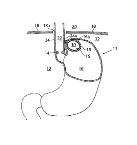
The present invention relates to an apparatus for treating Gastroesophageal
Reflux
.. Disease (GERD).
BACKGROUND
Gastroesophageal Reflux Disease (GERD), or acid reflux disease, is a chronic
condition resulting in mucosal damage in the oesophagus produced by the
recurring
occurrence of acid reflux in the'oesophagus. This is commonly due to transient
or
permanent changes in the barrier between the oesophagus and the stomach. This
can
be due to incompetence of the lower esophageal sphincter (LES), transient LES
relaxation, impaired expulsion of gastric reflux from the esophagus, or a
hiatal
hernia.
Gastroesophageal Reflux Disease can be treated in a number of different ways.
Treatments include, but are not limited to, both medical and surgical
treatments. A
standard surgical treatment, which sometimes is preferred over longtime use of
medication, is Nissen fundoplication surgery, in which the upper curve of the
stomach (the fundus) is wrapped around the LES to strengthen the sphincter and
prevent acid reflux and to repair a hiatal hernia. The procedure is often done
laparoscopically.
Another surgical treatment which has been used is the Anglechik prosthesis, in
which
a device formed like a horseshoe is placed around the oesophagus above the
cardia.
The intended effect is to prevent the cardia from slipping up into the thorax
cavity.
However, this device has a number of complications, including migrating
through
.. and damaging the oesophagus.
From experience with implantation of medical devices, it is known that sutures
between an implanted device and human tissue will not hold over the long term.
For
long term implantation of a device, there are two possibilities to keep the
device in
place. A first solution has been to suture human tissue to human tissue, to
thereby
.. keep the device in place. A second approach has been to provide sutures
holding a
CA 02749792 2011-07-14
WO 2009/096874
PCT/SE2009/000059
2
device in place in the short term and to allow in-growth of human tissue into
the
device for holding the device in place over the long term.
A problem with providing an implantable device associated with the oesophagus
is
that the outer surface of the oesophagus is only comprised of oesophagus
muscle
tissue, which is very easy to damage or migrate through. This is probably one
reason
why the Anglechik prosthesis described above has resulted in many
complications,
such as migration.
The stomach, on the other hand, has a serosa on its outside, thereby providing
a
much stronger membrane for suturing. Thus, suturing a device directly to the
stomach wall provides a better result than suturing an implanted device to the
oesophagus.
Today, there exists a need for a long term treatment of GERD that is more
effective
than prior treatments and which does not result in any severe complications.
SUMMARY OF THE INVENTION
It is an object of the present invention to overcome, or at least reduce, some
of the
problems associated with existing surgical treatments of Gastroesophageal
Reflux
Disease (GERD).
It is another object of the present invention to provide an apparatus for
treating
gastroesophageal reflux disease.
These objects and others are obtained by apparatus described in the appended
claims.
Thus, by providing an apparatus for the treatment of acid reflux disease
including an
implantable movement restriction device having an outer surface that includes
a
biocompatible material, wherein the movement restriction device is adapted to
rest
with at least a part of its outer surface against the patient's stomach fundus
wall, in a
position between the patient's diaphragm and the fundus wall, such that
movement of
the cardiac notch of the patient's stomach towards the patient's diaphragm is
restricted, an apparatus for treating Gastroesophageal Reflux Disease is
obtained.
The movement restriction device has a size of at least 125 mm3 and a
circumference
CA 02749792 2016-07-15
32004-3
3
of at least 15 mm and restricts movement of the cardiac notch of the patient's
stomach
towards the patient's diaphragm thereby preventing the cardia from sliding
through the
patient's diaphragm opening into the patient's thorax, maintaining the
supporting pressure
against the patient's cardia sphincter muscle exerted from the patient's
abdomen. Fixation
device are adapted to secure the movement restriction device in said position.
By adapting the outer surface of the implanted movement restriction device to
rest against the
wall of the fundus, there is a minimal risk of complications, such as
migration of damage to
tissue, because the fundus is less fragile than the oesophagus.
In a first embodiment of the invention, the fixation device comprises sutures
or staples that
attach together portions of the fundus stomach wall that enclose the movement
restriction
device to secure the movement restriction device in said position. I.e., the
movement
restriction device is at least partly placed in an invaginated space. Thus, by
affixing the
implantable movement restriction device indirectly in this manner, no suturing
between the
movement restriction device and tissue is required, which, in turn, further
reduces the risk for
complications. Keeping the movement restriction device in place in this manner
has resulted
in an elastic suspension with improved long term properties.
The fixation device, such as sutures or staples, may attach together portions
of the fundus
stomach wall so at to substantially or completely invaginate the movement
restriction device
from either inside or outside of the patient's stomach wall. Where the
movement restriction
device is placed on the outside of the patient's stomach wall, the movement
restriction device
is invaginated by the fundus stomach wall such that the stomach cavity is
substantially
reduced, by a volume substantially exceeding the volume of the movement
restriction device.
In a second embodiment of the invention, the fixation device comprises an
implantable first
fixation device that attach the movement restriction device in said position
to the fundus wall.
a second fixation device that secures, indirectly or directly, the movement
restriction device to
the oesophagus close to the patient's angle of His, and a third fixation
device that secures,
indirectly or directly, the
CA 02749792 2011-07-14
WO 2009/096874
PCT/SE2009/000059
4
movement restriction device to the patient's diaphragm muscle or associated
muscles. Any of the first, second and third fixation devices may be comprised
of a
plurality of sutures or staples. The first fixation device may comprise a
tissue growth
promoting structure for long term attachment of the movement restriction
device to
the stomach wall. The tissue growth promoting structure may be sutured to the
stomach wall with a relatively large contact surface towards the stomach. The
relatively large surface of the structure, such as a net, will allow for in-
growth of
human tissue for holding the movement restriction device in place over the
long
term. The tissue growth promoting structure may comprise sutures or staples
that
attach the net like structure to the fundus stomach wall.
In addition to invaginating the movement restriction device in accordance with
the
first embodiment of the invention, the second fixation device can be used to
secure,
indirectly or directly, the movement restriction device to the oesophagus
close to the
patient's angle of His, and the third fixation device may be used to secure,
indirectly
or directly, the movement restriction device to the patient's diaphragm muscle
or
associated muscles.
At least a part of the movement restriction device may be made of a material
which
is destructible or not destructible by stomach acid.
The movement restriction device may be inflatable and adapted to be inflated
with a
gel or fluid. A fluid or gel receiving member for receiving fluid to inflate
the
movement restriction device may be provided.
The movement restriction device may include a homogenous material and may be a
solid body.
The movement restriction device may include an enclosure wall defining a
chamber.
The movement restriction device may have a rigid, elastic or flexible outer
wall.
Where the outer wall is rigid, it is rigid enough to maintain non-deformed
when
subject to forces created by stomach movements. Where the movement restriction
device is invaginated, in accordance with the first embodiment described
above, the
CA 02749792 2016-07-15
32004-3
movement restriction device preferably comprises a body adapted to be at least
partly
invaginated by the patient's stomach fundus wall and having an outer surface
that includes a
biocompatible material. A substantial part of the outer surface of the body is
adapted to rest
against the stomach wall in said position between the patient's diaphragm and
the portion of
5 the lower part of the invaginated stomach fundus wall. Suitably, the body
is made of a
material softer than 25 or 15 Shore.
In accordance with a first general design of the body, the body has a maximum
circumference
as seen in a plane perpendicular to an axis through the body.1 he
circumferences of the body
as seen in other planes perpendicular to said axis are equal to the maximum
circumference or
decrease as seen along said axis in the direction from the maximum
circumference. For
example, the body may be substantially egg shaped, spherically shaped, or
substantially
shaped like an egg with an indented middle section or like a bent egg.
In accordance with a second general design of the body, the circumference of
the body as seen
in a plane perpendicular to an axis through the body increases and decreases
at least two times
as the plane is displaced along said axis, or decreases and increases at least
one time as the
plane is displaced along said axis. For example, the body may be substantially
shaped like a
kidney.
Preferably, the body is dimensioned with a size larger than the intestinal
outlet from the
stomach. The body may have a smallest outer diameter of 30 or 40mm or larger
and may have
a smallest outer circumference of 150, 110, 90, 70, 50 or 30 mm.
Suitably, the body has rounded contours without too sharp edges that would be
damaging to
the patient's stomach wall, and has a generally smooth outer surface for
resting against the
fundus wall.
The body is implantable either inside or outside of the patient's stomach and
is adapted to be
attached to the patient's stomach wall by surgery. The body may be
CA 02749792 2011-07-14
WO 2009/096874
PCT/SE2009/000059
6
changeable to assume a slender form having a smaller diameter than that of a
trocar
for laparoscopic use, whereby the body when changed to said slender form can
be
pushed or pulled through the trocar. The body may include a flexible outer
wall
defining a chamber filled with a fluid, such as a gel, allowing the body to
pass
through such a trocar. Alternatively, the body may include an elastic
compressible
material, allowing the body to pass through a trocar.
The body may be hollow and include at least two separate pieces adapted to be
inserted into the hollow body, and further adapted to be put together to one
unitary
piece inside the body, thereby allowing the body to pass through a trocar for
laparoscopic use. Alternatively, the body may include an outer wall and a
hollow
compressed inner part, for being filled with a fluid or gel after insertion
into the
patient's body.
The body may include a chamber with an injection port, wherein the chamber of
the
body is filled with a fluid through the injection port.
The body may include at least one holding device adapted to be used for
pushing or
pulling the body through a trocar for laparoscopic use. The holding device is
adapted
to hold a prolongation of the body that is adapted to be held by a surgical
instrument.
More specifically, the holding device is adapted to hold a thread or band
inserted
through the holding device. Where the body comprises an outer wall the holding
device is at least partly placed inside the outer wall of the body.
In an advantageous embodiment, the body is adjustable in size and invaginated
in the
patient's fundus stomach wall. As a result, the body stretches the patient's
stomach
fundus wall when the size thereof is increased, thereby creating satiety in a
patient
also suffering from obesity. At least two implantable adjustable stretching
devices
may be provided to stretch different parts of the patient's stomach wall, to
thereby
treat obesity by efficiently affecting the patient's appetite. The two
stretching devices
are suitably regulated from outside of the patient's body, whereby a first of
the
stretching devices is regulated at a first time to stretch a first part of the
patient's
CA 02749792 2011-07-14
WO 2009/096874
PCT/SE2009/000059
7
stomach wall and a second of the stretching devices is regulated at a second
time to
stretch a second part of the patient's stomach wall.
The stretching device may be hydraulically regulated. In this case, a
subcutaneously
implantable hydraulic reservoir connected to the hydraulic regulated
stretching -
device may be provided, whereby the hydraulic regulated stretching device is
non-
invasively regulated by manually pressing the hydraulic reservoir. Further,
the
movement restriction device suitably includes an inflatable body, and a pump
and a
chamber in fluid contact with the body are provided, wherein the pump
regulates the
hydraulic reservoir by pumping fluid or air from the body to the chamber.
The apparatus may include an implantable stimulation device that sends out
stimulation pulses to the cardia muscle to stimulate the cardia muscle and
thereby
further close the cardia to additionally prevent reflux disease. The
stimulation device
is comprised of at least one conductor and at least one electrode that
receives the
stimulation pulses and applies them to the cardia muscle to thereby stimulate
the
cardia muscle. The at least one electrode may also be kept in place by the
stomach-
oesophagal sutures or invagination in the stomach wall. The stimulation pulses
may
be sent as a train of pulses, wherein the pulse train is repeated with a time
break in
between, the break extending the break between each pulse in the pulse train.
The
stimulation device may include an electronic circuit and an energy source
preferably
adapted to incorporate the electronic circuit and energy source. In one
embodiment,
the stimulation of the cardia with the stimulation device is made with energy
pulses
to increase the sphincter tonus so that the cardia completely closes and a
control
device for controlling the stimulation device is operable by the patient to
set the
stimulation device into operation, in which operational state the stimulation
device
continuously alternates at a time when the patient does not swallow between an
operation mode in which the cardia sphincter is stimulated with said energy
pulses
and a rest mode in which the cardia is not stimulated.
The stimulation device preferably comprises at least one sensor for sensing a
physical parameter of the patient or a functional parameter of the movement
restriction device and an internal control unit for controlling the
stimulation device.
CA 02749792 2011-07-14
WO 2009/096874
PCT/SE2009/000059
8
Normally, the internal control unit controls the stimulation device in
response to
information from the sensor.
A sensor sensing a contraction wave of the oesophagus, or any other parameter
correlated to food intake, sends the information to the internal control unit
and the
internal control unit then ceases the stimulation in response to such
information from
the sensor.
The stimulation device may, at any time, be controlled by the patient.
The present invention further relates to abdominal surgical methods of
treating a
reflux disease.
According to a first method a reflux disease in a patient is treated by
implanting a
movement restriction device that, when implanted in a patient, restricts the
movement of the stomach notch in relation to the diaphragm muscle preventing
the
cardia to slide up through the diaphragm hiatus opening. The method comprises
the
steps of inserting a needle or a tube like instrument into the abdomen of the
patient's
body; using the needle or tube like instrument to fill the patient's abdomen
with gas;
placing at least two laparoscopic trocars in the patient's body; inserting a
camera
through one of the laparoscopic trocars into the patient's abdomen, inserting
at least
one dissecting tool through one of said at least two laparoscopic trocars;
dissecting
an area of the stomach; introducing the device into the abdominal cavity;
placing the
device on the outside of the stomach fundus wall; and creating a pouch in the
stomach fundus wall for the device; and in-vaginating the device in the pouch
by
providing sutures or staples to the stomach fundus wall, thereby preventing
the cardia
from sliding through the patient's diaphragm opening into the patient's
thorax, so as
to maintain the pressure support from the patient's abdomen that supports the
patient's cardia sphincter muscle.
A second abdominal method of treating a reflux disease for the same purpose
uses
the initial steps as the first method comprises creating a hole in the stomach
fundus
wall; introducing a movement restriction device into the abdominal cavity;
introducing the device through the hole and into the stomach; placing the
device on
CA 02749792 2011-07-14
WO 2009/096874
PCT/SE2009/000059
9
the inside of the stomach fundus wall; creating a pouch on the outside of the
stomach
cavity for the device placed on the inside of the stomach fundus wall, and in-
vaginating the device in the pouch by providing sutures or staples to the
stomach
fundus wall, preventing the cardia from sliding through the patient's
diaphragm
opening into the patient's thorax, so as to maintain the supporting pressure
from the
patient's abdomen that supports the patient's cardia sphincter muscle.
A third abdominal method of treating a reflux disease in a patient includes
implanting a movement restriction device for the same purpose as previously
disclosed methods and comprises the steps of surgically incising an opening in
the
patient's abdominal wall; dissecting an area of the patient's stomach;
introducing
the movement restriction device through the abdominal incision; and attaching
the
device to the stomach fundus wall, thereby preventing the cardia from sliding
through the patient's diaphragm opening into the patient's thorax, so as to
maintain
the supporting pressure from the patient's abdomen that supports the patient's
cardia
sphincter muscle. According to first alternative, the method includes placing
the
device on the outside of the stomach fundus wall; creating a pouch in the
stomach
fundus wall for the device; and in-vaginating the device in the pouch by
providing
sutures or staples to the stomach fundus wall, thereby preventing the cardia
from
sliding through the patient's diaphragm opening into the patient's thorax, so
as to
maintain the supporting pressure from the patient's abdomen that supports the
patient's cardia sphincter muscle. According to a second alternative, the
method
includes creating a hole in the stomach fundus wall; introducing the movement
restriction device through the hole and into the stomach; placing the device
on the
inside of the stomach fundus wall; creating a pouch on the stomach fundus wall
for
the device, and in-vaginating the device in the pouch by providing sutures or
staples
to the stomach fundus wall, preventing the cardia from sliding through the
patient's
diaphragm opening into the patient's thorax, so as to maintain the supporting
pressure from the patient's abdomen that supports the patient's cardia
sphincter
muscle.
The methods further comprise affixing the device to the stomach fundus wall by
providing sutures or staples and/or affixing the stomach fundus wall to the
lower part
CA 02749792 2011-07-14
WO 2009/096874
PCT/SE2009/000059
of the patient's esophagus by providing sutures or staples; and/or affixing
the
stomach fundus wall to the patient's diaphragm muscle or associated muscles.
The
methods can further comprise the provision of an apparatus for regulating the
reflux
treatment device from the outside of the patient's body; and operating said
apparatus
5 to regulate the reflux treatment device. The regulation of the reflux
treatment device
can include changing the volume of the filling body when implanted. For this
purpose, the methods can include the provision of an injection type syringe
comprising a fluid for injection into an implanted filling body; and injecting
volume
of fluid into filling body. Preferably, the methods comprise enclosing the
device in
10 the pouch. In one embodiment, the method admits the pouch being at least
partly
open, whereby the pouch can exhibit only one opening, or the pouch can exhibit
two
openings and to extend non-circumferentially around the stomach. It is
generally
preferable that the volume of the pouch is more than 15 millilitres
A further laparoscopic abdominal method of treating a reflux disease comprises
inserting a needle or a tube like instrument into the abdomen of the patient's
body;
using the needle or tube like instrument to fill the patient's abdomen with
gas;
placing at least two laparoscopic trocars in the patient's body; inserting a
camera
through one of the laparoscopic trocars into the patient's abdomen; inserting
at least
one dissecting tool through one of said at least two laparoscopic trocars;
dissecting
an area of the stomach; creating a pouch from the stomach fundus wall for the
device; closing the pouch by providing sutures and staples; introducing a
injecting
member comprising an injectable filling material; and injecting the filling
material
into the pouch, thereby creating a filling body that, fills a volume in the
patient's
abdomen that is close to and above the patient's cardia when the patient is in
a
standing position in order to prevent the cardia from sliding through the
patient's
diaphragm opening into the patient's thorax, so as to maintain pressure in the
patient's abdomen supporting the patient's cardia sphincter muscle.
A further surgical abdominal method of treating a reflux disease comprises
cutting an
opening in the skin to enter the patients abdomen dissecting an area of the
stomach;
creating a pouch from the stomach fundus wall for the device; closing the
pouch by
providing sutures and staples; introducing a injecting member comprising an
CA 02749792 2011-07-14
WO 2009/096874
PCT/SE2009/000059
11
injectable filling material; and injecting the filling material into the
pouch, thereby
creating a filling body that, fills a volume in the patient's abdomen that is
close to
and above the patient's cardia when the patient is in a standing position in
order to
prevent the cardia from sliding through the patient's diaphragm opening into
the
patient's thorax, so as to maintain pressure in the patient's abdomen
supporting the
patient's cardia sphincter muscle.
The recited further methods can include creating the pouch on the outside of
the
stomach fundus wall, with the filling body placed against the inside of the
stomach
fundus wall, or alternatively, the methods include creating a hole in the
stomach
fundus wall and the pouch is created on the inside of the stomach fundus wall,
with
the filling body placed against the outside of the stomach fundus wall. The
recited
further methods preferably also include affixing the stomach fundus wall to
the lower
part of the patient's esophagus by providing sutures or staples and/or
affixing the
stomach fundus wall to the patient's diaphragm muscle or associated muscles.
It is
also generally preferable that the volume of the pouch is more than 15
millilitres. The
filling material is preferably capable of undergoing a curing process from
fluid
material to a semi-solid or solid material. Such a curing process is
preferably
triggered by an increase in temperature from ambient temperature to body
temperature. A suitable such material, well-known to persons skilled in the
art, is a
thermocurable polysiloxane which (in the presence of a crosslinker and a
catalyst)
can undergo a crosslinking reaction under the influence of heat.
A still further laparoscopic abdominal method of treating a reflux disease in
a patient
by implanting a movement restriction device that, when implanted in a patient,
restricts the movement of the stomach notch in relation to the diaphragm
muscle
preventing the cardia to slide up through the diaphragm hiatus opening,
comprises
the steps of inserting a needle or a tube like instrument into the abdomen of
the
patient's body; using the needle or tube like instrument to fill the patient's
abdomen
with gas; placing at least two laparoscopic trocars in the patient's body;
inserting a
camera through one of the laparoscopic trocars into the patient's abdomen;
inserting
at least one dissecting tool through one of said at least two laparoscopic
trocars;
dissecting an area of the stomach; creating a hole in the stomach fundus wall;
CA 02749792 2011-07-14
WO 2009/096874
PCT/SE2009/000059
12
introducing a movement restriction device into the abdominal cavity;
introducing the
device through the hole and into the stomach; placing the device on the
outside of the
stomach fundus wall; fixating the device placed on the outside of the stomach
fundus
wall, and preventing the cardia from sliding through the patient's diaphragm
opening
into the patient's thorax, so as to maintain the supporting pressure from the
patient's
abdomen that supports the patient's cardia sphincter muscle. The method can
further
comprise the step of affixing the device to the stomach fundus wall by
providing
sutures or staples.
The present invention also relates to a laparoscopic instrument for providing
a
movement restriction device to be invaginated in the stomach fundus wall of a
human patient to treat reflux disease, suitable for use with any of the
mentioned
laparoscopic methods. The instrument generally comprises an elongated member
having a proximal end and a distal end, the elongated member having a diameter
less
than that of a laparoscopic trocar to be introduced into the patients abdomen
during a
laparoscopic operation; a stomach pushing device for pushing the stomach
fundus
wall to create a tube like shaped portion of the stomach fundus wall
protruding into
the normal stomach cavity, said pushing device comprising the movement
restriction
device to be invaginated by the stomach fundus wall in the tube like shaped
portion
thereof. The pushing device comprises a vacuum sucking device to suck the
stomach
fundus to assist the instrument in forming the tube like shaped portion of the
stomach
fundus wall together with the pushing device. The vacuum sucking device
comprises a vacuum passageway leading from the proximal to the distal end of
the
instrument and at the end portion of the instrument, which includes the
pushing
device. The vacuum passageway is divided up in multiple small openings adapted
to
suck the stomach wall portion to become adherent to the pushing device to
further
form the tube like stomach wall portion. The instrument further comprises an
insertion device adapted to introduce the movement restriction device into the
tube
like shaped stomach portion. The instrument can further comprise at least one
clamping device for holding the opening of the tube like portion substantially
closed
by clamping together stomach to stomach in said opening, wherein the
instrument is
adapted to place the at least one clamping device at the opening in such a way
that it
allows later suturing of the opening. The instrument can further comprise an
inflation
CA 02749792 2011-07-14
WO 2009/096874
PCT/SE2009/000059
13
device for inflating the movement restriction device before or after the
suturing. The
instrument can further comprise a suturing device adapted to suture the
opening of
the tube like portion with stomach to stomach sutures for creating at least
partly a
closed space enclosing the movement restriction device, wherein the instrument
is
adapted to be withdrawn leaving the movement restriction device at least
partly
invaginated in the stomach fundus wall. The suturing device can comprise a
first and
second suture positioning member provided on the elongated member to be
located
in the stomach at the distal end thereof. The instrument further comprises an
operation device adapted to adjust the first and second suturing member in a
position
.. in which the first and second suture positioning members are in front of
each other
with the stomach wall on both sides of the open end of the cup like portion,
and
adapted to suture the open end of the cup like portion of the fundus wall with
a row
of stomach to stomach sutures. The suturing device preferably comprises an
operable
reloadable multi-suturing device, which is reloadable with sutures from
outside of
the patient's body and which is adapted to suture the open end of the cup like
portion
of the fundus wall with said row of stomach to stomach sutures, wherein the
row of
sutures comprises two or more sutures or staples to be sutured simultaneously.
The
suturing device can also comprise multiple sutures for suturing two or more
sutures
simultaneously.
The present invention also relate an intraluminar method of treating a reflux
disease
in a patient by implanting a device comprising an implantable movement
restriction
device that, when implanted in a patient, restricts the movement of the
stomach notch
in relation to the diaphragm muscle preventing the cardia to slide up through
the
diaphragm hiatus opening. The method comprises the steps of introducing a
gastroseope in the esophagus and into the stomach of the patient; introducing
an
instrument in the esophagus and into the stomach of the patient, said
instrument
being integrated in said gastroscope or separate from the same; providing, by
means
of said of instrument, a pouch from the stomach fundus wall for accommodating
the
device and invaginating the device in the pouch with sutures or stables to the
stomach fundus wall, thereby preventing the cardia from sliding through the
patient's
diaphragm opening into the patient's thorax, so as to maintain the supporting
pressure against the patient's cardia sphincter muscle exerted from the
patient's
CA 02749792 2011-07-14
WO 2009/096874
PCT/SE2009/000059
14
abdomen. In a first alternative, the method comprises the steps of introducing
the
device into the stomach by means of the instrument; placing the device on the
inside
of the stomach fundus wall, using said instrument; creating, by means of said
instrument, a pouch in a portion of the stomach fundus wall on the outside of
the
stomach cavity, the device placed resting against the inside of the stomach
fundus
wall; and invaginating the device in the pouch by with sutures or stables to
the
stomach fundus wall. In a second alternative, the method comprises the steps
of
creating, by means of said instrument, a pouch of a portion of the stomach
fundus
wall; introducing the device by means of the instrument into the pouch; and
invaginating the device with sutures or stables to the stomach fundus wall.
According
to this alternative, the method comprises inflating the device to its filling
volume,
preferably by injecting a filling fluid into the device so it obtains its
filling volume.
The filling fluid can thereby be a curable fluid of the characteristics
earlier describes,
such as the aforedescribed thermocurable polysiloxanes. In a third
alternative, the
method comprises the steps of creating a hole in the stomach fundus wall;
introducing the device into the stomach by means of the instrument; moving the
device through the hole and placing it on the outside of the stomach fundus
wall;
creating, by means of said instrument, a pouch of a portion of the stomach
fundus
wall on the inside of the stomach cavity, with the device placed against the
outside of
the stomach fundus wall; invaginating the device in the pouch with sutures or
stables
to the stomach fundus wall; and sealing the hole with sutures or staples.. In
a fourth
alternative, the method comprises the steps of creating a hole in the stomach
fundus
wall; creating, by means of said instrument, a pouch of a portion of the
stomach
fundus wall on the inside of the stomach cavity; introducing the device into
the
stomach by means of the instrument; moving the device through the hole and
placing
it on the outside of the stomach fundus wall; introducing the device by means
of the
instrument into the pouch; invaginating the device with sutures or stables to
the
stomach fundus wall; and sealing the hole with sutures or staples. Also this
method
can comprise inflating the device to its filling volume, preferably by
injecting a
filling fluid into the device so it obtains its filling volume. The filling
fluid can have
all the previously described characteristics. The method also comprises
affixing the
device to the stomach fundus wall by providing sutures or staples; and/or
affixing the
CA 02749792 2011-07-14
WO 2009/096874
PCT/SE2009/000059
stomach fundus wall to the lower part of the patient's oesophagus by providing
sutures or staples; and/or affixing the stomach fundus wall to the patient's
diaphragm
muscle or associated muscles. The method can also comprise the provision of an
apparatus for regulating the reflux treatment device from the outside of the
patient's
5 body; and operating said apparatus to regulate the reflux treatment
device.
Regulation of the reflux treatment device preferably includes changing the
volume of
the filling body when implanted. For this purpose, the method can comprise the
provision of an injection type syringe comprising a fluid for injection into
an
implanted filling body; and injecting volume of fluid into filling body. The
filling
10 fluid can be curable fluid of a nature as discussed in previous
sections. The pouch
created by the method can enclose the movement restriction device, or it can
be at
least partially open, in one example it has only one opening, and according to
another
example it exhibits two openings and extends non-circumferentially around the
stomach. Preferably, the volume of the pouch is more than 15 millilitres. It
is
15 generally preferably in the method that the gastroscope and the
instrument are
integrated. The method can further include inflating the stomach with gas. In
a
special embodiment of the method, the instrument generates vacuum when
providing
the pouch from the stomach fundus wall.
The invention also relates to a method of restoring the location of the cardia
and the
fundus in a patient suffering from a reflux disease comprising introducing an
elongated instrument having at least one flexible part into oesophagus of a
patient;
activating a holding device by the instrument, said activated holding device
having
larger crossectional area than said instrument; holding the distal esophagus
or
stomach with said holding device; moving and pushing the instrument in a
distal
direction so the cardia and the stomach wall, or part of fundus, incorrectly
located
above diaphragm, or its associated muscles, slide back in a position below the
diaphragm, or its associated muscles. In a first alternative, the method
comprises
expanding the holding device radially above the cardia in the oesophagus and
using
the device to push the cardia and the stomach wall or part of fundus below the
diaphragm or its associated muscles. In a second alternative, the method
comprises
releasing a balloon member at the proximal end of the instrument in the lower
part of
the stomach, and using the member to push the instrument against a lower wall
part
CA 02749792 2011-07-14
WO 2009/096874
PCT/SE2009/000059
16
of the stomach so the cardia and the fundus or part of fundus slide below the
diaphragm or its associated muscles. In a third alternative, the method
comprises
locating the distal end of the instrument at the level of the diaphragm or its
associated
muscles; expanding the member in radial direction; attaching the member to the
stomach wall; and pushing the instrument in a distal direction so the cardia
and the
stomach wall or, part of fundus, slide below the diaphragm or its associated
muscles.
The method according to all recited alternatives can comprise the employment
of an
affixing member as part of the holding device in the distal part of the
instrument
capable providing sutures or stables or other invasive mechanical members for
affixing the esophagus and stomach wall to the instrument, preferably, the
method
comprises the employment of a suturing member in the distal part of the
instrument
capable providing sutures or stables for suturing the stomach fundus wall to
the
lower part of the esophagus above said cardia,
The invention is also directed to a surgical gastroscopic instrument for
treating a
patient suffering from hiatal hernia, wherein a portion of the patient's
stomach passes
through the hiatus of the diaphragm muscle with the patient's cardia placed
above the
diaphragm muscle in the thorax. The instrument comprises: (i) an elongated
member
having a proximal end and a distal end, said elongated member having a
diameter
less than that of the patient's esophagus and being flexible, thereby allowing
introducing the flexible elongated member with its distal end first from the
throat
into the esophagus; (ii) a holding device secured to the elongated member and
operable between an activated state, in which it is adapted to engage and hold
the
patient's esophagus or stomach, and an inactivated state, in which it is
adapted to be
released from the esophagus or stomach; (iii) an operation device for
operating the
holding device to shift between said activated and inactivated states from
outside the
patient's body; and (iv) a handle connected to the elongated member at the
proximal
end thereof to be held manually for moving the elongated member distally,
wherein
the holding device, when operated by the operation device in its activated
state, is
adapted to engage and hold the esophagus or stomach strong enough to allow the
elongated member, when manually moved, to move and reintroduce the cardia back
in the distal direction to a position below the diaphragm muscle. The operable
holding device of the instrument is preferably adapted to radially expand
relative to
CA 02749792 2011-07-14
WO 2009/096874
PCT/SE2009/000059
17
the elongated member from said inactivated state to said activated state, such
that
said holding device, when radially expanded, engages and holds the stomach or
esophagus by force and friction. Alternatively, the operable holding device
comprises at least one introducing member adapted to invasively introduce into
the
wall of the stomach or esophagus to secure the holding device on the esophagus
or
the stomach, when the holding device is in its activated state. In both
alternatives, the
operable holding device is adapted to engage and hold the esophagus proximal
to the
cardia or at the cardia, or to engage and hold the stomach at a position in
the hiatus or
distal thereto, when the cardia is above the diaphragm muscle. According to
another
alternative, the elongated member of the instrument comprises a first suture
positioning member, which is situated in the esophagus when the holding device
is in
its activated state holding the esophagus or stomach, and a second suture
positioning
member at the distal end of the elongated member. The operation device of the
instrument is adapted to bend the flexible elongated member around the stomach
notch into a position in which the first suture positioning member is above
the cardia
and the first and second suture positioning members are in front of each other
with
the fundus wall and esophagus wall moved together by the first and second
suture
positioning members. The instrument further comprises a reloadable multi-
suturing
device for suturing together the patient's esophagus proximal to the cardia
with the
fundus stomach wall by a row of sutures, where the fundus wall and esophagus
wall
are moved together by the first and second suture positioning members, said
row of
sutures comprising two or more sutures to be sutured simultaneously by the
multi-
suturing device.
The invention further comprises another embodiment of a surgical gastroscopic
instrument for treating a human patient suffering from hiatal hernia, This
instrument
comprises: (i) an elongated member having a proximal end and a distal end,
said
elongated member having a diameter less than that of the patient's esophagus
and
being flexible, thereby allowing introducing the flexible elongated member
with its
distal end first from the patient's throat into the esophagus;
(ii) a first suture positioning member provided on the elongated member such
that it
is situated in the esophagus proximal to the cardia when the elongated member
has
been introduced in the esophagus with the distal end of the elongated member
CA 02749792 2011-07-14
WO 2009/096874
PCT/SE2009/000059
18
situated in the stomach; (iii) a second suture positioning member provided on
the
elongated member at the distal end thereof; (iv) an operation device adapted
to bend
the flexible elongated member around the stomach notch into a position in
which the
first and second suture positioning members are in front of each other with
the
fundus wall and esophagus wall moved together by the first and second suture
positioning members; and (v) a re-loadable multi-suturing device for suturing
together the patient's esophagus proximal to the cardia with the fundus
stomach wall
by a row of sutures, where the fundus wall and esophagus wall are moved
together
by the first and second suture positioning members, the multi-suturing device
being
reloadable with sutures from outside the body to apply further rows of sutures
in
front or back of the first row of sutures, wherein said row of sutures
comprises two
or more sutures to be sutured simultaneously by the multi-suturing device. The
instrument further comprises a holding device secured to the elongated member
and
operable between an activated state, in which it is adapted to engage and hold
the
patient's esophagus or stomach, and an inactivated state, in which it is
adapted to be
released from the esophagus or stomach. The holding device is operable by the
operation device to shift between said activated and inactivated states from
outside
the patient's body, and a handle connected to the elongated member at the
proximal
end thereof to be held manually for moving the elongated member distally,
wherein
the holding device, when operated by the operation device in said activated
state, is
adapted to engage and hold the esophagus or stomach strong enough to allow the
elongated member, when manually moved, to move and reintroduce the cardia back
in the distal direction to a position below the diaphragm muscle. In one
alternative,
the operable holding device is adapted to radially expand relative to the
elongated
member from said inactivated state to said activated state, such that the
holding
device, when radially expanded, engages and holds the stomach or esophagus by
force and friction. In another alternative, the operable holding device
comprises at
least one introducing member adapted to invasively introduce into the wall of
the
stomach or esophagus to secure the holding device on the esophagus or the
stomach,
when the holding device is in its activated state. In both alternatives, the
operable
holding device is adapted to engage and hold the esophagus proximal to the
cardia or
CA 02749792 2011-07-14
WO 2009/096874
PCT/SE2009/000059
19
at the cardia, or to engage and hold the stomach at a position in the hiatus
or distal
thereto, when the cardia is above the diaphragm muscle.
The invention further comprises still another embodiment of a surgical
gastroscopic
instrument for providing a movement restriction device to be invaginated in
the
stomach fundus wall of a human patient to treat reflux disease. The instrument
comprises (i) an elongated member having a proximal end and a distal end, the
elongated member having a diameter less than that of the patient's esophagus
and
being flexible, thereby allowing introduction of the flexible elongated member
with
its distal end first through the patient's throat, esophagus and into the
stomach to the
fundus wall; (ii) an operable stomach penetration device provided the
elongated
member at the distal end thereof for penetrating the stomach fundus wall to
create a
hole in the stomach fundus wall, to allow introduction of the elongated member
through the hole; (iii) an operable special holding device provided on the
elongated
member proximal to the penetration device, when penetrating said stomach wall,
to
hold the elongated member in a position in which the elongated member extends
through the stomach fundus wall and is prevented from moving through the hole
in
the proximal direction, wherein the special holding device includes an
expandable
member expandable at least radially substantially perpendicular to the
elongated
member to abut against the fundus wall on the outside thereof; and
(iv) an insertion device for inserting the movement restriction device through
the
hole in the stomach fundus wall to the outside thereof to be invaginated in
the fundus
wall. The instrument can further comprise a forming device provided on the
elongated member proximal to the special holding device to abut against the
fundus
wall on the inside thereof. The forming device together with the special
holding
device is adapted to form the stomach fundus wall in a cup like shape, whereby
the
special holding device is retractable relative to the forming device to pull
the
stomach wall against the forming device to form said cup like shaped portion
of the
stomach. The instrument can further comprise a suturing device adapted to
suture the
open end of the cup like portion of the fundus wall with stomach to stomach
sutures
to create a space that is at least in part enclosed by a portion of the fundus
wall. The
suturing device preferably comprises multiple sutures for suturing two or more
CA 02749792 2011-07-14
WO 2009/096874
PCT/SE2009/000059
sutures simultaneously. The suturing device is adapted to suture the open end
of the
cup like portion of the fundus wall before the movement restriction device is
inserted
by the insertion device through the hole of the fundus wall. The instrument
can
further comprise an inflation device for inflating the movement restriction
device
5 after being introduced by the insertion device through the hole of the
fundus wall, or
for inflating the movement restriction device after being introduced by the
movement
restriction device introduced through the hole of the fundus wall. The
suturing device
can comprise an operable reloadable multi-suturing device, which is reloadable
with
sutures from outside the patient's body and which is adapted to suture the
open end
10 of the cup like portion of the fundus wall with a row of stomach to
stomach sutures,
wherein the row of sutures comprises two or more sutures or staples to be
sutured
simultaneously. In another alternative, the instrument comprises an inflatable
movement restriction device, the penetration device comprises a wire adapted
to be
introduced through the hole in the stomach fundus wall and to be advanced at
least
15 up to the abdominal wall or be passed therethrough. The wire serves as a
guide for a
hydraulic tube, which is connected to the inflatable movement restriction
device and
which is connectable to an injection port to be placed subcutaneously for
filling the
inflatable movement restriction device with a fluid and adjusting the amount
thereof,
when the movement restriction device has been inserted by the insertion device
20 through the hole of the fundus wall.
The invention further comprises still yet another embodiment of a surgical
gastroscopic instrument for providing an inflatable movement restriction
device to be
invaginated in the stomach fundus wall of a human patient to treat reflux
disease.
The instrument comprises: (i) an elongated member having a proximal end and a
distal end, the elongated member having a diameter less than that of the
patient's
esophagus and being flexible, thereby allowing introduction of the flexible
elongated
member with its distal end first through the patient's throat, esophagus and
into the
stomach to the fundus wall; and (ii) an operable stomach penetration device
provided
on the elongated member at the distal en thereof for penetrating the stomach
fundus
wall to create a hole in the stomach fundus wall, to allow introduction of the
elongated member through the hole, wherein the penetration device includes a
wire
CA 02749792 2011-07-14
WO 2009/096874
PCT/SE2009/000059
21
to be introduced through the hole in the stomach fundus wall and to be
advanced at
least up to the abdominal wall or be passed therethrough, said wire serving as
a guide
for a hydraulic tube, which is connected to the inflatable movement
restriction device
and which is connectable to an injection port to be placed subcutaneously for
filling
the inflatable movement restriction device with a fluid and adjusting the
amount
thereof, and wherein at least one of the wire and tube can be pulled to move
the
movement restriction device inflated with fluid towards the fundus stomach
wall to
be placed on the inside of the stomach fundus wall where the movement
restriction
device is to be invaginated in the fundus wall. Preferably, at least one of
the wire and
tube when pulled to move the inflated movement restriction device towards the
fundus stomach wall allow a portion of the stomach fundus wall to move to form
a
cup like shaped portion of the stomach protruding out from the normal stomach
cavity. The instrument can further comprise an operable forming device having
a
cup like shape for forming the cup like shaped portion of the stomach. The
instrument can further comprise a suturing device adapted to suture the open
end of
the cup like portion of the fundus wall with stomach to stomach sutures to
create a
space that is at least in part enclosed by a portion of the fundus wall. The
suturing
device can comprises multiple sutures for suturing two or more sutures
simultaneously. The suturing device can be adapted to suture the open end of
the
cup like portion of the fundus wall before the movement restriction device is
inserted
through the hole of the fundus wall. The movement restriction device,
preferably is
inflatable, further comprising an inflation device for inflating the movement
restriction device after being introduced through the hole of the fundus wall.
The
instrument can further comprise a first and second suture positioning member
provided on the elongated member situated in the stomach at the distal end
thereof,
and an operation device adapted to adjust the first and second suturing member
in a
position in which the first and second suture positioning members are in front
of each
other with the stomach wall on both sides of the open end of the cup like
portion, and
adapted to suture the open end of the cup like portion of the fundus wall with
a row
of stomach to stomach sutures. The suturing device can comprise an operable re-
loadable multi-suturing device, which is reloadable with sutures from outside
the
patient's body and which is adapted to suture the open end of the cup like
portion of
CA 02749792 2011-07-14
WO 2009/096874
PCT/SE2009/000059
22
the fundus wall with said row of stomach to stomach sutures, wherein the row
of
sutures comprises two or more sutures or staples to be sutured simultaneously.
The invention further comprises still yet another embodiment of a surgical
gastroscopic instrument providing a movement restriction device to be
invaginated in
the stomach fundus wall of a human patient to treat reflux disease. The
instrument
comprising: (i) an elongated member having a proximal end and a distal end,
the
elongated member having a diameter less than that of the patient's esophagus
and
being flexible such that introduction of the flexible elongated member with
its distal
end first through the patient's throat, esophagus and into the stomach to the
fundus
wall is allowed; (ii) an operable stomach pushing device for pushing the
stomach
fundus wall to create a cup like shaped portion of the stomach fundus wall
protruding out from the normal stomach cavity, said pushing device including
the
movement restriction device to be invaginated by the stomach fundus wall in
the cup
like shaped portion thereof; and (iii) a suturing device adapted to suture the
opening
of the cup like shaped portion of the stomach fundus wall with stomach to
stomach
sutures to enclose at least in part the movement restriction device. The
instrument
further can comprise a forming device provided on the elongated member
proximal
to the pushing device to pull the fundus wall on the inside thereof. The
forming
device together with the pushing device is adapted to form the stomach fundus
wall
in an optimal cup like shape, wherein the pushing device is pushed to form
said cup
like shaped portion of the stomach. The instrument can further comprise first
and
second suture positioning member provided on the elongated member situated in
the
stomach at the distal end thereof; and an operation device adapted to adjust
the first
and second suturing member in a position in which said first and second suture
positioning members are in front of each other with the stomach wall on both
sides of
the open end of the cup like portion, and adapted to suture the open end of
the cup
like portion of the fundus wall with a row of stomach to stomach sutures. The
suturing device can comprise an operable re-loadable multi-suturing device,
which is
reloadable with sutures from outside the patient's body and which is adapted
to
suture the open end of the cup like portion of the fundus wall with said row
of
stomach to stomach sutures, wherein the row of sutures comprises two or more
sutures or staples to be sutured simultaneously. The suturing device can also
CA 02749792 2016-07-15
32004-3
23
comprise multiple sutures for suturing two or more sutures simultaneously. The
instrument
can further comprise an inflating device for inflating the movement
restriction device after the
suturing. The forming device can preferably comprise a vacuum sucking device
to suck the
stomach fundus to help the instrument to form the cup like shaped portion of
the stomach
fundus wall together with the pushing device.
The embodied gastroscopic instruments as described in previous section can
comprise an
optical device for examining the inside the esophagus or the stomach. For this
purpose, the
instruments can further comprise electrical wires extending along the
elongated member, and
the optical device comprises a camera placed distally on the elongated member
and connected
to the wires, which lead out from the patient's body for external exposure of
images from the
camera. The instruments can further comprise a light source placed distally on
the elongated
member for illuminating the inside of the esophagus or stomach. The optical
device can
suitably comprise optical fibers placed along the elongated member and leading
out from the
patient's body for external examination of the inside of the esophagus or
stomach.
The present invention further relates to an apparatus for treating a reflux
disease and obesity.
This apparatus comprises a movement restriction device and the fixation
devices, adjustment
device, wireless remote control function, wireless energy transmitter and
further features as
described earlier with an apparatus for treating a reflux disease. In addition
the apparatus for
combined treatment of a reflux disease comprises at least one operable
stretching device that,
when implanted in the patient, stretches a part of the patient's stomach wall,
to thereby treat
obesity by affecting the patient's appetite; and an operation device for
operating the stretching
device when implanted to stretch the stomach wall portion such that satiety is
created. The
stretching device may be kept in contact with the stomach wall by stomach-to-
stomach sutures
or staples, in a position in which the stretching device is capable of
stretching the stomach
wall. Specifically, the stretching device may be invaginated by the stomach
wall by means of
stomach-to-stomach sutures or staples. The stretching device may be adapted to
be placed in
the stomach cavity. To this end, the stretching device may be adapted to be
inserted into the
stomach cavity via a gastroscope or intraluminar instrument, and be adapted to
be attached to
the stomach wall by surgery. Alternatively, the stretching device may be
adapted to be placed
on the outside of the stomach. In an embodiment, the stretching device
comprises a first
CA 02749792 2016-07-15
32004-3
24
engaging member adapted to engage a first part of the stomach wall and a
second engaging
member adapted to engage a second part of the stomach wall close to but spaced
from the first
stomach part. The operation device is adapted to operate the first and second
engaging
member to move away from each other to stretch the stomach wall portion
between the first
and second parts of the stomach such that satiety is created. At least one of
the first and
second engaging members may be adapted to at least in part be invaginated by
the stomach
wall by stomach-to-stomach sutures or staples holding the engaging member in
place. In
addition, at least one of the first and second engaging members may be adapted
to be kept in
place by sutures or staples between the engaging member and the stomach wall.
Suitably, at
least one of the first and second engaging members comprises a tissue growth
promoting
structure, preferably a net like structure, adapted to be in contact with the
stomach wall to secure
long term attachment of the stretching device to the stomach wall. In another
embodiment, the
stretching device comprises at least one expandable body adapted to be
invaginated by a portion
of the patient's stomach wall, and the operation device comprises a fluid
reservoir, which is in
fluid communication with a chamber of the body. The operation device is non-
invasively
operable to distribute fluid from the fluid reservoir to the chamber of the
body to expand the
body such that the stomach wall portion is stretched, when the body is
invaginated. The fluid
reservoir may be operated by manually pressing it. The operation device may
comprise a
reverse servo, wherein a small volume of fluid in the fluid reservoir is
compressed with a
higher force and the chamber of the body creates a movement of a larger total
volume with
less force per unit of volume. The fluid reservoir may be placed
subcutaneously or in the
abdomen, and may be regulated by moving a wall of the reservoir, for example
by a motor.
Alternatively, a pump may be provided for pumping fluid or air from the
reservoir to the
body's chamber. The term "reversed servo means" encompasses the definition of
an device
that is controlled with a higher force and a small stroke i.e. for example
movement of a small
amount of fluid with a high force controls a larger amount of fluid moving by
means of very
smaller force, but may alternatively or additionally encompass the definition
of a mechanism
that transfers a strong force acting on a moving element having a short stroke
into a small
CA 02749792 2011-07-14
WO 2009/096874
PCT/SE2009/000059
force acting on another moving element having a long stroke. The reversed
servo
means is preferably used when manual control of the device through intact skin
is
possible. In another embodiment the apparatus for treating a reflux disease
and
obesity comprises a large chamber in contact with one or more smaller
chambers.
5 .. The chambers are adapted to communicate with fluid or air being
distributed
between the chambers. A reversed servo for distributing fluid between the
chambers
may be provided, wherein a small volume of fluid in the large chamber is
compressed with a higher force and the smaller chamber creates a movement of a
larger total volume with less force per unit of volume. The large chamber may
be
10 adapted to be invaginated in the patient's fundus stomach wall to also
treat reflux
disease by restricting movement of the cardiac notch towards the diaphragm
muscle
of the patient, whereas the small chambers function as stretching devices to
treat
obesity. The large chamber may distribute fluid or air to the small chambers
to cause
them to expand and stretch the stomach fundus wall. In another embodiment, the
15 stretching device comprises a mechanical stretching device, wherein a
motor for
mechanically regulating the stretching device may be provided. The
mechanically
regulated stretching device may be adapted to engage a first part of the
stomach wall
and a second part of the stomach, wherein the mechanically regulated
stretching
device comprises a joint mechanism adapted to be moved by the operation
device.
20 .. Alternatively, the stretching device may comprise a first engaging
member adapted
to engage a first part of the stomach wall and a second engaging member
adapted to
engage a second part of the stomach wall close to but spaced from the first
stomach
part, wherein the mechanical stretching device regulates the distance between
the
first and second parts of the stomach wall. As an alternative, the hydraulic
means
25 .. described above may be used for regulating such a mechanical stretching
device by
the hydraulic distribution of fluid or air. The stretching device may be non-
invasively
adjustable postoperatively. The operation device for operating the stretching
device
may in its simplest form comprise a subcutaneous switch adapted to be non-
invasively operated by manually pressing the switch for the operation of the
stretching device. At least two operable stretching devices adapted to stretch
at least
two different portions of the stomach wall may be provided, wherein the
apparatus is
adapted to be postoperatively and non-invasively regulated. Specifically, the
CA 02749792 2011-07-14
WO 2009/096874
PCT/SE2009/000059
26
apparatus may be regulated from time to time such that at a first time one of
the
stretching devices stretches one of the portions of the stomach wall and at a
second
time the other of the stretching devices stretches the other portion of the
stomach
wall. In another embodiment, the stretching device comprises a body adapted to
fill
out a volume defined by wall portions of the stomach. The body suitably has
rounded
contours without too sharp edges that would be damaging to the patient's
stomach
wall. Where the body is to be invaginated it may have varying circumference to
better be kept in place invaginated by stomach wall portions of the patient.
The body
may be shaped like an egg or like a kidney. Generally, any kind of mechanical
construction may be used. Any mechanical construction driven mechanically or
hydraulically or any pneumatic construction may be used. Any motor or any pump
or
moving material changing form when powered may be used to achieve the simple
goal of stretching a part of the stomach wall by moving at least two part s of
the
stomach wall away from each other. Any kind of hydraulic operation may be
used. It
will be appreciated that instead of hydraulic operation; pneumatic operation
can be
used, wherein air instead of hydraulic fluid is moved between a reservoir and
a
chamber formed by the stretching device. Preferably the reservoir has a
locking
position to keep it in the desired position if it is handled by the patient.
To compress
the reservoir it preferably stays compressed and releases after pressing
again. Any
kind of hydraulic solution may be used for the stretching device. The
hydraulic
solution may be driven by both mechanically and powered with any motor or pump
as well as manual. Of course, just expanding an in-vaginated part of the
stomach also
stretches away the stomach wall which also may be achieved both mechanically,
hydraulically, pneumatically and both being powered with a motor or pump or by
manual force.
The present invention also provides a system for a combined treatment of a
reflux
disease and obesity treatment system comprising an apparatus for treating
obesity as
described above. The system may comprise a subcutaneous electric switch
adapted to
manually and non-invasively control a function of the apparatus for treating
obesity.
The system may comprise a hydraulic device having a hydraulic reservoir,
wherein
the apparatus for treating obesity is adapted to non-invasively be regulated
by
manually pressing the hydraulic reservoir. The system may comprise a wireless
CA 02749792 2011-07-14
WO 2009/096874
PCT/SE2009/000059
27
remote control for controlling a function of the apparatus. The wireless
remote
control comprises at least one external signal transmitter and an internal
signal
receiver may be provided to be implanted in the patient. The wireless remote
control
is adapted to transmit at least one wireless control signal for controlling
the
apparatus. The wireless control signal may comprise a frequency, amplitude, or
phase modulated signal or a combination thereof, and an analogue or a digital
signal,
or a combination of an analogue and digital signal. Alternatively, the
wireless control
signal comprises an electric or magnetic field, or a combined electric and
magnetic
field. The remote control may transmit a carrier signal for carrying the
wireless
control signal. The carrier signal may comprise digital, analogue or a
combination of
digital and analog signals. The remote control may transmit an electromagnetic
carrier wave signal for carrying the digital or analog control signal. The
system may
comprise a wireless energy transmitter for non-invasively energizing the
apparatus
with wireless energy. The energy transmitter transmits energy by at least one
wireless energy signal. The wireless energy signal may comprise a wave signal
selected from the following: a sound wave signal, an ultrasound wave signal,
an
electromagnetic wave signal, an infrared light signal, a visible light signal,
an ultra
violet light signal, a laser light signal, a micro wave signal, a radio wave
signal, an x-
ray radiation signal and a gamma radiation signal. Alternatively, the wireless
energy
signal comprises an electric or magnetic field, or a combined electric and
magnetic
field. The wireless energy transmitter may transmit a carrier signal for
carrying the
wireless energy signal. The carrier signal may comprise digital, analogue or a
combination of digital and analog signals. The system may comprise an energy-
transforming device for transforming the wireless energy from a first form
into a
second form energy. The energy-transforming device may directly during energy
transfer operate the apparatus with the second form energy. The second form
energy
may comprise a direct current or pulsating direct current, or a combination of
a direct
current and pulsating direct current. The second form energy may comprise an
alternating current or a combination of a direct and alternating current. An
accumulator may be provided, wherein the second form energy is used at least
partly
to charge the accumulator. The energy of the first or second form may comprise
magnetic energy, kinetic energy, sound energy, chemical energy, radiant
energy,
CA 02749792 2011-07-14
WO 2009/096874
PCT/SE2009/000059
28
electromagnetic energy, photo energy, nuclear energy or thermal energy. One of
the
energy of the first form and the energy of the second form may be non-
magnetic,
non-kinetic, non-chemical, non-sonic, non-nuclear or non-thermal. The system
may
comprise an energy source adapted to power the apparatus. The energy source
may
comprise an internal energy source adapted to receive energy from an external
energy source transmitting energy in a wireless mode. The internal energy
source is
charged by the energy in the wireless mode.
The system may comprise a feedback device for sending information from inside
the
patient's body to the outside thereof to give feedback information related to
a
functional parameter. The system may comprise a sensor sensing a parameter,
such
as a functional parameter of the system, which is correlated to the transfer
of energy
for charging an internal energy source. An internal control unit may be
provided for
controlling the operation device of the apparatus in response to the sensor
sensing a
functional parameter. Alternatively, sensor senses a physical parameter of the
patient.
The physical parameter may be one of body temperature, blood pressure, blood
flow,
heartbeats and breathing. The physical parameter sensor may be a pressure or
motility sensor, or a sensor sensing measure, bending, stretching or food
intake. The
internal control unit may control the operation device in response to the
sensor
sensing the physical parameter. An internal control unit may be provided for
receiving information from the sensor. The operation device of the apparatus
may
comprise a motor or a pump. Specifically, the operation device may comprise an
electric motor. The operation device may be electrically powered, may be a
hydraulic
operation device or may be a pneumatic operation device. The transmitted
energy,
directly in its wireless form may affect the operation device to create
kinetic energy
to operate the stretching device of the apparatus during energy transfer. The
system
may comprise a feedback device for sending information from inside the
patient's
body to the outside thereof to give feedback information related to a
functional
parameter. The system may comprise an external data communicator and an
implantable internal data communicator communicating with the external data
communicator, wherein the internal communicator is adapted to feed data
related to
the apparatus for treating obesity or the patient back to the external data
communicator or the external data communicator feeds data to the internal data
CA 02749792 2011-07-14
WO 2009/096874
PCT/SE2009/000059
29
communicator. The system may comprise implantable electrical components
including at least one voltage level guard and/or at least one constant
current guard.
The present invention also provides methods of treating a patient suffering
from both
a reflux disease and obesity. The methods can be performed together with or in
conjunction with earlier described abdominal or intraluminar methods of
treating a
reflux disease. The invention provides methods listed below:
a) A method for surgically treating an obese patient, the method comprising
the steps
of:
cutting an opening in the abdominal wall of the patient,
dissecting an area around the stomach,
placing an apparatus for treating obesity as described above, engaging the
stomach
wall of the patient, and
suturing the stomach wall.
The method may further comprise the additional step of:
postoperatively regulating the stretching device to stretch a part of the
stomach wall
to affect the appetite of the patient, wherein the step of regulating the
stretching
device is controlled from outside the patient's body.
The method may further comprise the additional steps of:
placing an additional apparatus for treating obesity as described above,
engaging the
stomach wall of the patient,
stretching a first part of the stomach wall by means of the apparatus for
treating
obesity, and
stretching a second part of the stomach wall by means of the additional
apparatus for
treating obesity.
b) A method for surgically placing an apparatus for treating obesity in a
patient via a
laparoscopic abdominal approach, the method comprising the steps of:
inserting a needle or a tube like instrument into the abdomen of the patient's
body,
using the needle or a tube like instrument to fill the patient's abdomen with
gas
thereby expanding the patient's abdominal cavity,
CA 02749792 2011-07-14
WO 2009/096874
PCT/SE2009/000059
placing at least two laparoscopic trocars in the patient's body,
inserting a camera through one of the laparoscopic trocars into the patient's
abdomen,
inserting at least one dissecting tool through one of the at least two
laparoscopic
5 trocars and dissecting an intended placement area of the patient, and
placing an apparatus for treating obesity as described above, engaging the
stomach
wall.
c) A method of using the system for treating obesity as described above,
comprising
the step of regulating the stretching device postoperatively to stretch a
portion of the
10 stomach wall to affect the appetite of the patient, wherein the step of
regulating the
stretching device is performed non-invasively. The stretching device comprises
a
mechanical or hydraulic stretching device. The hydraulic stretching device may
comprise a reservoir, for moving gel or gas or fluid to or from the stretching
device.
The reservoir may be placed subcutaneously for being reached by the patients
hand
15 for moving fluid manually to or from the stretching device. The
stretching device
may be powered by an internal energy source for stretching or releasing the
stretching device, wherein by means of a control device controlling the power
from
an internal control unit or from the outside the patient's body. A wireless
energy
transmitter for wireless transfer of energy powers the operation device to get
the
20 stretching device to directly during energy transfer cause the
stretching device to
stretch the stomach wall. A wireless energy transmitter for wireless transfer
of
energy charges the internal energy source. A reversed servo may be provided,
wherein moving, in a closed hydraulic system, a small amount of fluid, a
larger
movement of fluid is achieved in a second larger closed hydraulic system,
wherein
25 the small amount of fluid is moved with by a higher force per area unit
than the large
volume. An invaginated stretching device in the fimdus stomach wall of the
patient is
adapted to be adjustable, wherein the stretching device placed invaginated in
the
stomach fimdus wall is adapted to be adjusted and stretching the stomach
fundus wall
thereby creating satiety.
CA 02749792 2011-07-14
WO 2009/096874
PCT/SE2009/000059
31
The method may further comprise sending feedback information from inside the
body to the outside thereof to give feedback related to the functional
parameters of
the device. Alternatively, the method may further comprise sending feedback
information from inside the body to the outside thereof to give feedback
related to
the physical parameters of the patient. The functional parameter of the device
may be
correlated to the transfer of energy for charging the internal energy source.
The
device is programmable from outside the patient's body.
The method may further comprise the steps of:
sensing a physical parameter of the patient or a functional parameter of the
device,
and
sending sensing information to a control unit adapted for regulating the
stretching
device.
The method may further comprise the steps of:
sensing a physical parameter of the patient or a functional parameter of the
device,
and
sending sensing information to a control unit adapted for regulating the
charging of
the internal energy source.
The method may further comprise subcutaneously placing a reversed servo having
a
small control reservoir and moving a small volume from the control reservoir
with a
higher force per area unit, creating a larger movement of the stretching
device with
less force per area unit.
The method may further comprise performing the non-invasive regulation by
manually pressing a subcutaneous switch.
The method may further comprise performing the non-invasive regulation by a
wireless remote control.
The method may further comprise performing the non-invasive regulation by a
wireless energy transmitter.
CA 02749792 2011-07-14
WO 2009/096874
PCT/SE2009/000059
32
The method may further comprise powering the apparatus for treating obesity by
an
internal energy source.
The method may further comprise powering the apparatus for treating obesity by
an
external energy source transmitting wireless energy, wherein the energy source
comprises an external energy source transmitting wireless energy.
The method may further comprise transmitting wireless energy from an external
energy source to charge a rechargeable internal energy source.
d) A method of using an apparatus as described above, wherein the stretching
device
comprises a main body including a large chamber in contact with one or more
smaller reservoirs/chambers adapted to stretch the stomach wall, wherein the
chambers are adapted to communicate with fluid or air being moved between the
chambers.
e) A method of using an apparatus as described above, wherein the large
chamber are
adapted to, with its main volume to be the stretching device's most important
volume
and wherein, the small chambers are as the stretching devices stretching the
stomach
wall to treat obesity, wherein the main chamber is communicating with fluid or
gel to
the small chambers causing the stretching effect in the stomach fundus wall
thereby
treating obesity.
0 A method of using an apparatus as described above, comprising treating
reflux
disease by invaginating the large chamber with its main volume in the fundus
stomach wall thereby restricting movement of the stomach notch towards the
diaphragm muscle of the patient, and stretching the stomach fundus wall using
the
small chambers, communicating with fluid or air from the large chamber to the
small
chambers causing a stretching effect in the stomach fundus wall thereby
treating
obesity.
In another aspect, the invention relates an apparatus to treat a reflux device
and/or
obesity of a patient having a stomach with a food cavity. The apparatus
generally
comprises at least one volume filling device adapted to be at least
substantially
invaginated by a stomach wall portion of the patient, wherein the volume
filling
=
CA 02749792 2011-07-14
WO 2009/096874
PCT/SE2009/000059
33
device is adapted to be placed on the outside of the stomach wall, so that the
volume
of the food cavity is reduced in size by a volume substantially exceeding the
volume
of the volume filling device, wherein the surface of the volume filling device
comprises a biocompatible material, wherein a substantial part of the surface
of the
volume filling device is adapted to rest against the outside of the stomach
wall, and
wherein the volume filling device has a maximum circumference of at least 30
millimeters. The apparatus preferably comprises a volume filling device
comprising
an inflatable device expandable to an expanded state. The inflatable device,
preferably has an inlet port for a fluid or a gel and is adapted to be
connected to a
gastroscopic instrument. The inlet port preferably comprises a fluid
connection
adapted to interconnect the volume filling device and the gastroscopic
instrument.
The volume filling device has an elongated shape. Alternatively, the volume
filling
device has a rounded shape, or a bent or curved shape. The volume filling
device,
preferably comprises an elastic material. The volume filling device,
preferably
comprises a bio-compatible material. Preferably, the volume filling device
comprises
silicone. The volume filling device can be provided with a coating of one or
multiple
layers, such as a Parylene coating, a polytetrafluoroethylene coating, or a
polyurethane coating. The volume filling device comprises a fluid that is
adapted to
be transformed into solid state or fixed form. In one example the fluid is
liquid
polyurethane. In another example, the fluid is isotonic. In another example,
the fluid
comprises large molecules to prevent diffusion. In another example, the fluid
comprises iodine molecules. The volume filling device comprises a homogenous
material. The volume filling device can be a solid body, it can comprise an
enclosure
wall defining a chamber, it can comprise a rigid outer surface, it can
comprise an
elastic outer surface, and it can comprise a flexible outer surface. The
volume filling
device preferably has a maximum circumference of at least 50 millimeters, more
preferably a circumference of at least 80 millimeters. The volume filling
device,
preferably has a volume of between 0.00001 and 0.001 m3, more preferably a
volume
of between 0.00001 and 0.0002 m3. Preferably, the volume filling device is
defon-nable to a maximum diameter, so as to be inserted into a laparoscopic
trocar.
The volume filling device is adapted to be kept in place by stomach-to-stomach
sutures or staples to invaginate the device in the stomach wall. The stomach-
to-
CA 02749792 2011-07-14
WO 2009/096874
PCT/SE2009/000059
34
stomach sutures or staples are provided with fixation portions exhibiting a
structure
adapted to be in contact with the stomach wall to promote growth in of human
tissue
to secure the long term placement of the volume filling device attached to the
stomach wall. The structure preferably comprises a net like structure. The
volume
.. filling device is adapted to be non-invasively adjustable postoperatively.
In one
embodiment, the volume filling device has varying circumference to be better
adapted to be kept in place invaginated in the stomach wall of the patient.
The
apparatus according to can further comprise a stretching device placed outside
the
stomach wall and adapted to stretch a part of the stomach wall, thereby
affecting the
.. patient's appetite, the apparatus further comprising a fluid connection
interconnecting the stretching device and the volume filling device. In one
embodiment, the volume filling device is adapted to be placed outside the
stomach
wall via a gastroscopic instrument, hi one embodiment, the volume filling
device
comprises at least two interconnectable portions, and wherein the volume
filling
device is adapted to be placed outside the stomach wall as separate portions.
In one
embodiment, an outer surface layer of the volume filling device comprises
polyurethane, Teflon , or PTFE, or a combination thereof. In one embodiment,
the
volume filling device is adapted to be destroyed by acid, preferably
hydrochloric
acid. In one embodiment, the volume filling device comprises gel, preferably,
the gel
has a shore value of less than 15. In one embodiment, the volume filling
device
comprises an attachment device adapted to co-operate with a gripping
instrument. In
one embodiment, the volume filling device is adapted to be completely
invaginated
by the stomach wall of the patient. The apparatus can further comprise a
fixating
device adapted to fixate the food intake reducing device to the stomach wall
to keep
the volume filling device in place, when the volume filling device is
implanted. The
volume filling device has a volume of less than 0.0002 m3, preferably, a
volume of
between 0.0001 and 0.001 m3. In one embodiment, the volume filling device is
adapted to be un-destroyed by acid, preferably hydrochloric acid. Preferably,
the
volume filling device has a circumference of at least 120 mm, more preferably,
of at
.. least 150 mm, still more preferably of at least 180 mm, and even more
preferably, a
circumference of at least 220 mm. The volume filling device, preferably
comprises a
flexible non-elastic material. The apparatus preferably comprises a volume
filling
81596522
device which comprises a fixation device adapted to be involved in the
fixation of the device
to the stomach wall. In one embodiment, the volume filling device comprises
two or more
fixation devices adapted to be involved in the fixation of the device to the
stomach wall. In
one embodiment, the volume filling device comprises a holding device adapted
to be able to
5 be held by an instrument and simplify the implantation of the device. In
one embodiment, the
volume filling device comprises two or more holding devices adapted to be able
to be held by
an instrument and simplify the implantation of the device. In one embodiment,
the volume
filling device comprises at least one tube connected to the device. In one
embodiment, the
volume filling device comprises an injection port for connecting to said tube.
10 The so described apparatus to treat a reflux device and/or obesity of a
patient comprising at
least one volume filling device can be combined with any feature disclosed in
previous
sections, such as, but not limited to the stretch cardia stimulation devices.
It is also conceived
that the so described apparatus to treat a reflux device and/or obesity of a
patient comprising
at least one volume filling device can be implanted by generally employing the
abdominal or
15 intraluminar methods as outlined in previous sections of this document
and that suitable
features of the earlier described movement restriction device can be employed
with the
volume filling device.
According to one aspect of the present invention, there is provided an
apparatus for treating
reflux disease of a human patient, the apparatus comprising an implantable
movement
20 restriction device adapted to be completely invaginated by the patient's
stomach fundus wall
and having an outer surface that includes a biocompatible material, wherein a
substantial part
of the outer surface of the movement restriction device is adapted to rest
against the stomach
fundus wall, without restricting food passage in the food passageway when
implanted and in
function, and without injuring the stomach fundus wall in a position between
the patient's
25 diaphragm and the cardia sphincter, such that movement of the cardiac
notch of the patient's
stomach towards the patient's diaphragm is restricted, when the movement
restriction device
is implanted, to thereby prevent the cardia from sliding through the patient's
diaphragm
opening into the patient's thorax, wherein the movement restriction device is
adapted to
Date Recue/Date Received 2020-12-02
81596522
35a
contact directly or indirectly the diaphragm muscle to prevent such sliding of
the cardia
through the patient's diaphragm, so as to maintain the supporting pressure
against the patient's
cardia sphincter muscle exerted from the patient's abdomen, the movement
restriction device
having a size, when implanted and in function: of at least 125 mm3 and a
circumference of at
least 15 mm, and, such that the functional movement restriction device can be
completely
invaginated by the stomach fundus of the human patient.
It should be noted that any embodiment or part of embodiment or feature or
method or
associated system or part of system described herein may be combined in any
combination.
BRIEF DESCRIPTION OF THE DRAWINGS
The present invention will now be described in more detail by way of non-
limiting examples,
and with reference to the accompanying drawings, in which:
Figs. 1A-C are schematic views of various embodiments of an apparatus for
treating
Gastroesophageal Reflux Disease implanted in a human patient.
Figs. 2A-B are schematic views of various embodiments of an apparatus for
treating
Gastroesophageal Reflux Disease implanted in a human patient.
Date Recue/Date Received 2020-12-02
CA 02749792 2011-07-14
WO 2009/096874 PC T/SE2009/000059
36
Figs. 3A-B are schematic views of various embodiment of an apparatus for
treating
Gastroesophageal Reflux Disease implanted in a human patient.
Figs. 4A-D are schematic views of embodiments of an apparatus for treating
Gastro-
esophageal Reflux Disease and obesity implanted in a human patient.
Fig. 5 is a schematic view of an embodiment of an apparatus for treating
Gastro-
esophageal Reflux Disease implanted in a human patient.
Figs. 6A-D and 7-9 show alternative shapes of a movement restriction device
for
treating Gastroesophageal Reflux Disease adapted to be implanted in a human
patient.
Fig. 10 is an overall view of a patient with an implanted movement restriction
device
for treating Gastroesophageal Reflux Disease.
Figs. 11 ¨ 27 are schematic views of various ways of powering an apparatus for
treating Gastroesophageal Reflux Disease.
Figs. 28 ¨34 are schematic views of various ways of arranging the hydraulic or
pneumatic powering of an apparatus of the invention for treating
Gastroesophageal
Reflux Disease.
Fig. 35 is a flowchart illustrating steps performed when implanting a movement
restriction device for treating Gastroesophageal Reflux Disease.
Figs 36-41 shows methods for restoring the location of the cardia and the
fundus in a
patient suffering from reflux disease.
Figs. 42-46 show different shapes and features of a reflux treatment device
comprised in an apparatus according to the invention,
Figs. 47a-d show a deflated inflatable reflux treatment device comprised in an
apparatus according to the invention and an instrument for placing the reflux
treatment device on the outside of the stomach wall of the patient.
RECTIFIED SHEET (RULE 91)
CA 02749792 2011-07-14
WO 2009/096874
PCT/SE2009/000059
37
Figs. 48a-i illustrate different steps of invaginating the inflatable device
of Fig. 47a
on the outside of a stomach wall of a patient,
Fig. 49 shows an embodiment wherein the reflux treatment apparatus is also
adapted
to treat obesity.
Figs. 50-51 show an embodiment wherein the reflux treatment apparatus adapted
also for treating obesity
Figs. 52a-h illustrate different steps of invaginating the inflatable device
of Fig. 47a
on the inside of a stomach wall of a patient,
Figs 53 a-c show an instrument for creating an invagination of the wall of the
stomach.
Figs 54-55 show an abdominal method for treating reflux disease.
DETAILED DESCRIPTION
Fig. lA is a schematic view depicting an apparatus 11, including a movement
restriction device 10 of a biocompatible material, for treating reflux
disease, in
accordance with the invention, implanted in a human patient. In Fig. 1A, the
device
10 is invaginated in the fundus. The device 10 comprises a body 13 having an
outer
surface 15 suitable for resting against a portion of the outside wall 16a of
the
stomach fundus wall 16 in a position between the patient's diaphragm 18 and at
least
a portion of the lower part of the invaginated stomach fundus wall 16. Thus,
with the
device 10 invaginated in this fashion, movement of the cardiac notch of the
patient's
stomach towards the patient's diaphragm is restricted, thereby the cardia is
prevented
from sliding through the patient's diaphragm opening into the patient's thorax
20 and
the supporting pressure against the patient's cardia sphincter muscle exerted
from
the patient's abdomen is maintained
The body 13 is inflatable and adapted to be inflated with a gel or fluid. A
fluid or gel
receiving member for receiving fluid to inflate said movement restriction
device may
RECTIFIED SHEET (RULE 91)
CA 02749792 2016-07-15
32004-3
38
be provided. Alternatively, the body 13 includes a homogenous material and be
a solid body.
Alternatively, the body 13 includes an outer wall in the form of an enclosure
wall defining a
chamber. The outer wall may be rigid, elastic or flexible. Where the outer
wall is rigid, it is
rigid enough to maintain non-deformed when subject to forces created by
stomach
movements.
The body 13 of the movement restriction device 10 can be affixed to the wall
16a of the
fundus 16 in a number of different ways. In the embodiment shown in Fig. 1A,
the device 10
is invaginated in the fundus stomach wall from outside the stomach. After
invagination, a first
fixation device consisting of a number of stomach-to-stomach sutures or
staples 22a is applied
to keep the invagination in tact in the short term. This allows the growth of
human tissue to
keep the invagination intact over the long term.
There may optionally be a second fixation device consisting of a number of
sutures or staples
22b that are provided between the wall 16a of the fundus 16 and the wall 24a
of the
oesophagus 24 to hold the device 10 in said position between the patient's
diaphragm 18 and
at least a portion of the lower part of the invaginated stomach fundus wall
16a. Thus, the
device 10 is affixed in this position by this second fixation apparatus. A
direct or indirect
affixation of the device 10 to the diaphragm muscle 18 or associated muscles
may be
provided. As an alternative, a direct or indirect affixation of the device 10
to the oesophagus
His can be provided. Alternatively, or additionally, there may be a third
fixation device in the
form of sutures or staples 22c provided between the wall 16a of the fundus 16
and the
diaphragm 18 to hold the device 10 in said position.
Figure 1B shows an embodiment substantially similar to the one shown in Figure
1A. In
figure 1B the body 13 and invagination are, in addition to the affixation 22,
fixed by means of
sutures and/or staples 22c between the reflux body 13 and the diaphragm 18, to
hold the
device in position above the cardia 14.
Figure 1C shows another embodiment substantially similar to the one shown in
figure 1A. In
figure 1C the reflux treatment device is held in place by stomach-to-stomach
sutures or staples
22a that connects the wall 16a of the fundus 16 to the wall 16a of the fundus
16. In addition
CA 02749792 2016-07-15
32004-3
39
the reflux treatment device 10 is held in place by sutures 22b or staples from
the wall 16 of the
fundus 16a to the wall of the esophagus 24a, and by sutures or staples from
the wall of the
fundus 16a to the diaphragm.
An alternative embodiment of an apparatus 17 for the treatment of reflux
disease in
accordance with the invention is depicted in Fig. 2A. This embodiment is, in
many aspects,
similar to the one described above with reference to Fig. 1A-C. Thus, a
movement restriction
device 10 is shown implanted in a human patient and invaginated in the fundus.
However, in
the embodiment shown in Fig. 2A, the device 10 is invaginated from the inside
of the
stomach, instead of from outside of the stomach, as in Fig. 1A-C. The movement
restriction
device 10 comprises a body 13 adapted to rest against a portion of the inside
wall of the
stomach fundus wall 16 in a position between the patient's diaphragm 18 and at
least a portion
of the lower part of the invaginated stomach fundus wall 16. In this
embodiment, the body 13
is situated above the cardia area 14 of a standing human or animal mammal
patient. The body
13 of the device 10 is shaped to rest against the wall 16a of the fundus 16,
and further, has an
outer surface 15 suitable to rest against this fundus wall. Thus, with the
device 10 invaginated
in this fashion as described above in connection with Fig. 1A, movement of the
cardiac notch
of the patient's stomach towards the patient's diaphragm is restricted,
thereby the cardia is
prevented from sliding through the patient's diaphragm opening into the
patient's thorax 20
and the supporting pressure against the patient's cardia sphincter muscle
exerted from the
patient's abdomen is maintained.
After invagination, a number of stomach-to-stomach sutures or staples 33a
comprising a first
fixation device are applied from inside the stomach 16 to keep the
invagination in tact in the
short term. This allows the growth of human tissue, keeping the invagination
in tact over the
long term. Additional sutures or staples 22b comprising a second fixation
device can be
provided between a wall portion 16b of the fundus 16 forming part of the
invagination of the
device 10 and the wall 24a of the oesophagus 24 to hold the device 10 in said
position.
Similarly, a third fixation device in the form of sutures or staples 22c can
be provided between
another wall
CA 02749792 2011-07-14
WO 2009/096874
PCT/SE2009/000059
portion 16c of the fundus 16 forming part of the invagination of the device 10
and
the diaphragm 18 to hold the device 10 in said position.
An alternative embodiment is shown in Figure 2B. This embodiment is in many
aspects similar to the one described with reference to Fig 2A. However, here
the
5 .. sutures and staples 22b and 33a are all connected to the fixator of the
reflux
treatment device 10. This embodiment lacks stomach-to-diaphragm sutures or
staples.
An alternative an apparatus 19 for the treatment of reflux disease is depicted
in Fig.
3A. This alternative is in many aspects similar to the ones described above
with
10 .. reference to Figs. 1A-C and 2 A-B. Thus, a movement restriction device
10 is shown
implanted in a human patient. The device 10 comprises a body 13 adapted to
rest
against a portion of the stomach fundus wall 16 in a position between the
patient's
diaphragm 18 and stomach fundus wall 16. However, in this alternative, the
device
10 is not invaginated in the stomach 16. Instead, the affixation of the device
10
15 .. comprises an attachment structure 10a, preferably a net like-structure
that is adapted
to be in contact with the fundus stomach wall 16a to promote the growth of
human
tissue to secure long term placement of the reflux disease treatment device
attached
to the stomach wall. In the short term, a first fixation device in the form of
sutures or
staples 44a may be provided between the attachment structure 10a and the
fundus
20 .. wall 16a to keep the attachment structure 10a in place.
The attachment structure 10a may be adapted for a second fixation device in
the form
of sutures or staples 44b that are provided between the wall 16a of the fundus
16 and
the wall 24a of the oesophagus 24 to hold the device 10 in said position
between the
patient's diaphragm 18 and stomach fundus wall 16. Similarly, the attachment
25 structure 10a may also be adapted for a third fixation device in the
form of sutures or
staples 44c that are provided between the wall 16a of the fundus 16 and the
diaphragm 18, again, to hold the device 10 in said position.
An alternative embodiment is shown in Figure 3B. This embodiment is in many
aspects similar to the one described with reference to Fig 3A. In this
embodiment,
30 the reflux treatment device 10 is, like in figure 2A-B invaginated from
the inside of
CA 02749792 2011-07-14
WO 2009/096874
PCT/SE2009/000059
41
the stomach. The attachment structure 10a is positioned on the wall 16a of the
fundus
16 above and around the invagination created by the reflux treatment device
10.
A alternative embodiment of an apparatus 21 for treatment of reflux disease in
accordance with the invention is depicted in Fig. 4A. This embodiment is in
many
aspects similar to the one described above with reference to Fig. 1A-C. In
Fig. 4A, a
view of a device 10 for treatment of reflux disease in accordance with the
invention
is shown implanted in a human patient. In Fig. 4A, the movement restriction
device
is again invaginated in the fundus 16. The device 10 comprises a body 13
having
an outer surface 15 suitable for resting against a portion of the outside wall
16a of the
10 stomach fundus wall 16 in a position between the patient's diaphragm 18
and at least
a portion of the lower part of the invaginated stomach fundus wall 16. The
body 13 is
shaped to rest against the outside wall 16a of the fundus 16. Thus, with the
device
10 invaginated in this fashion, movement of the cardiac notch of the patient's
stomach towards the patient's diaphragm is restricted, thereby the cardia is
prevented
.. from sliding through the patient's diaphragm opening into the patient's
thorax 20 and
the supporting pressure against the patient's cardia sphincter muscle exerted
from
the patient's abdomen is maintained.
In the embodiment of Fig. 4A, as in the embodiment of Fig. 1A, after
invagination of
the device 10 in the fundus 16, a first fixation device consisting of a number
of
stomach-to-stomach sutures or staples 22a is applied to keep the invagination
in tact
in the short term. A second fixation device consisting of a number of sutures
or
staples 22b is provided to hold the device 10 in said position between the
patient's
diaphragm 18 and at least a portion of the lower part of the invaginated
stomach
fundus wall 16. Additionally, a third fixation device in the form of sutures
or staples
.. 22c may be provided between the wall 16a of the fundus 16 and the diaphragm
18,
again, to hold the device 10 in said position.
In the embodiment depicted in Fig. 4A, the size of the movement restriction
device
10 can be regulated while being implanted. The device 10 is associated with a
hydraulic reservoir 52 connected to the device 10 by a lead 52b, whereby a non-
CA 02749792 2011-07-14
WO 2009/096874
PCT/SE2009/000059
42
invasive regulation can be performed by manually pressing the reservoir 52.
The
device 10 is, in turn, connected to one or more smaller chambers 10b.
Furthermore, the embodiment above may alternatively be used to also treat
obesity.
The apparatus may, in this embodiment, be adapted to treat obesity by using
the
volume of the movement restriction body 13 to contain a fluid, and further
using one
or more smaller chambers 10b connected to the body 13 with a pump to be filled
with fluid to stretch the fimdus wall to create satiety. The small chambers
10b are
also adapted to be invaginated to in the fundus stomach wall, and when filled
with
fluid, an expansion occurs that results in human sensor feedback creating
satiety. By
placing the small hydraulic reservoir/pump subcutaneously in the patient, the
patient
is able to pump hydraulic fluid to fill the small chambers to feel full on
request.
An alternative embodiment is shown in figure 4B. This embodiment is
substantially
similar to the one shown in figure 4A but differs in how the reflux treatment
device
10 and chambers 10b are controlled. Here, the chambers 10b are not controlled
by a
subcutaneous pump but a powered internal control unit 56. The internal control
unit
56 comprises means for the patient to control the device 10 in how it shall be
used
regarding treatment of reflux and/or obesity. It may also comprise means of
supplying power to the device.
The internal control unit 56 may comprise a battery 70, an electric switch 72,
a
motor/pump 44, a reservoir 52, an injection port 1001. An energy transmission
device 34 with a remote control is adapted for controlling and powering the
device.
The items being selected depending on the circumstances, e.g. if the device is
electrically, hydraulically, pneumatically or mechanically operated.
The control unit may receive input from any sensor 76, specially a pressure
sensor.
Any type of sensor may be supplied. The internal control unit 56 preferable
includes
intelligence in forms of a FPGA or MCU or ASIC or any other circuit, component
or
memory (For a more extensive description see below under "system").
Figure 4C shows essentially the same as figure 4A with the difference that
there is
one small chamber 10b instead of two small chambers as in 4A. Figure 4C shows
the
CA 02749792 2011-07-14
WO 2009/096874
PCT/SE2009/000059
43
small chamber 10b in its empty state whereas figure 4D shows the small
chambers
10b when it has been filled and enlarged to create satiety.
Yet an alternative embodiment of an apparatus 23 for the treatment of reflux
disease
in accordance with the invention is depicted in Fig. 5A. This embodiment is,
again,
in many aspects similar to the one described above with reference to Fig. 1A-
C.
Thus, as in the embodiment of Fig. 1A, a movement restriction device 10, which
is
invaginated in the fundus, is comprised of a body 13 having an outer surface
15
suitable for resting against a portion of the outside wall 16a of the stomach
fundus
wall 16 in a position between the patient's diaphragm 18 and at least a
portion of the
lower part of the invaginated stomach fundus wall 16. The body 13 of the
device 10
is shaped to rest against the outside wall 16a of the fundus 16 and has a
generally
smooth outer surface 15 suitable for resting against this fundus wall. And,
again,
after invagination of the device 10 in the fundus 16, a first fixation device
consisting
of a number of stomach-to-stomach sutures or staples 22a is applied to keep
the
invagination in tact in the short term. A second fixation device consisting of
a
number of sutures or staples 22b applied between the wall 16a of the fundus 16
and
the wall 24a of the oesophagus 24 is provided to hold the device 10 in said
position.
In the alternative embodiment shown in Fig. 5A, the apparatus 23 further
comprises a
stimulation device 26 for sending out stimulation pulses adapted to stimulate
the
cardia muscle to further close the cardia to additionally prevent reflux
disease. The
apparatus 23 comprises at least one conductor 26a and at least one electrode
26b
adapted to receive the stimulation pulses.
The stimulation device 26 preferably comprises an electronic circuit and an
energy
source, which in the preferred embodiment is provided in the device 10.
The stimulation device 26 preferably sends stimulation pulses as a train of
pulses,
wherein the pulse train is adapted to be repeated with a time break in
between, the
break extending the break between each pulse in the pulse train.
Figure 5B shows essentially the same embodiment as in figure 5A, with the
addition
of an internal control unit 56, a remote control 28 and an external energy
CA 02749792 2011-07-14
WO 2009/096874
PCT/SE2009/000059
44
transmission device 34. The internal control unit 56 is connected to the
stimulation
device with a power lead 56b.The internal control unit 57 may comprise a
battery 70
and an electric switch 72 and other components described below under "system".
The reflux disease treatment device 10 can, in accordance with one embodiment
of
the present invention, be formed as a generally egg shaped body, as is shown
in Fig.
6A. The reflux disease treatment device 10 can, in accordance with another
embodiment of the present invention, also be formed as an egg or sphere shaped
body with an indent in its middle, as is shown in Fig. 6B. The reflux disease
treatment device 10 can, in accordance with yet another embodiment of the
present
invention, further be formed as a slightly bent egg shaped body as shown in
Fig. 6C.
The reflux disease treatment device 10 can, in accordance with a further
embodiment
of the present invention, be formed as a generally spherically-shaped body, as
shown
in Fig. 6D.
As discussed above, the reflux treatment device 10 is fixed in a position
which is
above the esophagus in a standing patient. To enable this, one embodiment of
the
reflux treatment shown in Figure 7 comprises a fixator 10d that may, for
example,
serve as an attachment point for sutures or staples. The fixator may be a loop
or a
ridge with or without holes or have any other shape that makes it suitable for
fixating
the reflux treatment device 10.
Figure 8 show an embodiment of the reflux treatment device 10 where it is
adjustable
by a hydraulic means, and 10e is an injection port where hydraulic fluid can
be in
order to expand the device. Alternatively, in one embodiment the reflux
treatment
device 10 can be inflated from a small size to a larger size during a surgical
procedure where it is advantageous that the device is initially of small size,
for
example during a laparoscopic procedure. In such an embodiment, any filling
material, solid, liquid or gas many injected trough the injection port 10e in
order for
the reflux treatment device 10 to achieve its final shape.
CA 02749792 2011-07-14
WO 2009/096874
PCT/SE2009/000059
Figure 9 shows an embodiment where the reflux treatment device 10 has a sunken
ridge 10f adapted to being held with a surgical tool. This is to be used, for
example,
during a surgical procedure when the reflux treatment device is implanted.
When the reflux disease treatment device 10 is generally spherical, whereby it
can be
5 made to wholly or partly encompass the esophagus, the inner diameter D of
the
reflux disease treatment device 10, is preferably such that it can encompass
the
esophagus and at least a part of the fundus so that the device does not rest
directly
against the wall of the esophagus when implanted.
The movement restriction device 10 may take any form that enables the device
10 to
10 rest in a position in which movement of the cardiac notch of the
patient's stomach
towards the patient's diaphragm is restricted, thereby the cardia is prevented
from
sliding through the patient's diaphragm opening into the patient's thorax and
the
supporting pressure against the patient's cardia sphincter muscle exerted from
the
patient's abdomen is maintained
15 .
System
An energy and operation system, generally designated 28, to be incorporated in
the
apparatus according to the invention, will now be described with reference to
Figs.
10-27.
20 The system 28 shown in Fig. 10 comprises an internal energy source in
the form of
an implanted energy transforming device 30 adapted to supply energy consuming
components of the reflux disease treatment apparatus with energy via a power
supply
line 32. An external energy transmission device 34 includes a wireless remote
control transmitting a wireless signal, which is received by a signal receiver
which
25 may be incorporated in the implanted energy transforming device 30, or
be separate.
The implanted energy transforming device 30 transforms energy from the signal
into
electric energy which is supplied via the power supply line 32.
CA 02749792 2011-07-14
WO 2009/096874
PCT/SE2009/000059
46
The system 28 of Fig. 10 is shown in a more generalized block diagram form in
Fig.
11, wherein the patient's skin 36, generally shown by a vertical line,
separates the
interior of the patient 29 to the right of the line from the exterior to the
left of the
line.
Fig. 11 shows a simplified block diagram showing the movement restriction
device
10, the energy transforming device 30 powering the device 10 via power supply
line
32, and the external energy transmission device 34.
Fig. 12 shows an embodiment of the invention identical to that of Fig. 11,
except that
a reversing device in the form of an electric switch 38 operable by polarized
energy
also is implanted in the patient 29 for reversing the device 10. The wireless
remote
control of the external energy transmission device 34 transmits a wireless
signal that
carries polarized energy and the implanted energy transforming device 30
transforms
the wireless polarized energy into a polarized current for operating the
electric switch
38. When the polarity of the current is shifted by the implanted energy
transforming
device 30 the electric switch 38 reverses the function performed by the device
10.
Fig. 13 shows an embodiment of the invention identical to that of Fig. 11,
except
that an operation device 40 implanted in the patient for regulating the reflux
disease
treatment device 10 is provided between the implanted energy transforming
device
30 and the device 10. This operation device can be in the form of a motor 40,
such
as an electric servomotor. The motor 40 is powered with energy from the
implanted
energy transforming device 30, as the remote control of the external energy
transmission device 34 transmits a wireless signal to the receiver of the
implanted
energy transforming device 30.
Fig. 14 shows an embodiment of the invention identical to that of Fig. 11,
except that
it also comprises an operation device is in the form of an assembly 42
including a
motor/pump unit 78 and a fluid reservoir 46 is implanted in the patient. In
this case
the device 10 is hydraulically operated, i.e. hydraulic fluid is pumped by the
motor/pump unit 44 from the fluid reservoir 46 through a conduit 48 to the
device 10
to operate thedevice, and hydraulic fluid is pumped by the motor/pump unit 44
back
from the device 10 to the fluid reservoir 46 to return the device 10 to a
starting
CA 02749792 2011-07-14
WO 2009/096874
PCT/SE2009/000059
47
position. The implanted energy transforming device 30 transforms wireless
energy
into a current, for example a polarized current, for powering the motor/pump
unit 44
via an electric power supply line 50.
Instead of a hydraulically operated movement restriction device 10, it is also
envisaged that the operation device comprises a pneumatic operation device. In
this
case, pressurized air can be used for regulation and the fluid reservoir is
replaced by
an air chamber and the fluid is replaced by air.
In all of these embodiments the energy transforming device 30 may include a
rechargeable accumulator like a battery or a capacitor to be charged by the
wireless
energy and supplies energy for any energy consuming part of the apparatus.
The external energy transmission device 34 is preferably wireless and may
include a
remotely controlled control device for controlling the device 10 from outside
the
human body.
Such a control device may include a wireless remote control as well as a
manual
control of any implanted part to make contact with by the patient's hand most
likely
indirect for example a button to press placed under the skin.
Fig. 15 shows an embodiment of the invention comprising the external energy
transmission device 34 with its wireless remote control, the device 10, in
this case
hydraulically operated, and the implanted energy transforming device 30, and
further
comprising a hydraulic fluid reservoir 52, a motor/pump unit 44 and an
reversing
device in the form of a hydraulic valve shifting device 54, all implanted in
the
patient. Of course the hydraulic operation could easily be performed by just
changing the pumping direction and the hydraulic valve may therefore be
omitted.
The remote control may be a device separated from the external energy
transmission
or included in the same. The motor of the motor/pump unit 44 is an electric
motor.
In response to a control signal from the wireless remote control of the
external
energy transmission device 34, the implanted energy transforming device 30
powers
the motor/pump unit 44 with energy from the energy carried by the control
signal,
whereby the motor/pump unit 44 distributes hydraulic fluid between the
hydraulic
CA 02749792 2011-07-14
WO 2009/096874
PCT/SE2009/000059
48
fluid reservoir 52 and the device 10. The remote control of the external
energy
transmission device 34 controls the hydraulic valve shifting device 54 to
shift the
hydraulic fluid flow direction between one direction in which the fluid is
pumped by
the motor/pump unit 44 from the hydraulic fluid reservoir 52 to the device 10
to
operate the device 10, and another opposite direction in which the fluid is
pumped by
the motor/pump unit 44 back from the device 10 to the hydraulic fluid
reservoir 52 to
return the device 10 to a starting position.
Fig. 16 shows an embodiment of the invention identical to that of Fig. 15,
except that
an internal control unit 56 controlled by the wireless remote control of the
external
energy transmission device 34, an accumulator 58 and a capacitor 60 also are
implanted in the patient. The internal control unit 56 arranges storage of
electric
energy received from the implanted energy transforming device 30 in the
accumulator 58, which supplies energy to the device 10. In response to a
control
signal from the wireless remote control of the external energy transmission
device
34, the internal control unit 56 either releases electric energy from the
accumulator
58 and transforms the released energy via power lines 62 and 64, or directly
transforms electric energy from the implanted energy transforming device 30
via a
power line 66, the capacitor 60, which stabilizes the electric current, a
power line 68
and the power line 64, for the operation of thedevice 10.
The internal control unit is preferably programmable from outside the
patient's body.
In a preferred embodiment, the internal control unit is programmed to regulate
the
device 10 to stretch the stomach according to a pre-programmed time-schedule
or to
input from any sensor sensing any possible physical parameter of the patient
or any
functional parameter of the device.
In accordance with an alternative, the capacitor 60 in the embodiment of Fig.
16 may
be omitted. In accordance with another alternative, the accumulator 58 in this
embodiment may be omitted.
Fig. 17 shows an embodiment of the invention identical to that of Fig. 10,
except that
a battery 70 for supplying energy for the operation of thedevice 10 and an
electric
switch 72 for switching the operation of the device 10 also are implanted in
the
CA 02749792 2011-07-14
WO 2009/096874
PCT/SE2009/000059
49
patient. The electric switch 72 is operated by the energy supplied by the
implanted
energy transforming device 30 to switch from an off mode, in which the battery
70 is
not in use, to an on mode, in which the battery 70 supplies energy for the
operation
of the device 10.
Fig. 18 shows an embodiment of the invention identical to that of Fig. 16,
except that
an internal control unit 56 controllable by the wireless remote control of the
external
energy transmission device 34 also is implanted in the patient. In this case,
the
electric switch 72 is operated by the energy supplied by the implanted energy
transforming device 30 to switch from an off mode, in which the wireless
remote
control is prevented from controlling the internal control unit 56 and the
battery is
not in use, to a standby mode, in which the remote control is permitted to
control the
internal control unit 56 to release electric energy from the battery 70 for
the
operation of the device 10.
Fig. 19 shows an embodiment of the invention identical to that of Fig. 17,
except that
an accumulator 58 is substituted for the battery 70 and the implanted
components are
interconnected differently. In this case, the accumulator 58 stores energy
from the
implanted energy transforming device 30. In response to a control signal from
the
wireless remote control of the external energy transmission device 34, the
internal
control unit 56 controls the electric switch 72 to switch from an off mode, in
which
the accumulator 58 is not in use, to an on mode, in which the accumulator 58
supplies energy for the operation of the device 10.
Fig. 20 shows an embodiment of the invention identical to that of Fig. 18,
except that
a battery 70 also is implanted in the patient and the implanted components are
interconnected differently. In response to a control signal from the wireless
remote
control of the external energy transmission device 34, the internal control
unit 56
controls the accumulator 58 to deliver energy for operating the electric
switch 72 to
switch from an off mode, in which the battery 70 is not in use, to an on mode,
in
which the battery 70 supplies electric energy for the operation of the device
10.
Alternatively, the electric switch 72 may be operated by energy supplied by
the
accumulator 58 to switch from an off mode, in which the wireless remote
control is
CA 02749792 2011-07-14
WO 2009/096874
PCT/SE2009/000059
prevented from controlling the battery 70 to supply electric energy and is not
in use,
to a standby mode, in which the wireless remote control is permitted to
control the
battery 70 to supply electric energy for the operation of the device 10.
It should be understood that the switch should be interpreted in its broadest
5 embodiment. This means an FPGA or a DA converter or any other electronic
component or circuit may switch power on and off preferably being controlled
from
outside the patient's body or by an internal control unit.
Fig. 21 shows an embodiment of the invention identical to that of Fig. 17,
except that
a motor 40, a mechanical reversing device in the form of a gear box 74, and an
10 internal control unit 56 for controlling the gear box 74 also are
implanted in the
patient. The internal control unit 56 controls the gear box 74 to reverse the
function
performed by the device 10 (mechanically operated). Even simpler is to switch
the
direction of the motor electronically.
Fig. 22 shows an embodiment of the invention identical to that of Fig. 20
except that
15 the implanted components are interconnected differently. Thus, in this
case, the
internal control unit 56 is powered by the battery 70 when the accumulator 58,
suitably a capacitor, activates the electric switch 72 to switch to an on
mode. When
the electric switch 72 is in its on mode the internal control unit 56 is
permitted to
control the battery 70 to supply, or not supply, energy for the operation of
the device
20 10.
Fig. 23 schematically shows conceivable combinations of implanted components
of
the apparatus for achieving various communication options. Basically, there
are the
device 10, the internal control unit 56, motor or pump unit 44, and the
external
energy transmission device 34 including the external wireless remote control.
As
25 already described above the wireless remote control transmits a control
signal which
is received by the internal control unit 56, which in turn controls the
various
implanted components of the apparatus.
A feedback device, preferably in the form of a sensor 76, may be implanted in
the
patient for sensing a physical parameter of the patient, such as a contraction
wave in
CA 02749792 2011-07-14
WO 2009/096874
PCT/SE2009/000059
51
the oesophagus informing the patient is eating. The internal control unit 56,
or
alternatively the external wireless remote control of the external energy
transmission
device 34, may control the device 10 in response to signals from the sensor
76. A
transceiver may be combined with the sensor 76 for sending information on the
sensed physical parameter to the external wireless remote control. The
wireless
remote control may comprise a signal transmitter or transceiver and the
internal
control unit 56 may comprise a signal receiver or transceiver. Alternatively,
the
wireless remote control may comprise a signal receiver or transceiver and the
internal control unit 56 may comprise a signal transmitter or transceiver. The
above
transceivers, transmitters and receivers may be used for sending information
or data
related to the device 10 from inside the patient's body to the outside
thereof.
Alternatively, the sensor 76 may be arranged to sense a functional parameter
of the
device 10.
Where the motor/pump unit 44 and battery 70 for powering the motor/pump unit
44
are implanted, the battery 70 may be equipped with a transceiver for sending
information on the condition of the battery 70. To be more precise, when
charging a
battery or accumulator with energy feed back information related to said
charging
process is sent and the energy supply is changed accordingly.
Fig. 24 shows an alternative embodiment wherein the device 10 is regulated
from
outside the patient's body. The system 28 comprises a movement
restrictiondevice
10 connected to a battery 70 via a subcutaneous switch 80. Thus, the
regulation of
the device 10 is performed non-invasively by manually pressing the
subcutaneous
switch, whereby the operation of the device 10 is switched on and off. It will
be
appreciated that the shown embodiment is a simplification and that additional
components, such as an internal control unit or any other part disclosed in
the present
application can be added to the system.
Fig. 25 shows an alternative embodiment, wherein the system 28 comprises a
movement restriction device 10 in fluid connection with a hydraulic fluid
reservoir
52. Non-invasive regulation is performed by manually pressing the hydraulic
reservoir connected to the device 10.
CA 02749792 2011-07-14
WO 2009/096874
PCT/SE2009/000059
52
A further embodiment of a system to be incorporated in the apparatus according
to
the invention comprises a feedback device for sending information from inside
the
patient's body to the outside thereof to give feedback information related to
at least
one functional parameter of the movement restriction device or apparatus or a
physical parameter of the patient, thereby optimizing the performance of the
apparatus.
One preferred functional parameter of the device is correlated to the transfer
of
energy for charging the internal energy source.
In Fig. 26, an arrangement is schematically illustrated for supplying an
accurate
amount of energy to a system 28 implanted in a patient, whose skin 36 is
indicated
by a vertical line. A movement restriction device 10 is connected to an
implanted
energy transforming device 30, likewise located inside the patient, preferably
just
beneath the patient's skin 36. Generally speaking, the implanted energy
transforming device 30 may be placed in the abdomen, thorax, muscle fascia
(e.g. in
the abdominal wall), subcutaneously, or at any other suitable location. The
implanted energy transforming device 30 is adapted to receive wireless energy
E
transmitted from an external energy source 34a provided in the external energy
transmission device 34 located outside the patient's skin 36 in the vicinity
of the
implanted energy transforming device 30.
As is well known in the art, the wireless energy E may generally be
transferred by
means of any suitable Transcutaneous Energy Transfer (TET) device, such as a
device including a primary coil arranged in the external energy source 34a and
an
adjacent secondary coil arranged in the implanted energy transforming device
30.
When an electric current is fed through the primary coil, energy in the form
of a
voltage is induced in the secondary coil which can be used to operate a
movement
restriction device, e.g. after storing the incoming energy in an energy
storing device
or accumulator, such as a battery or a capacitor. However, the present
invention is
generally not limited to any particular energy transfer technique, TEl devices
or
energy storing devices, and any kind of wireless energy may be used.
CA 02749792 2011-07-14
WO 2009/096874
PCT/SE2009/000059
53
The amount of energy received inside the body to the device may be compared
with
the energy used by the device. The term used by the device is then understood
to
include also energy stored by the device. The amount of transferred energy can
be
regulated by means of an external control unit 34b controlling the external
energy
source 34a based on the determined energy balance, as described above. In
order to
transfer the correct amount of energy, the energy balance and the required
amount of
energy can be determined by means of an internal control unit 56 connected to
the
reflux disease treatment device 10. The internal control unit 56 may thus be
arranged
to receive various measurements obtained by suitable sensors or the like, not
shown,
measuring certain characteristics of the r10, somehow reflecting the required
amount
of energy needed for proper operation of the device 10. Moreover, the current
condition of the patient may also be detected by means of suitable measuring
devices
or sensors, in order to provide parameters reflecting the patient's condition.
Hence,
such characteristics and/or parameters may be related to the current state of
the
device 10, such as power consumption, operational mode and temperature, as
well as
the patient's condition reflected by, e.g., body temperature, blood pressure,
heartbeats and breathing.
Furthermore, an energy storing device or accumulator 58 may optionally be
connected to the implanted energy transforming device 30 for accumulating
received
energy for later use by the device 10. Alternatively or additionally,
characteristics of
such an accumulator, also reflecting the required amount of energy, may be
measured as well. The accumulator may be replaced by a battery, and the
measured
characteristics may be related to the current state of the battery, such as
voltage,
temperature, etc. In order to provide sufficient voltage and current to the
device 10,
and also to avoid excessive heating, it is clearly understood that the battery
should be
charged optimally by receiving a correct amount of energy from the implanted
energy transforming device 30, i.e., not too little or too much. The
accumulator may
also be a capacitor with corresponding characteristics.
For example, battery characteristics may be measured on a regular basis to
determine
the current state of the battery, which then may be stored as state
information in a
suitable storage means in the internal control unit 56. Thus, whenever new
CA 02749792 2011-07-14
WO 2009/096874
PCT/SE2009/000059
54
measurements are made, the stored battery state information can be updated
accordingly. In this way, the state of the battery can be "calibrated" by
transferring a
correct amount of energy, so as to maintain the battery in an optimal
condition.
Thus, the internal control unit 56 is adapted to determine the energy balance
and/or
the currently required amount of energy, (either energy per time unit or
accumulated
energy) based on measurements made by the above-mentioned sensors or measuring
devices on the reflux disease treatment device 10, or the patient, or an
energy storing
device if used, or any combination thereof. The internal control unit 56 is
further
connected to an internal signal transmitter 82, arranged to transmit a control
signal
reflecting the determined required amount of energy, to an external signal
receiver
34c connected to the external control unit 34b. The amount of energy
transmitted
from the external energy source 34a may then be regulated in response to the
received control signal.
Alternatively, sensor measurements can be transmitted directly to the external
control
unit 34b wherein the energy balance and/or the currently required amount of
energy
can be determined by the external control unit 34b, thus integrating the above-
described function of the internal control unit 56 in the external control
unit 34b. In
that case, the internal control unit 56 can be omitted and the sensor
measurements are
supplied directly to the internal signal transmitter 82 which sends the
measurements
over to the external signal receiver 34c and the external control unit 34b.
The energy
balance and the currently required amount of energy can then be determined by
the
external control unit 34b based on those sensor measurements.
Hence, the present solution employs the feed back of information indicating
the
required energy, which is more efficient than previous solutions because it is
based
on the actual use of energy that is compared to the received energy, e.g. with
respect
to the amount of energy, the energy difference, or the energy receiving rate
as
compared to the energy rate used by the device 10. The device 10 may use the
received energy either for consuming or for storing the energy in an energy
storage
device or the like. The different parameters discussed above would thus be
used if
relevant and needed and then as a tool for determining the actual energy
balance.
CA 02749792 2011-07-14
WO 2009/096874
PCT/SE2009/000059
However, such parameters may also be needed per se for any actions taken
internally
to specifically operate the device.
The internal signal transmitter 82 and the external signal receiver 34c may be
implemented as separate units using suitable signal transfer means, such as
radio, IR
5 (Infrared) or ultrasonic signals. Alternatively, the internal signal
transmitter 82 and
the external signal receiver 34c may be integrated in the implanted energy
transforming device 30 and the external energy source 34a, respectively, so as
to
convey control signals in a reverse direction relative to the energy transfer,
basically
using the same transmission technique. The control signals may be modulated
with
10 respect to frequency, phase or amplitude.
To conclude, the energy supply arrangement illustrated in Fig. 26 may operate
basically in the following manner. The energy balance is first determined by
the
internal control unit 56. A control signal reflecting the required amount of
energy is
also created by the internal control unit 56, and the control signal is
transmitted from
15 the internal signal transmitter 82 to the external signal receiver 34c.
Alternatively,
the energy balance can be determined by the external control unit 34b instead
depending on the implementation, as mentioned above. In that case, the control
signal may carry measurement results from various sensors. The amount of
energy
emitted from the external energy source 34a can then be regulated by the
external
20 .. control unit 34b, based on the determined energy balance, e.g. in
response to the
' received control signal. This process may be repeated intermittently at
certain
intervals during ongoing energy transfer, or may be executed on a more or less
continuous basis during the energy transfer.
The amount of transferred energy can generally be regulated by adjusting
various
25 transmission parameters in the external energy source 34a, such as
voltage, current,
amplitude, wave frequency and pulse characteristics.
A method is thus provided for controlling transmission of wireless energy
supplied to
an electrically operable reflux disease treatment device implanted in a
patient. The
wireless energy E is transmitted from an external energy source located
outside the
30 patient and is received by an internal energy receiver located inside
the patient, the
CA 02749792 2011-07-14
WO 2009/096874
PCT/SE2009/000059
56
internal energy receiver being connected to the device 10 for directly or
indirectly
supplying received energy thereto. An energy balance is determined between the
energy received by the internal energy receiver and the energy used for the
device
10. The transmission of wireless energy E from the external energy source is
then
controlled based on the determined energy balance.
A system is also provided for controlling transmission of wireless energy
supplied to
an electrically operable movement restriction device 10 implanted in a
patient. The
system is adapted to transmit the wireless energy E from an external energy
source
located outside the patient which is received by an implanted energy
transforming
device located inside the patient, the implanted energy transforming device
being
connected to the device 10 for directly or indirectly supplying received
energy
thereto. The system is further adapted to determine an energy balance between
the
energy received by the implanted energy transforming device and the energy
used for
the device 10, and control the transmission of wireless energy E from the
external
energy source, based on the determined energy balance.
The functional parameter of the device is correlated to the transfer of energy
for
charging the internal energy source.
In yet an alternative embodiment, the external source of energy is controlled
from
outside the patient's body to release electromagnetic wireless energy, and
released
electromagnetic wireless energy is used for operating the device 10.
In another embodiment, the external source of energy is controlling from
outside the
patient's body to release non-magnetic wireless energy, and released non-
magnetic
wireless energy is used for operating the device 10.
Those skilled in the art will realize that the above various embodiments
according to
Figs. 14-26 could be combined in many different ways. For example, the
electric
switch 38 operated polarized energy could be incorporated in any of the
embodiments of Figs. 12, 15-21, the hydraulic valve shifting device 54 could
be
incorporated in the embodiment of Fig. 24, and the gear box 74 could be
CA 02749792 2011-07-14
WO 2009/096874
PCT/SE2009/000059
57
incorporated in the embodiment of Fig. 33. It should be noted that the switch
simply
could mean any electronic circuit or component.
Wireless transfer of energy for operating the movement restriction device 10
has
been described to enable non-invasive operation. It will be appreciated that
the
.. device 10 can be operated with wire bound energy as well. One such example
is
shown in Fig. 26, wherein an external switch 84 is interconnected between the
external energy source 34a and an operation device, such as an electric motor
regulating the device 10, by means of power lines 86 and 88. An external
control
unit 34b controls the operation of the external switch to effect proper
operation of the
device 10.
Hydraulic or pneumatic powering
Figs. 28-31 show in more detail block diagrams of four different ways of
hydraulically or pneumatically powering a movement restriction device
according to
the invention.
Fig. 28 shows a system for treating reflux disease as described above with.
The
system comprises a device 10 and further a separate regulation reservoir 46, a
one
way pump 44 and an alternate valve 54.
Fig. 29 shows the device 10 and a fluid reservoir 46. By moving the wall of
the
regulation reservoir or changing the size of the same in any other different
way, the
adjustment of the device may be performed without any valve, just free passage
of
fluid any time by moving the reservoir wall.
Fig. 30 shows the device 10, a two way pump 44 and the regulation reservoir
46.
Fig. 31 shows a block diagram of a reversed servo system with a first closed
system
controlling a second closed system. The servo system comprises a regulation
reservoir 46 and a servo reservoir 90. The servo reservoir 90 mechanically
controls a
movement restriction device 10 via a mechanical interconnection 94. The device
10
has an expandable/contactable cavity. This cavity is preferably expanded or
contracted by supplying hydraulic fluid from the larger adjustable reservoir
92 in
CA 02749792 2011-07-14
WO 2009/096874
PCT/SE2009/000059
58
fluid connection with the device 10. Alternatively, the cavity contains
compressible
gas, which can be compressed and expanded under the control of the servo
reservoir
90.
The servo reservoir 90 can also be part of the device itself.
In one embodiment, the regulation reservoir is placed subcutaneous under the
patient's skin and is operated by pushing the outer surface thereof by means
of a
finger. This reflux disease treatment system is illustrated in Figs 32-c. In
Fig. 31, a
flexible subcutaneous regulation reservoir 46 is shown connected to a bulge
shaped
servo reservoir 90 by means of a conduit 48. This bellow shaped servo
reservoir 90
is comprised in a flexible movement restriction device 10. In the state shown
in Fig.
32, the servo reservoir 90 contains a minimum of fluid and most fluid is found
in the
regulation reservoir 46. Due to the mechanical interconnection between the
servo
reservoir 90 and the device 10, the outer shape of the device 10 is
contracted, e., it
occupies less than its maximum volume. This maximum volume is shown with
dashed lines in the figure.
Fig. 32 shows a state wherein a user, such as the patient in with the device
is
implanted, presses the regulation reservoir 46 so that fluid contained therein
is
brought to flow through the conduit 48 and into the servo reservoir 90, which,
thanks
to its bellow shape, expands longitudinally. This expansion in turn expands
the
device 10 so that it occupies its maximum volume, thereby stretching the
stomach
wall (not shown), which it contacts.
The regulation reservoir 46 is preferably provided with means 46a for keeping
its
shape after compression. This means, which is schematically shown in the
figure,
will thus keep the device 10 in a stretched position also when the user
releases the
regulation reservoir. In this way, the regulation reservoir essentially
operates as an
on/off switch for the reflux disease treatment system.
An alternative embodiment of hydraulic or pneumatic operation will now be
described with reference to Figs. 33and 34. The block diagram shown in Fig. 33
comprises with a first closed system controlling a second closed system. The
first
CA 02749792 2011-07-14
WO 2009/096874
PCT/SE2009/000059
59
system comprises a regulation reservoir 46 and a servo reservoir 90. The servo
reservoir 90 mechanically controls a larger adjustable reservoir 92 via a
mechanical
interconnection 94. A movement restriction device 10 having an
expandable/eontactable cavity is in turn controlled by the larger adjustable
reservoir
92 by supply of hydraulic fluid from the larger adjustable reservoir 92 in
fluid
connection with the device 10.
An example of this embodiment will now be described with reference to Fig. 34.
Like in the previous embodiment, the regulation reservoir is placed
subcutaneous
under the patient's skin and is operated by pushing the outer surface thereof
by
means of a finger. The regulation reservoir 46 is in fluid connection with a
bellow
shaped servo reservoir 90 by means of a conduit 48. In the first closed system
46,
48, 90 shown in Fig. 32a, the servo reservoir 90 contains a minimum of fluid
and
most fluid is found in the regulation reservoir 46.
The servo reservoir 90 is mechanically connected to a larger adjustable
reservoir 92,
in this example also having a bellow shape but with a larger diameter than the
servo
reservoir 90. The larger adjustable reservoir 92 is in fluid connection with
the device
10. This means that when a user pushes the regulation reservoir 46, thereby
displacing fluid from the regulation reservoir 46 to the servo reservoir 90,
the
expansion of the servo reservoir 90 will displace a larger volume of fluid
from the
larger adjustable reservoir 92 to the device 10. In other words, in this
reversed servo,
a small volume in the regulation reservoir is compressed with a higher force
and this
creates a movement of a larger total area with less force per area unit.
Like in the previous embodiment described above with reference to Figs. 32a-c,
the
regulation reservoir 46 is preferably provided with means 46a for keeping its
shape
after compression. This means, which is schematically shown in the figure,
will thus
keep the device 10 in a stretched position also when the user releases the
regulation
reservoir. In this way, the regulation reservoir essentially operates as an
on/off
switch for the reflux disease treatment system.
In Fig. 35, a flow chart illustrating steps performed when implanting a device
in
accordance with the present invention. First in a step 102, an opening is cut
in the
CA 02749792 2011-07-14
WO 2009/096874
PCT/SE2009/000059
abdominal wall. Next, in a step 104 an area around the stomach is dissected.
Thereupon, in a step 106 at least one movement restriction device in
accordance with
the invention is placed in contact with the stomach wall, in particular the
fundus
wall. The stomach wall is then sutured in a step 108.
5
Method for the restoration of the location of the cardia and the fundus
Figure 36 shows how an instrument 200 having at least one flexible part 201 is
introduced into the esophagus 24 of a patient that is suffering from a hiatal
hernia
202 where a part of the esophagus 24 and fundus 16 that is supposed to be
located
10 below the diaphragm 18 has moved through the hiatus opening 18a to a
position
above the diaphragm 18.
In figure 37 it is shown how, in a subsequent step, a member 203 having a
larger
cross sectional area than said instrument 200 is released from the instrument
200.
The member 203 is adapted as to have a cross-sectional that is larger than the
15 opening of the cardia 14. This can be achieved by radial expansion of
the member
203. The instrument 200 is then pushed in a distal direction so that the
cardia 14 and
the fundus 16, or part of fundus 16, incorrectly located above diaphragm 18,
slide
through the hiatus opening 18a back to a correct position below the diaphragm
18.
Figure 38 shows an alternative method to the one shown in Fig. 37 which is an
20 embodiment of the invention. In many aspects, this figure is similar to
fig 37. In
figure 38, the instrument 200 is adapted to release a balloon member 204 at
the end
205 of the instrument 200 in the lower part of the stomach 206, and using the
balloon
member 204 to push the instrument 200 against the lower wall part of the
stomach
207 so that the cardia 14 and the fundus 16 or part of fundus 16 slide through
the
25 hiatus opening 18a to a position below the diaphragm 18.
Figure 39 shows yet an alternative method which is an embodiment of the
invention.
Again, this figure is in many aspects similar to fig 37. However, in figure 39
the
method involves attaching the member 203 to the wall of the stomach 207 by a
fixation 208. As described above the instrument is then pushed in a distal
direction so
30 the cardia 14 and the fundus 16 or, part of fundus 16, slides below the
diaphragm 18.
CA 02749792 2011-07-14
WO 2009/096874
PCT/SE2009/000059
61
Figure 40 shows how the fundus 16 and cardia 14 is located in a position below
the
diaphragm 18 after having been pushed through the hiatal opening 18a by the
instrument 200.
Figure 41 shows a subsequent step of the method. After the fundus 16 and
cardia 14
has been pushed into its correct position below the diaphragm 18, the wall of
the
fundus 16a is affixed to the lower part of the oesophagus 24. This is carried
out by
using a member 209 in the proximal part 205 of the instrument 200 which is
capable
of providing sutures or staples 210. The fixation hinders the movement of the
cardia
14 and the fundus 16 to a postion above the diaphragm 18.
Other methods according to the invention are briefly described below.
A method of treating reflux disease of a patient comprises the step of
implanting a
reflux disease treatment system according to the invention into the patient's
body.
A method of using the system for treating reflux disease according to the
invention
comprises the step of regulating the device postoperatively to prevent reflux.
A method for surgically placing a movement restriction device according to the
invention in a patient comprises the steps of cutting an opening in the
abdominal wall
of the patient, dissecting the area around the stomach, placing a movement
restriction
device attached to the stomach wall, and suturing the stomach wall.
A method of using a reflux disease treatment system, postoperatively
controlled from
outside the body, regulating the device, comprises the steps of filling out a
volume
attached to a part of the stomach wall, and regulating the device from outside
the
patient's body to affect the reflux of the patient.
A method of using a movement restriction device comprises the steps of filling
out a
volume in a first part of the stomach wall by placing a first part of the
device, filling
out a volume in a second part of the stomach wall by placing a second part of
the
device, and regulating the devices from outside the patient's body to affect
the reflux
of the patient.
CA 02749792 2016-07-15
32004-3
62
A method of treating reflux disease in a patient comprises the steps of
inserting a needle or a
tube like instrument into the abdomen of the patient's body, using the needle
or tube like
instrument to fill the patient's abdomen with gas thereby expanding the
abdominal cavity,
placing at least two laparoscopic trocars in the patient's body, inserting a
camera through one
of the laparoscopic trocars into the patient's abdomen, inserting at least one
dissecting tool
through one of said at least two laparoscopic trocars and dissecting an
intended placement
area of at least one portion of the stomach of the patient, placing a movement
restriction
device according to the invention on the stomach fundus wall, invaginating the
device in the
stomach fundus wall, suturing the stomach wall to itself to keep the device in
place, suturing
the fundus of the stomach towards the lower part of the oesophagus, and
preventing the cardia
to slide up through the diaphragm into the thorax. Using the method and device
as described
herein will provide a treatment of Gastroesophageal Reflux Disease which is
very effective
and which does not suffer from complications such as damaging of tissue and
undesired
migration of non tissue into tissue.
The filling body of the device can be adapted to be pushed or pulled through a
trocar for
laparoscopic use, where the trocar has a diameter that is smaller than the
relaxed diameter of
the body. The filling body can include an outer wall and a hollow gas filled
inner part that
allow the body to pass through the trocar. Alternatively, the filling body can
include an outer
wall and a hollow fluid filled inner part that allow the body to pass through
the trocar. In this
latter case, the fluid can be a gel. The filling body can further include
multiple parts that can
be inserted into the trocar, and that can then be put together into one
unitary piece inside the
patient's body, allowing the tilling body to pass through the trocar. The
Filling body can
include an outer wall and a hollow compressed inner part that is filled with a
fluid or gel after
insertion into the patient's body. The can further include an injection port
that can be used to
fill the filling body with a fluid after insertion into the patient's body
through the injection
port.
The filling body of the device can be an elastic compressible material,
allowing the filling
body to pass through the trocar. The filling body can be made from a material
that is softer
than 25 Shore, or even 15 Shore.
CA 02749792 2011-07-14
WO 2009/096874
PCT/SE2009/000059
63
The filling body can also include an outer wall substantially taking the shape
of a
ball. The filling body can also include at least one holding device adapted to
be used
for pushing or pulling the filling body through a trocar for laparoscopic use.
The
holding device can be adapted to hold a prolongation of the device that is
adapted to
be held by a surgical instrument. The holding device can also hold a tread or
band
inserted through the holding device. The holding device can also be at least
partly
placed inside the outer wall of the filling body. The filling body of the
device can
preferably has a size that is larger than the intestinal outlet from the
stomach. to
avoid ileus if the ball, as a complication, should enter into the stomach.
Preferably,
the body has a smallest outer diameter between 30 mm and 40 mm or larger.
Preferably, the body has a smallest outer circumference between 30 mm and 150
mm.
Preferred embodiments of a device for treating reflux disease, a system
comprising a
device for treating reflux disease, and a method according to the invention
have been
described. A person skilled in the art realizes that these could be varied
within the
scope of the appended claims. Thus, although the different features have been
described in specific embodiments, it will be appreciated that they can be
combined
in different configurations when applicable. For example, although hydraulic
control
has been described in association with the device configuration of Fig. 4 A-B,
it can
also be applied to the device configurations of Figs. 2 A-B and 3A-B.
It is important that the implanted reflux treatment device is firmly kept in
place in the
stomach wall in which it is invaginated. To this end, the reflux treatment
device can
be provided with one or more through holes adapted for receiving sutures or
staples
used for fixation of the invagination. Such an embodiment is shown in Fig. 42,
where
the reflux treatment device 10 is provided with a row of holes 10i provided on
a
protruding flange-like protrusion on the reflux treatment device. In this
embodiment,
the row of holes extend along the longitudinal axis of the reflux treatment
device.
Fig. 43 illustrates how sutures 314 are provided so that they run through the
stomach
wall 12a and through the holes 10i. In this way, the reflux treatment device
is fixed
CA 02749792 2011-07-14
WO 2009/096874
PCT/SE2009/000059
64
in place in the pouch created from the stomach wall and will thus be prevented
from
sliding.
Although a plurality of holes is illustrated in the Fig. 42, it will be
appreciated that
one single hole is sufficient to obtain improved fixation of the reflux
treatment
device 10.
Fig. 44 illustrates a reflux treatment device provided with an inlet port 10h.
The
reflux treatment device is invaginated in the stomach wall and the inlet port
10h is
available for connection to a tube or the like from the abdominal area of the
patient.
Fig. 45 illustrates an invaginated reflux treatment device wherein, instead of
an inlet
port, a fixed tube lOg extends into the abdominal area of the patient.
Fig. 46 is a figure similar to Fig. 44 but also illustrating tunneling of a
connection
tube lOg in the stomach wall between the inlet port 10h and the reflux
treatment
device 10.
It has been shown that the shape of the reflux treatment device can take many
different forms. It will be appreciated that also the material of the reflux
treatment
device can vary. It is preferred that the reflux treatment device is provided
with a
coating, such as a Parylene, polytetrafluoroethylene (PTFE), or polyurethane
coating,
or a combination of such coatings, i.e., a multi-layer coating. This coating
or multi-
layer coating improves the properties of the reflux treatment device, such as
its
resistance to wear.
In one embodiment, the reflux treatment device comprises an inflatable device
expandable to an expanded state. In this case, the inflatable device is
provided with
an inlet port for a fluid and is adapted to be connected to a gastroscopic
instrument.
This embodiment will now be described in detail with reference to Figs. 47a-
47d.
An inflatable reflux treatment device in its non-expanded state is shown in
Fig. 47a.
It is essentially a balloon-like, deflated device 10 having an inlet port 10h.
In this
state, the inflatable device has a diameter of a few millimeters at the most,
allowing it
to be inserted into the stomach through the esophagus of the patient by means
of a
CA 02749792 2011-07-14
WO 2009/096874
PCT/SE2009/000059
gastroscopic, tube-like instrument 600, depicted in figure 47b. The instrument
comprises an outer sleeve 600a and an inner sleeve 600b which can be displaced
longitudinally relatively to the outer sleeve. The inner sleeve is provided
with a
cutter in the form of a cutting edge 615 at the distal end thereof. This
cutting edge
5 can be used for cutting a hole in the stomach wall, as will be explained
in detail in
the following.
When the instrument reaches a stomach wall, see Fig. 47c, the inner sleeve is
brought
forward from its position in the outer sleeve and into contact with the
stomach wall
12a. The cutting edge 615 of the inner sleeve then cuts a hole in the stomach
wall so
10 as to allow subsequent insertion of the reflux treatment device 10 into
and through
this hole, see Fig. 47d. In order to push the reflux treatment device through
the hole,
a piston 602 may be provided in the instrument. Thus, the instrument further
comprises a piston 602 adapted for pushing a deflated reflux treatment device
10 out
from a position in the inner sleeve, this position being shown in Fig. 47b, to
a
15 position outside of the inner sleeve, this being shown in Fig. 47d.
In order to protect the deflated reflux treatment device 10 from the cutting
edge 615
of the inner sleeve, a further protective sleeve (not shown) can be provided
around
the reflux treatment device.
An intraluminar method of invaginating a reflux treatment device 10 on the
outside
20 of the stomach wall 12a will now be described with reference to Figs.
48a-i. Initially,
an instrument 600, preferably a gastroscopic instrument, is inserted into the
mouth of
the patient, see Fig. 48a. The instrument comprises an injection device 601,
602 for
injecting either fluid or a device into the stomach of the patient. The
instrument 600
further comprises a control unit 606 adapted for controlling the operation of
the
25 instrument. To this end, the control unit 606 comprises one or more
steering devices,
in the embodiment shown in the figure in the form of two joysticks 603 and two
control buttons 604. A display 605 is provided for displaying the image
provided by
a camera (not shown) arranged at the outer end of the elongated member 607,
see
Figs. 48e-i. The camera may be assisted by a light source (not shown).
CA 02749792 2011-07-14
WO 2009/096874
PCT/SE2009/000059
66
The instrument is further inserted into the esophagus and into the stomach of
the
patient, see Fig. 48b. By means of the instrument 600, a hole 12b is created
in the
wall of the stomach 12. To this end, the instrument is provided with one or
more
cutters 615 at the distal end thereof, for example in the way described above
with
reference to Figs. 47a-d. These cutters can of course be designed in different
ways,
such as a toothed drum cutter rotating about the center axis of the tube-like
instrument. The instrument 600 is hollow providing a space for the reflux
treatment
device 10 in its deflated state.
After cutting a hole in the stomach wall, the distal end of the instrument 600
is
inserted into and through the hole 12b so that it ends up outside the stomach
wall
12a. This is shown in Fig. 48c, showing a side view of the stomach 12, and
Fig. 48d,
which is a sectional view through the stomach of Fig. 48c taken along the
lines Vd ¨
Vd. The deflated reflux treatment device 10 is then inserted in the abdominal
area.
The instrument 600 is adapted to create a "pocket" or "pouch" on the outside
of the
stomach 12 around the hole 12b in the stomach wall. Such an instrument and the
method of providing the pouch will now be described.
Figs. 48e-i show a gastroscopic or laparoscopic instrument for invaginating a
reflux
treatment device 10 in the stomach wall 12a of the patient by creating a pouch
of
stomach wall 12a material in which the reflux treatment device is placed. The
.. instrument, generally designated 600, and which may comprise the features
described above with reference to Figs. 47a-d, comprises an elongated member
607
having a proximal end and a distal end, the elongated member 607 having a
diameter
less than that of the patient's esophagus and being flexible such as to allow
introduction of the flexible elongated member 607 with its distal end first
through the
patient's throat, esophagus and into the stomach 12 to the stomach wall 12a.
The stomach penetration device or cutter 615 is provided on the elongated
member
607 at the distal en thereof for penetrating the stomach wall 12a so as to
create a hole
in the stomach wall 12a, to allow introduction of the elongated member 607
through
the hole. The stomach penetration device 615 could be adapted to be operable
for
retracting said stomach penetration device 615 after the stomach fundus wall
12a has
CA 02749792 2011-07-14
WO 2009/096874
PCT/SE2009/000059
67
been penetrated, for not further damaging tissue within the body. The
instrument
further comprises a special holding device 609 provided on the elongated
member
607 on the proximal side to the penetration device 615.
The elongated member further comprises an expandable member 611 which is
adapted to be expanded after the elongated member has penetrated the stomach
wall
12a and thereby assist in the creation of a cavity or pouch adapted to hold
the reflux
treatment device 610. The expandable member 611 may comprise an inflatable
circular balloon provided circumferentially around the distal end portion of
the
flexible elongated member 607.
The method steps when invaginating the reflux treatment device will now be
described in detail. After the instrument 600 has been inserted into the
stomach 12,
the stomach penetration device 615 is placed into contact with the stomach
wall 12a,
see Fig. 48e. The stomach penetration device or cutter 615 is then brought to
create
the hole 12b in the stomach wall, whereafter at least the expandable member
611 is
brought through the hole 12b in the stomach wall. The special holding device
609 is
in this step brought to a holding state wherein it expands radially so as to
form an
essentially circular abutment surface to the stomach wall 12a, see Fig. 48f.
In this
way, the insertion of the stomach penetration device 615 and the expandable
member
611 through the hole 12a in the stomach wall is limited to the position shown
in Fig.
48f.
The expandable member 611 is then expanded. In the case the expandable member
comprises a balloon or the like, air or other fluid is injected into it.
The part of the elongated member 607 comprising the expandable member 611 is
then retracted in the proximal direction, as indicated by the arrow in Fig.
48g,
thereby pulling the stomach wall 612 into a basket like structure created by
the
special holding device 609.
A suturing or stapling device 608 is further provided, either as a device
connected to
the elongated member 607 or as a separate instrument. The suturing or stapling
CA 02749792 2011-07-14
WO 2009/096874
PCT/SE2009/000059
68
member comprises a suturing or stapling end 613 which is adapted to close the
cavity
or pouch by means of stomach to stomach sutures or staples 14.
In a further step, illustrated in Fig. 48h, an inflatable reflux treatment
device 10 is
placed in its deflated state in the basket like structure. The reflux
treatment device 10
is then inflated to its inflated or expanded state, see Fig. 48i. This
inflation of the
reflux treatment device 10 can be accomplished by injecting a fluid or a gel
into the
deflated reflux treatment device. It can also be accomplished by injecting a
material
which is allowed to cure, thereby forming a solid device 10. Thus, the reflux
treatment device 10 shown in Figs. 48h and 48i can illustrate either a balloon-
like
device which is subsequently filled with fluid or gel or alternatively a
material which
is simply injected into the basket like structure formed by the stomach wall
12a.
The fluid which is used to fill the reflux treatment device 10 could be any
suitable
fluid suitable to fill the inflatable device 10, such as a salt solution. In
another
embodiment, when this fluid is a fluid which is adapted to be transformed into
solid
state, the fluid could be liquid polyurethane.
In order to minimize or entirely eliminate leakage, the fluid is iso-tonic,
i.e., it has
the same osmolarity as human body fluids. Another way of preventing diffusion
is to
provide a fluid which comprises large molecules, such as iodine molecules.
The stomach-to-stomach sutures or staples are preferably provided with
fixation
portions exhibiting a structure, such as a net like structure, adapted to be
in contact
with the stomach wall to promote growth in of human tissue to secure the long
term
placement of the reflux treatment device attached to the stomach wall.
After the inflatable device 10 has been inflated, partly or fully, the inlet
port 10b (not
shown in Figs. 48h and 48i) of the reflux treatment device 10, is sealed and
the
instrument 600 is retracted from the hole 12b, which is subsequently closed in
some
suitable way, such as by means of the instrument 600. The instrument is then
removed from the stomach 600 and the inflatable device 10 in its inflated or
expanded state is invaginated by a stomach wall portion of the patient on the
outside
CA 02749792 2011-07-14
WO 2009/096874
PCT/SE2009/000059
69
of the stomach wall. During one or more of the above described steps, the
stomach
may be inflated with gas, preferably by means of the gastroscopic instrument.
The reflux treatment device 10 described above with reference to Figs. 48a-i
has
been described as an inflatable reflux treatment device. It will be
appreciated that is
also can be an elastic reflux treatment device with an elasticity allowing
compression
so as to be inserted into a gastroscopic instrument and which expands to an
expanded
state after leaving the instrument.
The apparatus for treating reflux can have the additional functionality of
treating
obsesity. In such an embodiment, the reflux treatment device may be a volume
filling
device that fills a volume of the stomach and thereby creating satiety. An
embodiment having this function is shown in Fig. 49, wherein a combined reflux
treatment device and obesity treatment device 310 is invaginated in the
stomach wall
close to and at least partially above the patient's cardia 14 when the patient
is in a
standing position and is fixed to a position above the cardia area 14c by a
fixation,
such as sutures or staples 22. For example a direct or indirect fixation to
the
diaphragm muscle or associated muscles may be provided. As an alternative a
direct
or indirect fixation to the esophagus above and close to the angle of His can
be
provided. In this alternative embodiment, the combined device 310 rests in a
position
against stomach wall of the fundus when implanted and which also fills a
volume
above the cardia area 14c between the cardia and the diaphragm muscle so that
the
cardia is prevented from slipping up into the thorax cavity, whereby reflux
disease is
prevented.
Such a combined device 310 may be used for keeping electronics and/or an
energy
source and/or hydraulic fluid. Hydraulic fluid from that device may be
distributed to
several smaller inflatable device areas to vary the stretching area from time
to time
avoiding any possible more permanent stretching effect of the stomach wall.
Even
mechanically several stretching areas may be used.
Combination reflux treatment device ¨ stretching device
CA 02749792 2011-07-14
WO 2009/096874
PCT/SE2009/000059
In an alternative embodiment, which is shown in Fig. 50, the volume of an
inflatable
reflux treatment device 310 may be in fluid connection with one or more
preferably
smaller inflatable devices or chambers 10b. These chambers are adapted to
communicate with fluid or air being moved between the chambers.
5 Thus, the large chamber 310 is adapted to, with its main volume to be a
reflux
treatment device for reducing the size of the food cavity and for treating
reflux
disease and the one or several small chambers are adapted to function as the
inflatable devices to treat obesity, wherein the main chamber is adapted to
communicate with fluid or air to the small chambers causing a stretching
effect in the
10 stomach wall thereby further treating obesity.
Fig 51 show an embodiment with a combination of a volume filling device
invaginated in the central or lower portion of the stomach and a stretching
device
invaginated in the upper portion or fundus of the patient's stomach. These two
devices serve to treat obesity.
15 The volume filling device 399 fills a volume of the stomach creating
satiety. The
stretching device stretches the wall of the stomach. This stretches the tissue
setting
off a endogenous signaling that creates satiety. This mimics the stretching
effect of
filling the stomach with food. Thus, in Fig. 51 there is shown an adjustable
volume
filling device 399, which is invaginated in the stomach wall of a patient's
stomach
20 312. Additionally, an adjustable stretching device 350 with the
previously described
function is invaginated in the stomach fundus wall of the patient. It is
preferred that
the volume filling device 399 is substantially larger than the stretching
device 350.
The volume filling device 399 and the stretching device 350 can be adapted to
treat
reflux. In one embodiment, the volume filling device and the stretching device
are
25 positioned to prevent the cardia 14 from slipping upwards trough the
opening of the
hernia 18a to a position above the diaphragm 18.
The volume filling device 399 and the stretching device 350 are in fluid
communication with each other via a first fluid tube 352, in which a pump 354
is
provided. The pump 354 is under the control from an energy transforming device
CA 02749792 2011-07-14
WO 2009/096874
PCT/SE2009/000059
71
330, which is adapted to supply the pump 350 with energy via a power supply
line
356. The energy transforming device 330 is also connected to a sensor 319
provided
in the esophagus of the patient so that food intake can be detected.
The reflux treatment device 10 and the stretching device 350 are also in fluid
communication with each other via a second fluid tube 358, which preferably
has a
smaller cross-sectional area than the first fluid tube 352.
The operation of this arrangement is as follows. The volume filling device 399
functions as in the above described embodiments, i.e., it reduces the size of
the food
cavity of the patient's stomach 12. Additionally, when the stretching device
350 is
enlarged by pumping fluid from the volume filling device 10 and to the
stretching
device 350 by means of the pump 354, the stomach fundus wall is stretched,
creating
a feeling of satiety for the patient. Thus, for example when food intake is
detected by
means of the sensor 319, fluid is automatically pumped into the stretching
device 350
to increase the feeling of satiety and thereby limit the food intake.
When fluid has been injected into the stretching device 350, the internal
pressure
therein is higher than the internal pressure in the reflux treatment device
399. This
difference in pressure will create a flow of fluid in the second, preferably
narrower
tube 358 from the stretching device 350 to the reflux treatment device 399.
The flow
rate will be determined by among other things the difference in pressure and
the
cross-sectional area of the second tube 358. It is preferred that the second
tube is so
dimensioned, that the pressures in the volume filing device 399 and the
stretching
device 350 will return to equilibrium after 3 hours after fluid has been
injected into
the stretching device 350 to create the feeling of satiety.
In this embodiment, the function of the second tube 358 is to allow fluid to
return
from the stretching device 350 to the volume filling device 399. It will be
appreciated
that this function also can be performed by the pump 354 in the first tube 352
and
that the second tube 358 then can be omitted.
Method for placing an reflux treatment device on the inside of the stomach
wall:
CA 02749792 2011-07-14
WO 2009/096874 PC
T/SE2009/000059
72
In the following a method and an instrument for placing a reflux treatment
device on
the inside of the stomach wall will be described.
The invagination instrument described in Fig. 52a-1 generally designated 630,
comprises an elongated tube member 632 similar to the elongated member 607
described above with reference to Figs. 48a-i. Thus, it can be connected to a
control
unit 606, see Fig. 48a. The invagination instrument 630 further comprises a
perforated suction portion 634, which preferably is elongated. The suction
portion
634 exhibits a plurality of small holes 636, into which air will be sucked by
providing suction in the tube member 632. This suction effect will be used to
create
a "pocket" or "pouch" in a part of a stomach wall, generally designated 12a.
In other words, when the tip of the suction portion 634 is pressed against the
stomach
wall 12a, see Fig. 52a, a small recess will be formed therein. When the
suction
portion 634 is further pressed against the stomach wall 12a, see Fig. 52b, a
larger
recess will be formed. The part of the stomach wall 12a that forms the recess
will,
due to the suction effect, adhere to the suction portion 634 of the
invagination
instrument 630. As the suction portion 634 is further pressed into the stomach
wall
12a, see Fig. 52c, a deeper recess will be formed until the entire suction
portion 634
is embedded in the recess, see Fig. 18d.
The rim of the recess will at this stage be fixated by means of fixation
elements 638
and the suction portion be removed from the instrument, see Fig. 52e. A
compressed
elastic reflux treatment device 10 will subsequently be inserted into the
recess, see
Fig. 52f, for example in the way described above with reference to Fig. 47d.
This
compressed reflux treatment device is then expanded to its final shape, see
Fig. 52g,
where after the pouch is sealed by suturing or stapling by means of the
fixations
elements, see Fig. 52h.
All the alternatives described above with reference to Figs. 1-51 are also
applicable
to the embodiment described with reference to Figs. 52a-1, i.e., to the
embodiment
where the reflux treatment device is invaginated on the inside of the stomach
wall.
RECTIFIED SHEET (RULE 91)
CA 02749792 2011-07-14
WO 2009/096874
PCT/SE2009/000059
73
Figs 53 a-c show an instrument for creating an invagiation of the wall of the
stomach
that can either be placed on the outside of the wall of the stomach or on the
inside of
the wall of the stomach depending if the reflux treatment device is place on
the inside
or the outside of the wall. The instrument uses vacuum to such a portion of
the wall
of the stomach into the cup of the instrument.
It has been described how the reflux treatment device 10 is invaginated in the
stomach wall by means of a gastroscopic instrument. The gastroscopic
instrument
can be used for either placing the reflux treatment device on the outside of
the wall
of the stomach as shown in figure lA or on the inside of the stomach as shown
in
figure 2A. In the latter case, the instruments will be used to make an
incision in the
wall of the stomach from the inside of the stomach.
It will be appreciated that abdominal operation methods can be used as well.
Such
methods will now be described in with reference to Figs. 54-55.In figure 54 it
is
shown how the stomach is accessed by creating an incision 380 n the abdomen of
the
patient. In figure 55 it is shown how an instrument 381 is inserted into the
abdomen
of the patient. Any of the instruments and methods described can be selected
an
adapted for this purpose. Thus, for example, the reflux treatment device can
be
placed on the outside of the stomach as shown in figure 1A or on the inside as
shown
in figure 2A. In the later case an incision is made in the wall of the
stomach.
It is important that the implanted reflux treatment device is firmly kept in
place in the
stomach wall in which it is invaginated. To this end, the reflux treatment
device can
be provided with one or more through holes adapted for receiving sutures or
staples
used for fixation of the invagination. Such an embodiment is shown in Fig. 42,
where
the reflux treatment device 10 is provided with a row of holes 10i provided on
a
protruding flange-like protrusion on the reflux treatment device. In this
embodiment,
the row of holes extend along the longitudinal axis of the reflux treatment
device.
Fig. 43 illustrates how sutures 314 are provided so that they run through the
stomach
wall 12a and through the holes 10i. In this way, the reflux treatment device
is fixed
CA 02749792 2011-07-14
WO 2009/096874
PCT/SE2009/000059
74
in place in the pouch created from the stomach wall and will thus be prevented
from
sliding.
Although a plurality of holes is illustrated in the Fig. 42, it will be
appreciated that
one single hole is sufficient to obtain improved fixation of the reflux
treatment
device 10.
Fig. 44 illustrates a reflux treatment device provided with an inlet port 10h.
The
reflux treatment device is invaginated in the stomach wall and the inlet port
10h is
available for connection to a tube or the like from the abdominal area of the
patient.
Fig. 45 illustrates an invaginated reflux treatment device wherein, instead of
an inlet
port, a fixed tube lOg extends into the abdominal area of the patient.
Fig. 46 is a figure similar to Fig. 44 but also illustrating tunneling of a
connection
tube lOg in the stomach wall between the inlet port 10h and the reflux
treatment
device 10.
It has been shown that the shape of the reflux treatment device can take many
different forms. It will be appreciated that also the material of the reflux
treatment
device can vary. It is preferred that the reflux treatment device is provided
with a
coating, such as a Parylene, polytetrafluoroethylene (PTFE), or polyurethane
coating,
or a combination of such coatings, i.e., a multi-layer coating. This coating
or multi-
layer coating improves the properties of the reflux treatment device, such as
its
resistance to wear.
In one embodiment, the reflux treatment device comprises an inflatable device
expandable to an expanded state. In this case, the inflatable device is
provided with
an inlet port for a fluid and is adapted to be connected to a gastroscopic
instrument.
This embodiment will now be described in detail with reference to Figs. 47a-
47d.
An inflatable reflux treatment device in its non-expanded state is shown in
Fig. 47a.
It is essentially a balloon-like, deflated device 10 having an inlet port 10h.
In this
state, the inflatable device has a diameter of a few millimeters at the most,
allowing it
to be inserted into the stomach through the esophagus of the patient by means
of a
CA 02749792 2011-07-14
WO 2009/096874
PCT/SE2009/000059
gastroscopic, tube-like instrument 600, depicted in figure 47b. The instrument
comprises an outer sleeve 600a and an inner sleeve 600b which can be displaced
longitudinally relatively to the outer sleeve. The inner sleeve is provided
with a
cutter in the form of a cutting edge 615 at the distal end thereof. This
cutting edge
5 can be used for cutting a hole in the stomach wall, as will be explained
in detail in
the following.
When the instrument reaches a stomach wall, see Fig. 47c, the inner sleeve is
brought
forward from its position in the outer sleeve and into contact with the
stomach wall
12a. The cutting edge 615 of the inner sleeve then cuts a hole in the stomach
wall so
10 as to allow subsequent insertion of the reflux treatment device 10 into
and through
this hole, see Fig. 47d. In order to push the reflux treatment device through
the hole,
a piston 602 may be provided in the instrument. Thus, the instrument further
comprises a piston 602 adapted for pushing a deflated reflux treatment device
10 out
from a position in the inner sleeve, this position being shown in Fig. 47b, to
a
15 position outside of the inner sleeve, this being shown in Fig. 47d.
In order to protect the deflated reflux treatment device 10 from the cutting
edge 615
of the inner sleeve, a further protective sleeve (not shown) can be provided
around
the reflux treatment device.
An intraluminar method of invaginating a reflux treatment device 10 on the
outside
20 of the stomach wall 12a will now be described with reference to Figs.
48a-i. Initially,
an instrument 600, preferably a gastroscopic instrument, is inserted into the
mouth of
the patient, see Fig. 48a. The instrument comprises an injection device 601,
602 for
injecting either fluid or a device into the stomach of the patient. The
instrument 600
further comprises a control unit 606 adapted for controlling the operation of
the
25 instrument. To this end, the control unit 606 comprises one or more
steering devices,
in the embodiment shown in the figure in the form of two joysticks 603 and two
control buttons 604. A display 605 is provided for displaying the image
provided by
a camera (not shown) arranged at the outer end of the elongated member 607,
see
Figs. 48e-i. The camera may be assisted by a light source (not shown).
CA 02749792 2011-07-14
WO 2009/096874
PCT/SE2009/000059
76
The instrument is further inserted into the esophagus and into the stomach of
the
patient, see Fig. 48b. By means of the instrument 600, a hole 12b is created
in the
wall of the stomach 12. To this end, the instrument is provided with one or
more
cutters 615 at the distal end thereof, for example in the way described above
with
reference to Figs. 47a-d. These cutters can of course be designed in different
ways,
such as a toothed drum cutter rotating about the center axis of the tube-like
instrument. The instrument 600 is hollow providing a space for the reflux
treatment
device 10 in its deflated state.
After cutting a hole in the stomach wall, the distal end of the instrument 600
is
inserted into and through the hole 12b so that it ends up outside the stomach
wall
12a. This is shown in Fig. 48c, showing a side view of the stomach 12, and
Fig. 48d,
which is a sectional view through the stomach of Fig. 48c taken along the
lines Vd ¨
Vd. The deflated reflux treatment device 10 is then inserted in the abdominal
area.
The instrument 600 is adapted to create a "pocket" or "pouch" on the outside
of the
stomach 12 around the hole 12b in the stomach wall. Such an instrument and the
method of providing the pouch will now be described.
Figs. 48e-i show a gastroscopic or laparoscopic instrument for invaginating a
reflux
treatment device 10 in the stomach wall 12a of the patient by creating a pouch
of
stomach wall 12a material in which the reflux treatment device is placed. The
instrument, generally designated 600, and which may comprise the features
described above with reference to Figs. 47a-d, comprises an elongated member
607
having a proximal end and a distal end, the elongated member 607 having a
diameter
less than that of the patient's esophagus and being flexible such as to allow
introduction of the flexible elongated member 607 with its distal end first
through the
patient's throat, esophagus and into the stomach 12 to the stomach wall 12a.
The stomach penetration device or cutter 615 is provided on the elongated
member
607 at the distal en thereof for penetrating the stomach wall 12a so as to
create a hole
in the stomach wall 12a, to allow introduction of the elongated member 607
through
the hole. The stomach penetration device 615 could be adapted to be operable
for
retracting said stomach penetration device 615 after the stomach fundus wall
12a has
CA 02749792 2011-07-14
WO 2009/096874
PCT/SE2009/000059
77
been penetrated, for not further damaging tissue within the body. The
instrument
further comprises a special holding device 609 provided on the elongated
member
607 on the proximal side to the penetration device 615.
The elongated member further comprises an expandable member 611 which is
adapted to be expanded after the elongated member has penetrated the stomach
wall
12a and thereby assist in the creation of a cavity or pouch adapted to hold
the reflux
treatment device 610. The expandable member 611 may comprise an inflatable
circular balloon provided circumferentially around the distal end portion of
the
flexible elongated member 607.
The method steps when invaginating the reflux treatment device will now be
described in detail. After the instrument 600 has been inserted into the
stomach 12,
the stomach penetration device 615 is placed into contact with the stomach
wall 12a,
see Fig. 48e. The stomach penetration device or cutter 615 is then brought to
create
the hole 12b in the stomach wall, whereafter at least the expandable member
611 is
brought through the hole 12b in the stomach wall. The special holding device
609 is
in this step brought to a holding state wherein it expands radially so as to
form an
essentially circular abutment surface to the stomach wall 12a, see Fig. 48f.
In this
way, the insertion of the stomach penetration device 615 and the expandable
member
611 through the hole 12a in the stomach wall is limited to the position shown
in Fig.
48f.
The expandable member 611 is then expanded. In the case the expandable member
comprises a balloon or the like, air or other fluid is injected into it.
The part of the elongated member 607 comprising the expandable member 611 is
then retracted in the proximal direction, as indicated by the arrow in Fig.
48g,
thereby pulling the-stomach wall 612 into a basket like structure created by
the
special holding device 609.
A suturing or stapling device 608 is further provided, either as a device
connected to
the elongated member 607 or as a separate instrument. The suturing or stapling
CA 02749792 2011-07-14
WO 2009/096874
PCT/SE2009/000059
78
member comprises a suturing or stapling end 613 which is adapted to close the
cavity
or pouch by means of stomach to stomach sutures or staples 14.
In a further step, illustrated in Fig. 48h, an inflatable reflux treatment
device 10 is
placed in its deflated state in the basket like structure. The reflux
treatment device 10
is then inflated to its inflated or expanded state, see Fig. 48i. This
inflation of the
reflux treatment device 10 can be accomplished by injecting a fluid or a gel
into the
deflated reflux treatment device. It can also be accomplished by injecting a
material
which is allowed to cure, thereby forming a solid device 10. Thus, the reflux
treatment device 10 shown in Figs. 48h and 48i can illustrate either a balloon-
like
device which is subsequently filled with fluid or gel or alternatively a
material which
is simply injected into the basket like structure formed by the stomach wall
12a.
The fluid which is used to fill the reflux treatment device 10 could be any
suitable
fluid suitable to fill the inflatable device 10, such as a salt solution. In
another
embodiment, when this fluid is a fluid which is adapted to be transformed into
solid
state, the fluid could be liquid polyurethane.
In order to minimize or entirely eliminate leakage, the fluid is iso-tonic,
i.e., it has
the same osmolarity as human body fluids. Another way of preventing diffusion
is to
provide a fluid which comprises large molecules, such as iodine molecules.
The stomach-to-stomach sutures or staples are preferably provided with
fixation
portions exhibiting a structure, such as a net like structure, adapted to be
in contact
with the stomach wall to promote growth in of human tissue to secure the long
term
placement of the reflux treatment device attached to the stomach wall.
After the inflatable device 10 has been inflated, partly or fully, the inlet
port 10b (not
shown in Figs. 48h and 48i) of the reflux treatment device 10, is sealed and
the
instrument 600 is retracted from the hole 12b, which is subsequently closed in
some
suitable way, such as by means of the instrument 600. The instrument is then
removed from the stomach 600 and the inflatable device 10 in its inflated or
expanded state is invaginated by a stomach wall portion of the patient on the
outside
CA 02749792 2011-07-14
WO 2009/096874
PCT/SE2009/000059
79
of the stomach wall. During one or more of the above described steps, the
stomach
may be inflated with gas, preferably by means of the gastroscopic instrument.
The reflux treatment device 10 described above with reference to Figs. 48a-i
has
been described as an inflatable reflux treatment device. It will be
appreciated that is
also can be an elastic reflux treatment device with an elasticity allowing
compression
so as to be inserted into a gastroscopic instrument and which expands to an
expanded
state after leaving the instrument.
The apparatus for treating reflux can have the additional functionality of
treating
obsesity. In such an embodiment, the reflux treatment device may be a volume
filling
device that fills a volume of the stomach and thereby creating satiety. An
embodiment having this function is shown in Fig. 49, wherein a combined reflux
treatment device and obesity treatment device 310 is invaginated in the
stomach wall
close to and at least partially above the patient's cardia 14 when the patient
is in a
standing position and is fixed to a position above the cardia area 14c by a
fixation,
such as sutures or staples 22. For example a direct or indirect fixation to
the
diaphragm muscle or associated muscles may be provided. As an alternative a
direct
or indirect fixation to the esophagus above and close to the angle of His can
be
provided. In this alternative embodiment, the combined device 310 rests in a
position
against stomach wall of the fundus when implanted and which also fills a
volume
above the cardia area 14c between the cardia and the diaphragm muscle so that
the
cardia is prevented from slipping up into the thorax cavity, whereby reflux
disease is
prevented.
Such a combined device 310 may be used for keeping electronics and/or an
energy
source and/or hydraulic fluid. Hydraulic fluid from that device may be
distributed to
several smaller inflatable device areas to vary the stretching area from time
to time
avoiding any possible more permanent stretching effect of the stomach wall.
Even
mechanically several stretching areas may be used.
Combination reflux treatment device ¨ stretching device
CA 02749792 2011-07-14
WO 2009/096874
PCT/SE2009/000059
In an alternative embodiment, which is shown in Fig. 50, the volume of an
inflatable
reflux treatment device 310 may be in fluid connection with one or more
preferably
smaller inflatable devices or chambers 10b. These chambers are adapted to
communicate with fluid or air being moved between the chambers.
5 Thus, the large chamber 310 is adapted to, with its main volume to be a
reflux
treatment device for reducing the size of the food cavity and for treating
reflux
disease and the one or several small chambers are adapted to function as the
inflatable devices to treat obesity, wherein the main chamber is adapted to
communicate with fluid or air to the small chambers causing a stretching
effect in the
10 stomach wall thereby further treating obesity.
Fig 51 show an embodiment with a combination of a volume filling device
invaginated in the central or lower portion of the stomach and a stretching
device
invaginated in the upper portion or fundus of the patient's stomach. These two
devices serve to treat obesity.
15 The volume filling device 399 fills a volume of the stomach creating
satiety. The
stretching device stretches the wall of the stomach. This stretches the tissue
setting
off a endogenous signaling that creates satiety. This mimics the stretching
effect of
filling the stomach with food. Thus, in Fig. 51 there is shown an adjustable
volume
filling device 399, which is invaginated in the stomach wall of a patient's
stomach
20 312. Additionally, an adjustable stretching device 350 with the
previously described
function is invaginated in the stomach fundus wall of the patient. It is
preferred that
the volume filling device 399 is substantially larger than the stretching
device 350.
The volume filling device 399 and the stretching device 350 can be adapted to
treat
reflux. In one embodiment, the volume filling device and the stretching device
are
25 positioned to prevent the cardia 14 from slipping upwards trough the
opening of the
hernia 18a to a position above the diaphragm 18.
The volume filling device 399 and the stretching device 350 are in fluid
communication with each other via a first fluid tube 352, in which a pump 354
is
provided. The pump 354 is under the control from an energy transforming device
CA 02749792 2011-07-14
WO 2009/096874
PCT/SE2009/000059
81
330, which is adapted to supply the pump 350 with energy via a power supply
line
356. The energy transforming device 330 is also connected to a sensor 319
provided
in the esophagus of the patient so that food intake can be detected.
The reflux treatment device 10 and the stretching device 350 are also in fluid
communication with each other via a second fluid tube 358, which preferably
has a
smaller cross-sectional area than the first fluid tube 352.
The operation of this arrangement is as follows. The volume filling device 399
functions as in the above described embodiments, i.e., it reduces the size of
the food
cavity of the patient's stomach 12. Additionally, when the stretching device
350 is
enlarged by pumping fluid from the volume filling device 10 and to the
stretching
device 350 by means of the pump 354, the stomach fundus wall is stretched,
creating
a feeling of satiety for the patient. Thus, for example when food intake is
detected by
means of the sensor 319, fluid is automatically pumped into the stretching
device 350
to increase the feeling of satiety and thereby limit the food intake.
When fluid has been injected into the stretching device 350, the internal
pressure
therein is higher than the internal pressure in the reflux treatment device
399. This
difference in pressure will create a flow of fluid in the second, preferably
narrower
tube 358 from the stretching device 350 to the reflux treatment device 399.
The flow
rate will be determined by among other things the difference in pressure and
the
cross-sectional area of the second tube 358. It is preferred that the second
tube is so
dimensioned, that the pressures in the volume filing device 399 and the
stretching
device 350 will return to equilibrium after 3 hours after fluid has been
injected into
the stretching device 350 to create the feeling of satiety.
In this embodiment, the function of the second tube 358 is to allow fluid to
return
from the stretching device 350 to the volume filling device 399. It will be
appreciated
that this function also can be performed by the pump 354 in the first tube 352
and
that the second tube 358 then can be omitted.
CA 02749792 2011-07-14
WO 2009/096874
PCT/SE2009/000059
82
Method for placing an reflux treatment device on the inside of the stomach
wall:
In the following a method and an instrument for placing a reflux treatment
device on
the inside of the stomach wall will be described.
The invagination instrument described in Fig. 52a-1 generally designated 630,
comprises an elongated tube member 632 similar to the elongated member 607
described above with reference to Figs. 48a-i. Thus, it can be connected to a
control
unit 606, see Fig. 48a. The invagination instrument 630 further comprises a
perforated suction portion 634, which preferably is elongated. The suction
portion
634 exhibits a plurality of small holes 636, into which air will be sucked by
providing suction in the tube member 632. This suction effect will be used to
create
a "pocket" or "pouch" in a part of a stomach wall, generally designated 12a.
In other words, when the tip of the suction portion 634 is pressed against the
stomach
wall 12a, see Fig. 52a, a small recess will be formed therein. When the
suction
portion 634 is further pressed against the stomach wall 12a, see Fig. 52b, a
larger
recess will be formed. The part of the stomach wall 12a that forms the recess
will,
due to the suction effect, adhere to the suction portion 634 of the
invagination
instrument 630. As the suction portion 634 is further pressed into the stomach
wall
12a, see Fig. 52c, a deeper recess will be formed until the entire suction
portion 634
is embedded in the recess, see Fig. 18d.
The rim ofthe recess will at this stage be fixated by means of fixation
elements 638
and the suction portion be removed from the instrument, see Fig. 52e. A
compressed
elastic reflux treatment device 10 will subsequently be inserted into the
recess, see
Fig. 52f, for example in the way described above with reference to Fig. 47d.
This
compressed reflux treatment device is then expanded to its final shape, see
Fig. 52h,
where after the pouch is sealed by suturing or stapling by means of the
fixations
elements, see Fig. 52i.
All the alternatives described above with reference to Figs. 1-51 are also
applicable
to the embodiment described with reference to Figs. 52a-1, i.e., to the
embodiment
where the reflux treatment device is invaginated on the inside of the stomach
wall.
CA 02749792 2011-07-14
WO 2009/096874
PCT/SE2009/000059
83
Figs 53 a-c show an instrument for creating an invagiation of the wall of the
stomach
that can either be placed on the outside of the wall of the stomach or on the
inside of
the wall of the stomach depending if the reflux treatment device is place on
the inside
or the outside of the wall. The instrument uses vacuum to such a portion of
the wall
of the stomach into the cup of the instrument.
It has been described how the reflux treatment device 10 is invaginated in the
stomach wall by means of a gastroscopic instrument. The gastroscopic
instrument
can be used for either placing the reflux treatment device on the outside of
the wall
of the stomach as shown in figure 1 A or on the inside of the stomach as shown
in
figure 2A. In the latter case, the instruments will be used to make an
incision in the
wall of the stomach from the inside of the stomach.
It will be appreciated that abdominal operation methods can be used as well.
Such
methods will now be described in with reference to Figs. 54-55.In figure 54 it
is
shown how the stomach is accessed by creating an incision 380 n the abdomen of
the
patient. In figure 55 it is shown how an instrument 381 is inserted into the
abdomen
of the patient. Any of the instruments and methods described can be selected
an
adapted for this purpose. Thus, for example, the reflux treatment device can
be
placed on the outside of the stomach as shown in figure 1A or on the inside as
shown
in figure 2A. In the later case an incision is made in the wall of the
stomach.
25