Note: Descriptions are shown in the official language in which they were submitted.
WO 2012/045561 CA 02808782 2013-02-19 PCT/EP2011/066074
- 1 -
DIAGNOS T IC DEVICE
The invention relates to a method and a device for exposing
an active surface of a diagnostic element to body fluid for
the measurement of the concentration of an analyte compris-
ing a hollow guide needle penetrating through the skin of a
patient and housing the active surface and connection lines
to measuring means of the diagnostic element.
The monitoring of the level of endogenous analytes such as
glucose, lactate, creatinine or oxygen, in certain indi-
viduals, is vitally important for their health. Certain
substances such as glucose can also be administered for di-
agnostic stress-tests. In addition, monitoring of the level
of xenobiotics such as insulin, and certain drugs and their
metabolites is important for diagnosis of e.g. kidney and
liver function and can be vitally important for the choice
and correct dosing in drug treatment. For a chosen drug,
monitoring of its pharmacokinetics under treatment condi-
tions in a given patient can allow individualized optimiza-
tion of the treatment schedule and help to avoid poten-
tially serious drug-drug interactions. For such applica-
tions a reliable device which allows monitoring of analyte
concentration in body fluids such as e.g. subcutaneous in-
terstitial fluid for several hours to a few days is neces-
sary. To achieve acceptance from patients and for use in an
out-patient setting, convenience and minimal invasiveness
are extremely important features.
A convenient alternative to frequent blood sampling is to
measure the concentration of the analyte in dermal inter-
stitial fluid since the concentration of certain analytes
such as e.g. glucose is highly correlated between these two
fluid compartments (Bantle, et al., J Lab Clin Med 1997;
WO 2012/045561 CA 02808782 2013-02-19 PCT/EP2011/066074
- 2 -
130: 436-441, Boyne et al., Diabetes 2003; 52: 2790-2794).
Sensors for e.g. glucose monitoring in interstitial fluid
are known in the art, for example US Patent 6579690, pub-
lished June 17, 2003 by Bonnecaze et al., US Patent Appli-
cation 20070249922, published October 25, 2007 by Peyser et
al. (see also review by Heller and Feldman, Chemical Re-
views 2008, 108: 2482-2505). In patent applications various
embodiments of such sensor devices are described. One im-
portant feature of these devices as well as of devices
prior in the art is that the sensor is first implanted into
the body and in a second step, on the patient, has to be
connected to a control unit. Such a procedure, especially
with miniaturized components, needs a high level of skill
and the use of mounting tools is foreseen, but relatively
complicated for handling in several steps. These drawbacks
severely limit the acceptance and can easily lead to incor-
rect functioning. Fully implantable sensors including wire-
less transmitters avoid the problems of mounting together
the several components following implantation of the sen-
sor. On the other hand, their size necessitates a surgical
procedure for implantation with the associated inconven-
iences for the patient and needs qualified health care pro-
fessionals for the implantation. The damage inflicted on
the subcutaneous tissue upon implantation of the sensor is
dependent on the size and shape of the sensor or implanta-
tion guide and results in inflammatory tissue reactions
which can alter the performance of the sensor and even lead
to changes in the availability of analytes surrounding the
sensor. Therefore, for reliable measurements, minimal inva-
siveness is very important. This can only be achieved by
miniaturization of the implanted parts of the sensor and
optimization of the sensor shape and insertion means to
avoid tissue damage upon insertion as much as possible.
WO 2012/045561 CA 02808782 2013-02-19 PCT/EP2011/066074
- 3 -
Most sensors and insertion mechanisms of prior art are far
from optimal in this respect.
To circumvent the inherent handling problems with implant-
able sensors, several approaches were taken to e.g. with-
draw subcutaneous fluid by making holes into the skin by
lancing or with a laser beam, or to withdraw fluid with an
electric current. Since the volume which can be withdrawn
by these means is very small, usually below 1 pl, the de-
termination of analyte concentrations is technically diffi-
cult and not reliable and many factors, e.g. sweating can
lead to changes of the composition and to massively wrong
determinations.
In a patent application by Hadvary and Tschirky (EP1706019
(Al), published Oct. 17, 2006) a solution to overcome some
of the above mentioned problems was described by incorpo-
rating tailored functional elements such as sensor, implan-
tation means and measuring means into one single device
unit which is attached to the skin of the patient. The de-
scribed solution is however applicable only to rigid sen-
sors, which can be used directly for piercing the skin for
implantation. Most established technologies resulting in
rigid miniaturized sensors are based on core material that
is brittle at these dimensions, e.g. silicon, and therefore
is not suited for subcutaneous sensors. Other established
technologies for sensors are based e.g. on flexible plastic
substrates which can be inserted into the skin only with
the help of a rigid guide, which is then removed following
the implantation of the flexible sensor. In order to allow
removal of the rigid guide usually a U shaped cannula-type
guide is being used, as e.g. described in a patent applica-
tion by Huss, Stafford et. al. (CA2636034 (Al), published
Oct. 25, 2007) which limits the degree of possible minia-
WO 2012/045561 CA 02808782 2013-02-19PCT/EP2011/066074
- 4 -
turization since, besides manufacturing difficulties, a
very thin wall of the lateral side of the U inevitably re-
sults in a cutting edge and therefore in substantial tissue
damage.
The aim of the present invention is to overcome the current
problems with the insertion of miniaturized subcutaneous
sensors and other diagnostic elements needing a guide for
implantation.
According to the invention this is achieved in that after
its insertion the hollow guide needle is only partially re-
tracted, thereby exposing the active surface at the tip of
the diagnostic element to body fluid without interfering
with the connection lines of the diagnostic element. A de-
vice for performing this method has a hollow guide needle
accommodating loosely in its lumen the active surface and
the connection lines of the diagnostic element and means
for partially retracting the guide needle following the im-
plantation.
During partial retraction the guide needle is sliding over
the connection lines of the diagnostic element but the con-
necting elements to measuring means at the other end of the
connection lines remain outside of the needle and therefore
there is no need for a U shaped guide with a slit opening
allowing the entire retraction and removal of the guide.
Hollow needles with a smooth, cylindrical surface are mini-
mizing tissue damage and sensation upon insertion into the
skin. In contrast, miniaturized U shaped cannula-type
guides, because of the thin walls, inevitably result in
cutting edges like a scalpel which lead to substantial tis-
sue damage and bleeding. The problem with the removal of a
WO 2012/045561 CA 02808782 2013-02-19 PCT/EP2011/066074
- 5 -
hollow guide needle after implantation is overcome by par-
tial retraction of the guide needle allowing the exposure
of the active surface of the diagnostic element to tissue
fluid. A solution with pre-fabricated connection of fixedly
positioned conducting elements with other functional ele-
ments within the device is also disclosed leading to user-
friendly and safe operation even with miniaturized struc-
tures. Further, tailored functional elements such as means
for a controlled insertion of the guide needle with the di-
agnostic element first, and partial retraction of the guide
needle as a second step, in sequence, as well as means for
functional packaging which contribute to safe handling are
disclosed. In a preferred embodiment the diagnostic ele-
ments have an active surface and conducting part which con-
sists of a flexible plastic surface, less than 0.3 mm in
width, with a pre-fabricated connection between conducting
part and the other functional elements within the device,
and guide needles, insertion mechanism, control and measur-
ing means are all incorporated into one single device unit
which is attached to the skin of the patient. Further,
preferentially an insertion mechanism described by Hadvary
and Tschirky (EP1706019 (Al), published Oct. 17, 2006) is
used for insertion of the guide needle into the skin cir-
cumventing the need to move the diagnostic elements rela-
tive to all the other elements included in the device. This
allows a simpler construction and higher reliability with
safe performance as compared to moving elements or connec-
tions which have to be established by the user. Following
placing the device on the skin using the disclosed func-
tional packaging, which secures safe adhesion to the skin,
implantation of the sensor and start of the measurements
can be accomplished with one single and easy manipulation
step, such as pressing a release button. Such a construc-
tion allows also for an unprecedented miniaturization and
WO 2012/045561 CA 02808782 2013-02-19PCT/EP2011/066074
- 6 -
optimization of the design for the implanted part of the
diagnostic elements and of the guide needle, thus becoming
minimally invasive and therewith painless and of high reli-
ability. In addition, the partially retractable guide nee-
dle of the subject invention can accommodate many different
types of miniaturized diagnostic elements in an optimal
way.
The terms used in this specification are to be understood
according to the following definitions:
"Adhesive layer" for temporary wearing on the skin is made
of materials with strong adhesive properties, stretchabil-
ity and minimal allergenicity. This adhesive layer is fixed
on the flexible base of the device in such a way that it
does not interfere with its flexibility. Preferentially the
surface of the adhesive layer which is fixed to the skin is
significantly larger than its surface which is fixed to the
flexible base of the device. This can be accomplished e.g.
by an adhesive layer extending beyond the surface of the
base of the device or, preferentially by using a shape for
the adhesive surface to the skin similar to or only
slightly larger than the surface of the flexible surface of
the device but fixing it to the latter in such a way that
an outer annular zone is not fixed to the base of the de-
vice. Such a design is described in EP0825882 for a medical
device with a rigid base.
"Analyte" means any endogenous or exogenous substance the
concentration of which can be used to diagnose the health,
organ function, metabolic status, or drug metabolizing ca-
pacity of an individual. Examples of endogenous substances
are glucose, lactate, oxygen, creatinine, etc. Examples of
WO 2012/045561 CA 02808782 2013-02-19PCT/EP2011/066074
- 7 -
exogenous substances are drugs, metabolites of such drugs,
diagnostic substances (e.g. inulin) etc.
"Body fluid" is interstitial fluid or blood.
"Component with a flexible surface" is made up of a casing
which has preferentially a circular or oval footprint and
which has a flexible base. This base plate is constructed
in such a way that it can be deformed to a convex shape
with a protruding part e.g. like a cone or a gable (posi-
tion 1). An additional feature of this base is that it can
shoot from the convex shape into a flat shape (position 2)
with sufficient velocity and force that this movement can
provide the driving energy for implantation of the sensors.
Such a flexible surface can be achieved by appropriate seg-
mentation of the surface with hinge regions acting as
springs and/or by using elastic materials with the neces-
sary reversible stretching characteristics which moves e.g.
from a pre-stressed shape to adopt a flat, relaxed shape.
Means to position the flexible surface relative to the
guide needles in two defined positions consists of elements
which can bring about the deformation of the flexible sur-
face to a convex, pre-stressed shape and allow a rapid re-
lease from this position to adopt a flat, relaxed shape in
a coordinated way for the entire surface. This can be ac-
complished preferentially by several pin-shaped elements
protruding from the flexible surface and pushing onto a
sliding bolt mechanism, but other constructions using
screws, ramps, levers etc. are also possible.
Such a component with a flexible surface can be manufac-
tured by injection molding of suitable plastics but also by
using other materials like steel, composite or ceramic ma-
WO 2012/045561 CA 02808782 2013-02-19 PCT/EP2011/066074
- 8 -
terials, etc. The base of this element has an opening in
form of a hole or slit, preferentially in the center, as
opening for the guide needles. The guide needles are posi-
tioned axially to this base in such a way that in position
1 they are entirely covered up, whereas in position 2 they
protrude the base.
"Control and measuring means" contains all necessary elec-
tronics and software elements for all necessary functions
of the device like, but not limited to, initiating, con-
trolling and surveying the correct functioning of the de-
vice, feeding and controlling the diagnostic elements and
transforming sensor signals into analyte measurements,
storing, displaying and transmitting analyte measurements
online or batch-wise, interacting with external control de-
vices, preferentially wirelessly, and giving warning sig-
nals if the device is not functioning properly or if ana-
lyte measurements are not within a predefined range.
"Diagnostic element" is the functional element for the de-
termination of analyte concentrations and means, but is not
restricted to, any sensor, body fluid removal or microdi-
alysis system.
The tip of diagnostic element comprising the active surface
is in direct contact with the body fluid and exposes e.g. a
sensor, an opening or a semi-permeable/dialysis membrane
allowing the passage of the analyte from the body fluid to
a fluid passing through the diagnostic element by a tech-
nique known as micro-dialysis. The active surface is part
of an analytic or sample collecting system and is connected
to the other system elements within the non-implanted part
of the diagnostic element through a conducting part of the
diagnostic element.
WO 2012/045561 CA 02808782 2013-02-19 PCT/EP2011/066074
- 9 -
A sensor can contain one or more electrochemical, ion-
selective, sonar, or surface plasmon resonance probes with
electric or light conducting elements and can consist of
functionally similar or different elements which are selec-
tive for one or several analytes
The active surface of a sensor contains e.g. a probe at its
surface which provides some signal (e.g. electrochemical,
optic, thermometric, piezoelectric or magnetic) according
to the concentration of the analyte. The surface of the
sensor can be smooth or modeled in such a way that the sen-
sor is mechanically protected. In addition, the surface can
be increased by an appropriate geometry to increase the
signal generated by the sensor. A variety of methods for
the composition and structuring of suitable sensors has
been described in the literature. These include also meth-
ods which prevent the leakage of components of the sensor
while implanted into the skin and at the same time allow
the diffusion of the analytes of interest e.g. by the use
of suitable biocompatible polymers or by coating with semi-
permeable membranes.
In the case of electrochemical sensors the sensors are con-
structed as electrodes selective for the chosen analyte
e.g. glucose. In the case of optical sensors the active
surface can be constructed as optical fibers and can con-
tain also elements for the selective optical detection of
analytes in form of suitable coating and sensors and/or
measurement chambers. In the case of thermometric, piezo-
electric or magnetic sensors, the active surface is con-
structed in such a way that it can transduce the respective
signal in an optimal way.
An additional advantage of the present invention is that
several sensors can be exactly positioned relative to each
WO 2012/045561 CA 02808782 2013-02-19PCT/EP2011/066074
- 10 -
other, and an array can be constructed in such a way that
they form parts of one measuring system such as working
electrode and "counter electrode", or light source and
light collector.
In case of a micro-dialysis system the active surface is a
dialysis membrane forming the interface between the body
fluid and a dialysis fluid which is passed at the other
side of the membrane. In a preferred embodiment a micro-
dialysis probe consists of an outer and an inner tube, cov-
ered at the implantable tip by a dialysis membrane. The in-
ner tube is connected to a pump which delivers the dialysis
fluid and the outer tube is connected to an analysis or
collection element.
"Functional package" is designed to hold the rigid part of
the device by a releasable coupling mechanism and has a re-
movable cap to protect the active surface s of the sensors
or diagnostic element during storage in a defined environ-
ment, such as humidity and allows maintaining sterility.
The functional package has also a rim element allowing, af-
ter removal of the cap, the correct attachment of the rim
of the adhesive layer by pressing against the skin. Fur-
ther, the functional package protects the release/start
mechanism of the device against premature, unintended op-
eration and the release/start mechanism can be actuated
only following attachment of the device to the skin and re-
moval of the functional package.
"Guide needle" is a hollow needle with thin wall and an
outer diameter below lmm which accommodates loosely in its
lumen the active surface and part of the conductive part of
the diagnostic elements, and has a tip and configured and
being rigid enough to allow easy penetration of the skin.
WO 2012/045561 CA 02808782 2013-02-19PCT/EP2011/066074
- 11 -
Insertion into the skin can be achieved in a minimally in-
vasive and painless way if the diameter of this guide nee-
dle is very small, preferentially below 0.3 mm. The guide
needle is preferentially equal or shorter than the conduct-
ing part of the diagnostic element. Upon insertion of the
guide needle into the skin, containing the active surface
and part of the conductive part of the diagnostic elements,
the guide needle is only partially retracted, thereby ex-
posing the tip with the active surface of the diagnostic
element to the body fluid while the conducting part of the
diagnostic element remains within the guide needle.
"A sliding bolt mechanism" adapts upon a circular or linear
movement consecutively several fixed positions and consists
of elements which display a closed or open state, for exam-
ple a solid surface or a hole. The movement of the slide
mechanism is driven for example by a spring and actuated by
a release element, for example through pressing or releas-
ing a button or handle, or through a minimal turning move-
ment. Movement of the sliding bolt mechanism from the stor-
age position (position 1) to the next position (position 2)
upon an easy manipulation, e.g. by pressing a button actu-
ating a rapid release of a flexible surface from a pre-
stressed shape to adopt a flat, relaxed shape allows to ac-
tuate the movement of the sliding bolt mechanism to the
next position (position 3) e.g. upon releasing the button,
which actuates the partial retraction of the guide needle.
In the following a preferred embodiment of the invention is
described with reference to the accompanying drawings, in
which:
Fig. 1 shows a sectional view of the device in
ready-to-use mode
WO 2012/045561 CA 02808782 2013-02-19PCT/EP2011/066074
- 12 -
Fig. 2 shows a sectional view of the device in its
operational mode
Fig. 2a,b show enlargements indicated in Fig. 2
Fig. 3 is a diagrammatic representation of the
sliding bolt mechanism actuating insertion
of the guide needle into the skin and par-
tial retraction of the guide needle in a
consecutive way
Fig. 3a-c show enlargements of areas indicated in Fig.
3, with sectional views above on top and a
top views below
This embodiment is a diagnostic device which can be worn
and operated by the patient. The main aim of the present
invention is a solution for the use of miniaturized diag-
nostic elements having a support which is not suited for
direct insertion into the skin, e.g. flexprints or dialysis
membranes and avoiding slit guide needles, which cannot be
miniaturized below a certain limit. One aim of the present
invention is to insert the diagnostic elements into the
skin of a patient substantially without pain, thus avoiding
the natural reluctance of the patient to invasive proce-
dures and to reduce the reactions of the body to injury to
a minimum. Another aim is to maintain an exact positioning
of the active surface of the diagnostic elements relative
to the device, to the skin and to each other leading to
measurements with improved reliability. Further, immovable
connections between the active surface of the diagnostic
elements and the measuring equipment, which becomes possi-
ble according to the present invention, greatly improves
the reliability of the diagnostic elements and makes the
constructions much simpler. In addition, the necessary han-
dling by the patient is reduced to a minimum of easy ma-
nipulations, like the pressing of a knob, which do not re-
WO 2012/045561 CA 02808782 2013-02-19PCT/EP2011/066074
- 13 -
quire nimble fingers for implanting the diagnostic elements
and/or making the connections to the control and measuring
instruments.
In contrast to known sensor devices, in the present inven-
tive device the miniaturized active surface and the con-
ducting part of the diagnostic elements are implanted into
the skin within a needle with a smooth wall without a slit
with sharp edges at the necessary level of miniaturization.
Slit guide needles are generally used because they can be
removed leaving the active surface implanted. According to
the invention needles without a slit can be used if they
are only partially retracted leaving the active surface ex-
posed to body fluid. The guide needle housing the active
surface and part of the conducting part of the diagnostic
element is inserted into the skin by relaxing a pre-
stressed flexible surface which is attached to the skin by
means of an adhesive layer. After insertion into the skin
the guide needle is partially retracted, exposing the ac-
tive surface to the subcutaneous fluid.
In the ready-to-use state shown in Fig. 1, this flexible
surface projects beyond the tips of the guide needles. In
this position it holds the skin away from the tips when the
device is placed on a suitable body area, preferably the
abdomen, the thigh, the upper or the forearm, and by gentle
pressing is attached by means of the adhesive layer. To in-
sert the guide needles into the skin, the base plate is re-
leased from its pre-stressed position, preferentially by
pressing an actuation knob. This activates a mechanism re-
leasing the relaxation of the flexible surface into a flat
shape. The skin attached to this flexible surface is moved
relative to the guide needles and is penetrated by the
tips. It has been found that a construction according to
WO 2012/045561 CA 02808782 2013-02-19PCT/EP2011/066074
- 14 -
the present invention, with the flexible surface pre-
stressed to form a cone by radial segmentation or in form
of a gable, with a stretchable adhesive layer, can move the
skin with enough impulse that miniaturized guide needles of
even below 0.25 mm diameter can precisely be inserted into
the skin basically without sensation and with minimal dam-
age to the skin tissue. A construction which allows to op-
erate the implantation process by pressing on a release
mechanism like a knob vertically to the skin surface re-
sults in even better performance since adherence to the
skin and the exact geometric positioning of the implanted
parts of the sensors is greatly improved as compared e.g.
to a rotary movement. Following insertion of the guide nee-
dle into the skin, partial retraction is actuated preferen-
tially automatically and strictly consecutively, e.g. by a
sliding bolt mechanism as shown in Fig. 3. After partial
retraction of the guide needle the active surface of the
diagnostic element becomes exposed to subcutaneous fluid,
as depicted in Fig. 2 showing the device in operation mode.
A great advantage of the construction according to the pre-
sent invention compared to similar known devices is that no
slit guide needles have to be used and all connections to
the implanted parts of the diagnostic elements are rigid
and no new connections have to be established after inser-
tion - with known devices, such connections have to be es-
tablished after the implantation of the sensors and slit
guide needles cannot be sufficiently miniaturized without
resulting in cutting edges.
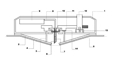
As shown in Fig.1 the diagnostic device has a casing having
a cylindrical side-wall 1, a disk-like flexible base plate
2 in the pre-stressed position enforced by pins 3 of the
flexible base plate, which is by means of an adhesive layer
4 attached to the skin 5. A guide needle 6 houses the ac-
WO 2012/045561 CA 02808782 2013-02-19PCT/EP2011/066074
- 15 -
tive surface 7 and the adjacent portion of the connection
lines or conductive part 8 of the diagnostic element which
forms a rigid connection to control and measuring means 9.
The guide needle 6 is fixed in a holder 10 at its end oppo-
site to the tip and kept in the lowered position by a slid-
ing bolt plate 11, which withholds also the pins 3 of the
flexible base plate. An actuation knob 12 actuates the im-
plantation and consecutive partial retraction of the guide
needle, described in more detail with reference to Fig. 3,
followed by starting the measuring process. In the ready-
to-use state the actuation knob and the holder of the guide
needle are pressed upwards against a stop by springs 13 and
14, respectively.
The base plate is preferentially annular or oval and has a
radial segmentation, preferably into 5 to 8 segments with a
spacing between them and a central concentric opening,
forming a cone upon central bending or alternatively it
consists of two segments with a diagonal slit, forming a
gable upon bending. The segments are attached to the cir-
cumference of the casing by springy hinge regions and are
in addition preferentially made of a flexible material. On
its underside, the flexible base plate has an annular or
oval adhesive layer for securing the device to the pa-
tient's skin with a concentric central opening or a diago-
nal slit, respectively similar to the base plate. This ad-
hesive layer is composed of three parts, a glue for fixing
to the flexible base plate, a textile providing the neces-
sary flexibility and a glue for fixing onto the skin. Suit-
able materials with low allergenic potential are commer-
cially available. The adhesive layer is protected during
storage with a suitable sheet. In this example, the adhe-
sive layer has a larger circumference than the device but
it could have also the same circumference if the attachment
WO 2012/045561 CA 02808782 2013-02-19PCT/EP2011/066074
- 16 -
to the base plate leaves an outer zone where it is not con-
nected to the housing.
Fig 2 shows the diagnostic device in the operational mode.
The flexible base plate 2 is depicted in the relaxed, i.e.
flat position. The pins 3 enforcing the pre-stressed posi-
tion of the flexible base plate in the ready-to-use mode
are now free in slits of the sliding bolt plate 11 and the
holder of the guide needle 10 has passed through a hole of
the sliding bolt plate by a slot and key construction and
is hold in place by a stop (not shown). The guide needles
and the active surfaces of the diagnostic elements protrude
through the opening or slit of the base plate and of the
adhesive layer and are inserted into the skin. A very im-
portant feature of the subject invention is that the con-
nections between the active surfaces of the diagnostic ele-
ments implanted into the skin and the other parts of the
device are stationary and therefore no connections have to
be made manually after the implantation process. In addi-
tion, the present invention obviates the removal of the
guide needle. As compared to similar devices of prior art
this is a big advantage for reliability, easy handling and
user acceptance.
The enlarged sectional view of Fig. 2a shows the holder of
the guide needle 10 which fixes and retracts the guide nee-
dle 6 in a geometrically well defined movement, sliding
over the conducting part of the diagnostic element 8. This
construction allows also the exact positioning of e.g. sen-
sor arrays in a geometrically well defined position.
Enlarged view of 2b shows the partially retracted guide
needle 6 and the active surface of the diagnostic element 7
which is directly exposed to the subcutaneous tissue. In
WO 2012/045561 CA 02808782 2013-02-19PCT/EP2011/066074
- 17 -
this example the conducting part of the diagnostic element
8 remains in the partially retracted guide needle and a
flexprint is used as substrate for the active surface and
conducting part of the diagnostic element. The active sur-
face of the diagnostic element holds an electrochemical
sensor 15 and the conducting part of the diagnostic element
holds an insulated electric conductor line 16. It is also
possible to place more than one sensor and conductor line
on the same and/or opposing faces of the flexprint sub-
strate.
Fig. 3 shows one embodiment of the means to bring the
flexible base plate from the ready-to-use position to the
position of the operation mode and consecutively to par-
tially retract the guide needle. This is in the described
embodiment a circular sliding bolt mechanism composed of
three pieces, a plate 11 with several slits, an actuation
knob 12 and a drive mechanism 21, e.g. a spring turning the
plate. In this figure a mechanism for a flexible baseplate
with four radial segments is shown but the principle of
this mechanism can be easily adapted to more radial seg-
ments, to two segments with a diagonal slit and to a linear
sliding bolt mechanism.
In the ready-to-use position the flexible base plate (not
shown) is pre-stressed by pins on the segments which are
restrained by the crosspieces 17 of the sliding bolt plate.
Following a first rotation of e.g. 300 these pins fall into
slits 18 and the baseplate thereby rapidly relaxes into a
flat position. The holder of the guide needle (not shown)
is pressed by a spring against the sliding bolt plate, has
a cylindrical shape fitting into the central hole of the
plate and has four wings which are restrained by the cross-
pieces 19 of the plate in the starting position and also
WO 2012/045561 CA 02808782 2013-02-19PCT/EP2011/066074
- 18 -
after the first rotation of the sliding bolt plate. Upon a
second rotation of e.g. again 30 these wings fall into
slits 20 and the holder of the guide needle is pressed by
the spring through the central hole of the sliding bolt
plate against a stop (not shown).
Consecutive actuation of the first and the second rotations
of the sliding bolt plate are accomplished by releasing the
drive mechanism 21 through pressing and then again releas-
ing the actuation knob 12. The actuation knob is in a slit
22; a narrowing 23 holds the sliding bolt plate in the
start position (Fig 3a), a protruding detent 24 stops the
first rotation of the sliding bolt plate (Fig 3b) and the
second rotation of the sliding bolt plate is stopped by the
end of the slit 22 (Fig 3c). The details how pressing and
again releasing the actuation knob actuates these rotations
consecutively are depicted in Figures 3A to 3C showing a
cross-section of the device casing 1, of the actuation knob
12 and of the sliding bolt plate 11 with the slit 22. Fur-
ther, a schematic horizontal cut at the level of the block-
ing interaction between actuation knob and sliding bolt
plate (broken line) is shown.
Fig 3a shows the actuation knob 12 and the sliding bolt
plate 11 in the starting position. The actuation knob has a
first rim 25 which is pressed by the spring 13 against the
cover of the casing 1. The sliding bolt plate is under ten-
sion by the drive mechanism 21 but a second rim of the ac-
tuation knob 26 is blocking against the narrowing 23. Upon
pressing the actuation knob the neck 27 between the first
and the second rim is moved to and the second rim out of
the plane of the narrowing and releases the first rotation
of the sliding bolt plate until the detent 24 hits the 1st
rim 25 and the rotation is stopped.
WO 2012/045561 CA 02808782 2013-02-19PCT/EP2011/066074
- 19 -
Fig. 3b shows the actuation knob 12 and the sliding bolt
plate 11 in the position stopped after the first rotation.
In this position the pre-stressed flexible base plate has
adopted a flat shape upon relaxation and the guide needle
is fully inserted into the skin. Upon releasing the actua-
tion knob it is pushed back to the starting position and
the first rim 25 is moved above the plane of the detent 24
releasing the second rotation of the sliding bolt plate un-
til the end of the slit 22 of the sliding bolt plate hits
the second rim of the actuation knob 26 and the rotation is
stopped.
Fig. 3c shows the actuation knob 12 and the sliding bolt
plate 11 in the final position, stopped after the second
rotation. In this position not only the flexible base plate
has adopted a flat shape but also the guide needle is par-
tially retracted exposing the active surface of the diag-
nostic element to the interstitial fluid of the skin and
the control and measuring means are activated by a switch
actuated at the end position of the second rotation (not
shown).
Upon reading this specification, various alternative em-
bodiments will become obvious to the skilled artisan. For
example, the implantation mechanism and the partial retrac-
tion of the guide needle could be achieved via numerous
chemical, mechanical, or electrical means. Further, a large
variety of diagnostic elements and sensor arrays as well as
control and measuring means can be accommodated with the
device. In addition a micro-dialysis system may be built
inserting a semi-permeable dialysis membrane into the skin
with a guide needle and exposing the dialysis membrane to
the subcutaneous fluid upon partial retraction of the guide
WO 2012/045561 CA 02808782 2013-02-19PCT/EP2011/066074
- 20 -
needle. The dialysate solution can be pumped through the
system with a micro-pump accommodated in the device and the
analytes in the dialysate analysed online in the device or
sampled for later analysis.
Preferred sensors for analytes fitting well with the speci-
fications of the subject device can be constructed follow-
ing state of the art procedures for electrochemical and op-
tical sensors. The construction of miniaturized electro-
chemical and optical sensors is greatly improved by the use
of matrix materials optimally suited for production by well
established methodologies but such materials are often not
suitable for direct implantation into the skin, e.g. be-
cause they are too flexible or can break if used directly
to penetrate the skin. Introduction of such sensors into
the skin can only be achieved with a guide needle and the
described partial retraction of the guide needle greatly
improves design and handling by the patient. It allows es-
tablishing permanent connections to the control and measur-
ing means during manufacturing: connections, esp. if done
following implantation by the patient are problematic with
miniaturized structures or almost impossible if conduction
of very low electrical or other signals or of fluid is nec-
essary. A slit guide needle often used allowing removal af-
ter implantation leads to important tissue damage and lim-
its miniaturization.
For the construction of electrochemical sensors silicon or
flexible substrates are ideal and technologically well es-
tablished but for both a guide needle is needed for implan-
tation. The use of flexprint technologies used for PCBs in
electronics is straight-forward by coating part of the ac-
tive surface with a suitable sensor e.g. for glucose and
manufacturing of flexprints is approaching a level of
WO 2012/045561 CA 02808782 2013-02-19PCT/EP2011/066074
- 21 -
miniaturization which makes it very suitable for diagnostic
elements. For the construction of optical sensors a wide
variety of methods can be optimally adapted for direct de-
termination of the analyte or for indirect monitoring using
suitable indicators. Such general methods can be coupled to
analyte-specific enzyme reactions or to specific binding to
receptors or antibodies. The current invention provides an
easy solution for establishing permanent connections to the
control and measuring means during manufacturing which is
very important for good performance of such miniaturized
electrical or light transmitting fibres.
The invention has been described with reference to a few
specific and preferred embodiments, techniques and applica-
tions. However, it will be apparent to one of ordinary
skill in the art that many variations and modifications and
adaptations to special applications and needs may be made
while remaining within the spirit and scope of the inven-
tion.