Note: Descriptions are shown in the official language in which they were submitted.
CA 02813796 2013-04-05
WO 2012/052230
PCT/EP2011/065771
1
ErbB3 binding antibody
Description
The present invention relates to an antibody, particularly a monoclonal
antibody, which binds to the ErbB3 receptor, wherein said binding reduces
ErbB3 receptor mediated signal transduction, and compositions comprising
such an antibody as well as methods using such an antibody.
Cancer is a disease characterized by uncontrolled proliferation of
transformed cells that invade and destroy adjacent tissues, and may spread
to distant anatomic sites through a process called metastasis1-4. In presence
of metastatic disease, cancer may cause death in a variable period of time
between a few months and some years1-4.
The most used drugs for cancer treatment are cytotoxic chemotherapeutic
agents (also called antiblastic agents or chemotherapeutic agents). These
drugs act by damaging DNA or inhibiting cell proliferation. In this way, they
kill all rapidly dividing cells, not only cancer cells, but also normal cells
that
are undergoing cell division. The lack of specificity of action of
chemotherapeutic drugs on cancer cells is responsible for considerable
toxicity following their administration. In the last decade, basic scientific
research has significantly increased our knowledge about molecular
mechanisms of cellular transformation and cell proliferation, leading to the
development of "molecularly targeted" drugs or "targeted therapies"5. They
refer to drugs that are specifically designed to act on cancer cells bearing
particular molecular and/or functional abnormalities. However, also targeted
therapies are associated with side effects, and in most cases they can block
tumor growth only temporarily.
ErbB3 receptor, also known as HER-3, belongs to the epidermal growth
factor receptor tyrosine kinase family (ErbB). This family of receptors
consists of four members: ErbB1 (HER1), ErbB2 (HER2), ErbB3 (HER3) and
CA 02813796 2013-04-05
WO 2012/052230
PCT/EP2011/065771
2
ErbB4 (HER4). Many studies have suggested a critical role for ErbB
receptors in cell survival, proliferation and differentiation, as well as in
malignant transformation'. The signal transduction mediated by tyrosine
kinase receptors is complex and involves the interaction with two categories
of ligands: epidermal growth factor (EGF) and EGF-like ligands (e.g. TGFa
and amphiregulin), Neuregulin (NRG), also defined Heregulin (HRG) or Neu
Differentiation Factor (NDF). Ligand binding to ErbB receptors induces the
formation of receptor homo- and heterodimers and activation of the intrinsic
kinase domain, resulting in phosphorylation on specific tyrosine residues
within the cytoplasmic tail. These phosphorylated residues serve as docking
sites for a range of proteins, the recruitment of which leads to the
activation
of intracellular signalling pathways. Generally, heterodimerization is
preferred over homodimerization; ErbB2 is the preferred heterodimerization
partner of the other ErbB receptors, including ErbB1 (activated by EGF or
EGF-like ligands), and ErbB3 and ErbB4 (activated by neuregulin, NRG).
The two major signaling pathways activated by ErbB receptors are Ras-Raf-
MAPK and PI3K-AKT pathways."
ErbB2 gene is amplified in 20 to 30% of breast cancers and is correlated with
a poor prognosis. In the same way, ErbB3 receptor has also been shown to
be overexpressed in breast cancer patients. High levels of expression of
both ErbB2 and ErbB3 receptors are associated with an aggressive biology
of tumor. In fact, upon NRG stimulation, ErbB2/ ErbB3 heterodimers deliver
the most potent and long-lasting proliferative intracellular signal among the
possible combinations of pairs of ErbB family members811. Several studies
have suggested an important role of ErbB3 receptor in progression of many
human tumor types, such as prostate cancer, melanoma, and gastric
carcinoma.
All together, experimental and clinical data indicate that ErbB3 plays an
essential role in tumor development and progression, suggesting that agents
targeting ErbB3 could provide a novel and promising approach toward the
treatment of some cancers.12-24
In spite of scientific progress and introduction into clinical practice of new
CA 02813796 2013-04-05
WO 2012/052230
PCT/EP2011/065771
3
chemotherapeutic agents and targeted therapies, cancer remains a disease
difficult to cure, responsible for about 13% of deaths worldwide.1-4
Consequently, there is an urgent need to develop new antitumor therapies,
more effective and possibly less toxic.
The inventor has found that specific ErbB3 inhibitors are able to induce
tumor regression. In particular, the monoclonal antibody, MP-RM-1, has
been used as an anti- ErbB3 inhibitor.
Thus, a first aspect of the present invention relates to an antibody or
fragment thereof which binds to the ErbB3 receptor and which comprises
a) a heavy chain amino acid sequence as encoded by SEQ ID NO: 1 or
at least the variable domain thereof or an amino acid sequence having
a sequence identity of at least 80% thereto
and/or
b) a light chain amino acid sequence as encoded by SEQ ID NO: 2 or
at least the variable domain thereof or an amino acid sequence having
a sequence identity of at least 80% thereto.
VH sequence (SEQ ID NO:1)
gacgtgcagctggtggagtctgggggagacttagtgaagcctggagggtccctgaaactctcctgtgtagtctctggat
tcactttcagtac
ctatggcatgtcttgggttcgccagactccagacaggaggctggagtgggtcgcaaccattagtcatggtgacggttat
acctactatcca
gacagtgtgaaggggcgattcaccatctccagagacaatgccaagaacaccctgcacctgcaaatgagcagtctgaagt
ctgagga
cacagccatgtattactgtgcaagacatggggattacgacgatgattactatgctatggactactggggtcaaggaacc
tcagtcaccgt
ctca
VL sequence (SEQ ID NO:2)
gatattgtgatgacccagtctccatcctccctgactgtgatagcaggagagaaggtcactatgagctgcaagtccagtc
agagtctgttaa
acagtggaaatcaaaagaactacttgacctggtaccaacagaaaccagggcagcctcctaaactgttgatctactgggc
atccactag
ggaatctggggtccctgatcgcttcacaggcagtggatctggaacagatttcactctcaccatcagcagtgtgcaggct
gaagacctggc
agtttattactgtcagaatgaatatacttatccgctcacgttcggtgctgggaccaagctggagctgaaacggg
The term "antibody" as used herein includes "fragments" or "derivatives",
which have at least one antigen binding site of the antibody and/or show the
CA 02813796 2013-04-05
WO 2012/052230
PCT/EP2011/065771
4
same biological activity.
Further, the antibody preferably comprises at least one heavy
immunoglobulin chain and at least one light immunoglobulin chain. An
immunoglobulin chain comprises a variable domain and optionally a constant
domain. A variable domain may comprise complementary determining
regions (CDRs), e.g. a CDR1, CDR2 and/or CDR3 region, and framework
regions.
As used herein, "sequence identity" between two polypeptide sequences,
indicates the percentage of amino acids that are identical between the
sequences, preferably over the entire length of the amino acid sequences as
encoded by SEQ ID NO: 1 and/or SEQ ID NO: 2. Preferred polypeptide
sequences of the invention have a sequence identity of at least 80%, more
preferably 85%, even more preferably 90%, 93%, 95%, 96%, 97%, 98% or
99%.
According to another preferred embodiment, antibodies of the invention
reduce ErbB3 receptor mediated signal transduction. Said reduction of
ErbB3 receptor mediated signal transduction is preferably caused by a
down-regulation of ErbB3. According to a further embodiment, down-
regulation of ErbB3 is preferably achieved by decreasing levels of ErbB3 on
the cell surface, i.e. preferably the antibody of the invention has the
ability to
decrease levels of ErbB3 on cell surfaces.
The antibody of the invention may have at least one antigen binding site, e.g.
one or two antigen binding sites.
The antibodies of the invention bind preferably to the extracellular domain of
ErbB3.
The antibody may be any antibody of natural and/or synthetic origin, e.g. an
antibody of mammalian origin. Preferably, the constant domain -if present- is
CA 02813796 2013-04-05
WO 2012/052230
PCT/EP2011/065771
a human constant domain. The variable domain is preferably a mammalian
variable domain, e.g. a humanized or a human variable domain.
Antibodies according to the invention may be polyclonal or monoclonal
5 antibodies. Monoclonal antibodies are preferred. In particular antibodies
of
the present invention are preferably selected from the group consisting of
recombinant antibodies, humanized or fully human antibodies, chimeric
antibodies, multispecific antibodies, in particular bispecific antibodies, or
fragments thereof.
Monoclonal antibodies may be produced by any suitable method such as
that of Kohler and Milstein "or by recombinant DNA methods. Monoclonal
antibodies may also be isolated from phage antibody libraries using
techniques described in Clackson et al.'
Humanized forms of the antibodies may be generated according to the
methods known in the art such as chimerization or CDR grafting. Alternative
methods for the production of humanized antibodies are well known in the
art and are described in, e.g., EP-Al 0 239 400 and WO 90/07861. Human
antibodies can also be derived by in vitro methods. Suitable examples
include but are not limited to phage display, yeast display, and the like.
According the present invention "chimeric antibody" relates to antibodies
comprising polypeptides from different species, such as, for example, mouse
and human. The production of chimeric antibodies is described, for example,
in WO 89/09622.
Monospecific antibodies are antibodies that all have affinity for the same
antigen. Multispecific antibodies are antibodies that have affinity for
several
antigens. A bispecific antibody has affinity for two different antigens.
The term antibody includes "fragments" or "derivatives", which have at least
one antigen binding site of the antibody. According to a preferred
CA 02813796 2013-04-05
WO 2012/052230
PCT/EP2011/065771
6
embodiment the antibody or fragment thereof may be a Fab fragment, a Fab'
fragment, a F(ab') fragment, a Fv fragment, a diabody, a ScFv, a small
modular immunopharmaceutical (SMIP), an affibody, an avimer, a
nanobody, a domain antibody and/or single chains.
"Avimer" relates to a multimeric binding protein or peptide engineered using,
for example, in vitro exon shuffling and phage display. Multiple binding
domains are linked, resulting in greater affinity and specificity compared to
single epitope immunoglobin domains.
"Nanobody" or single domain antibody relates to an antibody fragment
consisting of a single monomeric variable antibody domain.
"Affibody" molecules are small high affinity proteins being engineered to bind
specifically to a large number of target proteins.
The antibody of the invention may be preferably of the IgG1, IgG2, IgG3,
IgG4, IgM, IgA1, IgA2, IgAsec, IgD, and IgE antibody-type. It will be
appreciated that antibodies that are generated need not initially possess
such an isotype but, rather the antibody as generated can possess any
isotype and that the antibody can be isotype-switched.
The antibodies or antibody fragments of the invention are optionally
deimmunized for therapeutic purposes.
In an especially preferred embodiment of the invention, the antibody is MP-
RM-1, deposited at Deutsche Sammlung von Mikroorganismen und
Zellkulturen (DSMZ) on 15 October 2009 and designated DSM ACC3018.
According to a further preferred embodiment the antibody or fragment
thereof may be produced from the hybridoma cell line DSM ACC3018 or a
derivative thereof.
According to a preferred embodiment of the invention, the antibody or
CA 02813796 2013-04-05
WO 2012/052230
PCT/EP2011/065771
7
fragment produced from the hybridoma cell line DSM ACC3018 or a
derivative thereof is a humanized antibody or fragment thereof. Preferably,
this humanized antibody comprises at least one of the sequences 3-14 of the
present invention. According to an especially preferred embodiment, the
antibody is selected from the group cMP-RM-1 #1, cMP-RM-1 #2, cMP-RM-
1 #3, cMP-RM-1 #4, hMP-RM-1 #5, hMP-RM-1 #6, hMP-RM-1 #7, hMP-RM-
1 #8, hMP-RM-1 #9, hMP-RM-1 #10, hMP-RM-1 #11, hMP-RM-1 #12, hMP-
RM-1 #13, hMP-RM-1 #14, hMP-RM-1 #15, hMP-RM-1 #16, hMP-RM-1 #17,
hMP-RM-1 #18, hMP-RM-1 #19, hMP-RM-1 #20 (c, chimeric antibody; h,
humanized antibody).
An especially preferred embodiment relates to an antibody which is selected
from the group cMP-RM-1 #4, hMP-RM-1 #14, hMP-RM-1 #17 or hMP-RM-
1 #20.
Another preferred embodiment relates to the group of antibodies consisting
of hMP-RM-1 #6, hMP-RM-1 #7, hMP-RM-1 #8, hMP-RM-1 #9, hMP-RM-
1 #10, hMP-RM-1 #11, hMP-RM-1 #12, hMP-RM-1 #19 and hMP-RM-1 #20.
A particularly preferred group of antibodies comprises the antibodies hMP-
RM-1 #6, hMP-RM-1 #10 and hMP-RM-1 #20.
It will be apparent to those skilled in the art that the antibodies of the
invention can be further coupled to other moieties for, e.g., drug targeting
and imaging applications. Such coupling may be conducted chemically after
expression of the antibody or antigen to site of attachment or the coupling
product may be engineered into the antibody or antigen of the invention at
the DNA level.
Thus, for diagnostic purposes, the antibody or antibody fragment of the
invention may be labelled, i.e. coupled to a labelling group. Suitable labels
include radioactive labels, fluorescent labels, suitable dye groups, enzyme
labels, chromogenes, chemiluminescent groups, biotinyl groups,
predetermined polypeptide epitopes recognized by a secondary reporter etc.
CA 02813796 2013-04-05
WO 2012/052230
PCT/EP2011/065771
8
Those labelled antibodies or antibody fragments may be in particular used in
immunohistochemistry assays or for molecular imaging in vivo.
For therapeutic purposes, the antibody or antibody fragment of the invention
may be conjugated with a effector group, in particular a therapeutic effector
group such as a radioactive group or a cytotoxic group.
Labelling groups or effector groups may be attached by spacer arms of
various lengths to reduce potential steric hindrance.
According to another aspect, the present invention relates to a nucleic acid
molecule encoding the antibody of the invention or fragment thereof or a
nucleic acid capable of hybridizing thereto under stringent conditions. The
nucleic acid molecule of the invention encoding the above-described
antibody, antibody fragment or derivative thereof may be, e.g. DNA, cDNA,
RNA or synthetically produced DNA or RNA or recombinantly produced
chimeric nucleic acid molecule comprising any of those nucleic acid
molecules either alone or in combination. The nucleic acid molecule may
also be genomic DNA corresponding to the entire gene or a substantial
portion thereof or to fragments and derivatives thereof. The nucleotide
sequence may correspond to the naturally occurring nucleotide sequence or
may contain single or multiple nucleotide substitutions, deletions or
additions. In a particular preferred embodiment of the present invention, the
nucleic acid molecule is a cDNA molecule.
The term "hybridizing under stringent conditions" means that two nucleic
acid fragments hybridize with one another under standardized hybridization
conditions as described for example in Sambrook et al., "Expression of
cloned genes in E. coli" in Molecular Cloning: A laboratory manual (1989),
Cold Spring Harbor Laboratory Press, New York, USA. Such conditions are
for example hybridization in 6.0xSSC at about 45 C. followed by a washing
step with 2.0xSSC at 50 C, preferably 2.0xSSC at 65 C, or 0.2xSSC at
CA 02813796 2013-04-05
WO 2012/052230
PCT/EP2011/065771
9
50 C, preferably 0.2xSSC at 65 C.
Another aspect of the invention relates to a vector comprising a nucleic acid
molecule of the invention. Said vector may be, for example, a phage,
plasmid, viral or retroviral vector. Retroviral vectors may be replication
competent or replication defective. Preferably, the vector of the invention is
an expression vector wherein the nucleic acid molecule is operatively linked
to one or more control sequences allowing the transcription and optionally
expression in prokaryotic and/or eukaryotic host cells.
The invention further relates to a host comprising the vector of the
invention.
Said host may be a prokaryotic or eukaryotic cell or a non-human transgenic
animal. The polynucleotide or vector of the invention which is present in the
host may either be integrated into the genome of the host or it may be
maintained extrachromosomally.
The host can be any prokaryotic or eukaryotic cell, such as a bacterial,
insect, fungal, plant, animal, mammalian or, preferably, human cell.
Preferred fungal cells are, for example, those of the genus Saccharomyces,
in particular those of the species S. cerevisiae.
The invention additionally relates to a method for the preparation of an
antibody, comprising culturing the host of the invention under conditions that
allow synthesis of said antibody and recovering said antibody from said
culture.
A further aspect of the present invention relates to a pharmaceutical
composition comprising the antibody of the invention or a fragment thereof,
the nucleic acid molecule, the vector, the host of the invention or an
antibody
obtained by a method of the invention. The term "composition" as employed
herein comprises at least one compound of the invention. Preferably, such a
composition is a therapeutical/pharmaceutical or a diagnostic composition.
The diagnostic composition of the invention may be used for assessing the
CA 02813796 2013-04-05
WO 2012/052230
PCT/EP2011/065771
onset or the disease status of a hyperproliferative disease as defined herein.
The composition preferably comprises an pharmaceutically acceptable
carrier, diluent and/or excipient.
5
Examples of suitable pharmaceutical carriers, excipients and/or diluents are
well known in the art and include phosphate buffered saline solutions, water,
emulsions, such as oil/water emulsions, various types of wetting agents,
sterile solutions etc. Compositions comprising such carriers, excipients
10 and/or diluents can be formulated by well known conventional methods.
Administration of the suitable compositions may be effected by different
ways, e.g., by intravenous, intraperitoneal, subcutaneous, intramuscular,
topical, intradermal, intranasal or intrabronchial administration. Preferred
is
an intravenous, intramuscular and/or subcutaneous administration.
These pharmaceutical compositions can be administered to the subject at a
suitable dose. The dosage regimen can be determined by the attending
physician and clinical factors.
The compositions of the invention may be administered locally or
systemically. Preparations for parenteral administration include sterile
aqueous or non-aqueous solutions, suspensions, and emulsions. Examples
of non-aqueous solvents are propylene glycol, polyethylene glycol, vegetable
oils such as olive oil, and injectable organic esters such as ethyl oleate.
Aqueous carriers include water, alcoholic/aqueous solutions, emulsions or
suspensions, including saline and buffered media. Parenteral vehicles
include sodium chloride solution, Ringer's dextrose, dextrose and sodium
chloride, lactated Ringer's, or' fixed oils. Intravenous vehicles include
fluid
and nutrient replenishers, electrolyte replenishers (such as those based on
Ringer's dextrose), and the like. Preservatives and other additives may also
be present such as, for example, antimicrobials, anti-oxidants, chelating
agents, and inert gases and the like. Furthermore, the pharmaceutical
CA 02813796 2013-04-05
WO 2012/052230
PCT/EP2011/065771
11
composition of the invention may comprise further agents depending on the
intended use of the pharmaceutical composition.
According to an especially preferred embodiment the composition comprises
a further active agent, such as a further antibody or antibody fragment.
Preferably the composition of the invention is used in combination with at
least one further antineoplastic agent. Said combination is effective, for
example, in inhibiting abnormal cell growth. Many antineoplastic agents are
presently known in the art. In general the term includes all agents that are
capable of prevention, alleviation and/or treatment of hyperproliferative
disorders. Especially preferred are antineoplastic agents inducing apoptosis.
Preferably the antineoplastic agent is selected from the group consisting of
antibodies, small molecules, antimetabolites, alkylating agents, topo-
isomerase inhibitors, microtubule-targeting agents, kinase inhibitors, protein
synthesis inhibitors, immuno-therapeutics, hormones or analogs thereof,
and/or mTOR inhibitors.
Specific examples of antineoplastic agents which can be used in
combination with the antibodies provided herein include, for example,
gefitinib, lapatinib, sunitinib, pemetrexed, bevacisumab, cetuximab, imatinib,
alemtuzumab, trastuzumab, rituximab, erlotinib, bortezomib and the like, in
particular trastuzumab. Other specific antineoplastic agents to be used in the
compositions as described and claimed herein include for example,
chemotherapeutic agents such as Paclitaxel, Anthracyclines,
Fluoropirimidine, vinca alkaloids, platinum salts, in particular capecitabine,
daunorubicin, daunomycin, dactinomycin, doxorubicin, epirubicin, idarubicin,
esorubicin, bleomycin, mafosfamide, ifosfamide, cytosine arabinoside, bis-
chloroethylnitrosurea, busulfan, mitomycin C, actinomycin D, mithramycin,
prednisone, hydroxyprogesterone, testosterone, tamoxifen, dacarbazine,
procarbazine, hexamethylmelamine, pentamethylmelamine, mitoxantrone,
amsacrine, chlorambucil, methylcyclohexylnitrosurea,
melphalan,
CA 02813796 2013-04-05
WO 2012/052230
PCT/EP2011/065771
12
cyclophosphamide, 6-mercaptopurine, 6-thioguanine, cytarabine (CA), 5-
azacytidine, hydroxyurea, deoxycoformycin, 4-hydroxyperoxycyclophosphor-
amide, 5-fluorouracil (5-FU), 5-fluorodeoxyuridine (5-FUdR), methotrexate
(MTX), colchicine, taxol, vincristine, vinblastine, etoposide, trimetrexate,
teniposide, cisplatin and diethylstilbestrol (DES).
The compositions of the invention may be administered in combination with
a further therapeutic composition comprising an active agent as described
above and/or irradiation and/or radiotherapy.
According to a preferred embodiment, the compositions of the invention are
for the use in treating and/or preventing hyperproliferative diseases, in
particular neoplastic diseases or cancer. The compositions may also be
used for the manufacture of a medicament for treating and/or preventing
hyperproliferative diseases, in particular neoplastic diseases or cancer.
A hyperproliferative disease as defined herein includes any neoplasia, i.e.
any abnormal and/or uncontrolled new growth of tissue. The term
"uncontrolled new growth of tissue" as used herein may depend upon a
dysfunction and/or loss of growth regulation. A hyperproliferative disease
includes tumor diseases and/or cancer, such as metastatic or invasive
cancers.
The hyperproliferative disease is preferably selected from disorders
associated with, accompanied by or caused by ErbB3 expression,
overexpression or hyperactivity, such as cancer, in particular melanoma,
breast cancer, ovarian cancer, renal carcinoma, gastrointestinal/colon
cancer, lung cancer, clear cell carcinoma of the kidney and/or prostate
cancer. In particular, for these tumors, it has been demonstrated a role of
ErbB3 in promoting cancer development and growth, and, thus, the inhibition
of this protein could give certain benefits.
The invention further relates to a method of treating a disease wherein the
CA 02813796 2013-04-05
WO 2012/052230
PCT/EP2011/065771
13
antibody of the invention is administered to a mammal and wherein said
disease is correlated directly or indirectly with an abnormal level of
expression or activity of ErbB3.
A further aspect of the present invention relates to a method of inhibiting
EGF-like ligand mediated phosphorylation of ErbB3 in a subject, comprising
administering to the subject an antibody or antigen binding portion thereof of
as described above, in an amount sufficient to inhibit EGF-like mediated
phosphorylation of ErbB3. "Phosphorylation of ErbB3" refers to the
phosphorylation of amino acid residues, preferably tyrosine residues.
Yet another aspect of the present invention is directed to a method of
diagnosing a cancer associated with ErbB3 in a subject, comprising
(a) contacting ex vivo or in vivo cells from the subject with an antibody
or antigen binding portion thereof of any one of the preceding claims and
(b) measuring the level of binding to ErbB3 on the cells, wherein ab-
normally high levels of binding to ErbB3 indicate that the subject has a can-
cer associated with ErbB3.
In terms of the present invention, "abnormally high" means higher binding
levels of ErbB3 compared to a healthy subject having no cancer.
Preferably the subject is an animal, more preferably a mammalian and in
particular preferably a human.
CA 02813796 2013-04-05
WO 2012/052230
PCT/EP2011/065771
14
BRIEF DESCRIPTION OF THE DRAWINGS
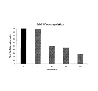
Figure 1 shows that MP-RM-1 reduces the expression of ErbB3 receptor on
the surface of breast cancer cells
Figure 2 shows that MP-RM-1 downregulates ErbB3 receptor in breast
cancer cells.
Figure 3 shows that MP-RM-1 is able to reduce ErbB3 receptor half-life in
MDA-MB-435 human breast cancer cells.
Figure 4 shows that MP-RM-1 is able to reduce ErbB3 receptor half-life in
SKBR-3 human breast cancer cells.
Figure 5 shows that the effect MP-RM-1 on the reduction of the ErbB3
receptor half-life is blocked by the lysosome inhibitor chloroquine.
Figure 6 shows that MP-RM-1 inhibits the phosphorylation of ErbB3 and
AKT induced by the receptor ligand NRG-1 in MDA-MB-435 human breast
cancer and IR-8 human melanoma cells.
Figure 7 shows that ligand-induced phosphorylation of ErbB3 and AKT is
inhibited by MP-RM-1 in a time-dependent fashion.
Figure 8 shows that MP-RM-1 is able to antagonize the ligand-induced
activation of ErbB3 and AKT.
Figure 9 shows that MP-RM-1 is rapidly (30 minutes) internalized into cells.
Figure 10 compares the effect of MP-RM-1 and Trastuzumab on ligand-
induced activation of ErbB3 and AKT in human breast and prostate cancer
cells.
CA 02813796 2013-04-05
WO 2012/052230
PCT/EP2011/065771
Figure 11 shows that MP-RM-1 is able to inhibit basal ErbB3 and AKT
phoshphorylation in MET-amplified MKN-45 human gastric cancer cells.
Figure 12 shows that MP-RM-1 inhibits ligand-induced proliferation in MDA-
5 MB-435 human breast cancer cells.
Figure 13 shows that MP-RM-1 inhibits the growth of DU145 human
prostate cancer xenografts
10 Figure 14 shows that as soon as 4 hours after injection into mice, MP-RM-
1
induces ErbB3 downregulation and inhibits AKT activation in IR-8 human
melanoma xenografts..
Figure 15 shows that treatment of IR-8 human melanoma cells with cMP-
15 RM-1 #1, hMP-RM-1 #6, hMP-RM-1 #10, hMP-RM-1 #20 MP-RM-1 antibody
variants reduces the expression of ErbB3 receptor on cell surface.
Figure 16 shows that the humanized variant hMP-RM-1 #6 is rapidly (30
minutes) internalized into the cells.
Figure 17 shows the effect of the chimeric variant cMP-RM-1 #1, and the
humanized variants hMP-RM-1 #6, hMP-RM-1 #10, hMP-RM-1 #20 antibody
variants on ErbB3 half-life in IR-8 human melanoma cells.
Figure 18 shows the inhibitory effect of cMP-RM-1 #1, hMP-RM-1 #6, hMP-
RM-1 #10 and hMP-RM-1 #20 antibody variants on the phosphorylation of
ErbB3 and AKT in human ovarian (A) and gastric (B) cancer cells.
Figure 19 shows the inhibitory effect of cMP-RM-1 #1, hMP-RM-1 #6, hMP-
RM-1 #10 and hMP-RM-1 #20 antibody variants on human melanoma (A) or
gastric cancer (B) cell colony formation in soft agar.
Figure 20 shows that tumor xenografts of mice treated with cMP-RM-1 #1,
CA 02813796 2013-04-05
WO 2012/052230
PCT/EP2011/065771
16
hMP-RM-1 #6, hMP-RM-1 #10 and hMP-RM-1 #20 antibody variants grow
significantly less than those of control mice.
Examples
Example 1: Production of the monoclonal antibody MP-RM-1.
Four-weeks old Balb/c mice were immunized by intraperitoneal injection of
live NIH/3T3 cells transfected with the human ErbB3 receptor. Seven days
later, mice were given an additional intraperitoneal injection of the
immunogen. After additional seven days, mice were boosted intravenously
with the immunogen, and spleens were removed for cell fusion 3 days later.
Somatic cell hybrids were prepared by fusion of immune splenocytes with
the murine nonsecreting myeloma cell line NS-1. Hybridoma supernatants
were selected on the basis of differential reactivity with LTR-ErbB3
transfected cells, but not with LTR-neo NIH/3T3. All positive hybridoma cell
colonies were cloned twice by limiting dilution and further characterized. The
selected monoclonal antibody, named MP-RM-1 (isotype IgG2a) was found
to specifically recognize the extracellular domain of the ErbB3 receptor.
The hybridoma murine cell line producing MP-RM-1 antibody was deposited
at the DSMZ (Deutsche Sammlung von Mikroorganismen und Zellkulturen)
and designated DSM ACC3018.
Example 2: Effect of MP-RM-1 on ErbB3 receptor expression on the
surface of breast cancer cells.
Materials and methods: MDA-MB-435 human breast cancer cells were
maintained on ice for 30 minutes with 10 pg/ml of MP-RM-1 and then
returned to 37 C. At the indicated times, cells were trypsinized and stained
with a fluorescein-labeled goat anti-mouse IgG antibody and analyzed by
FAGS.
CA 02813796 2013-04-05
WO 2012/052230
PCT/EP2011/065771
17
Results: MP-RM-1 decreases ErbB3 receptor expression on the cell surface
in a time-dependent manner (Figure 1).
Example 3: Effect of MP-RM-1 on downregulation of the ErbB3 receptor
in breast cancer cells.
Materials and methods: MDA-MB-435 human breast cancer cells were
grown in 0.2% FBS DMEM for 24 hours and then incubated in the presence
or absence of 10 pg/ml of MP-RM-1 for 15, 60, 120 and 240 minutes. At the
end of the incubation periods, cells were lysed and analyzed for ErbB3 and
AKT protein levels by Western blotting with anti-ErbB3 and anti-AKT. The
same filter was reprobed with anti-PLC -1 for a loading control.
Results: MP-RM-1 induces downregulation of ErbB3 receptor after 120
minutes (Figure 2).
Example 4: Effect of MP-RM-1 on ErbB3 receptor half-life in breast
cancer cells.
Materials and methods: MDA-MB-435 human breast cancer cells were
grown in 0.2% FBS DMEM for 24 hours and then chased with the protein
synthesis inhibitor cycloheximide at 10 pg/ml with or without MP-RM-1. Cells
were lysed and analyzed for ErbB3 and AKT protein levels by Western
blotting with anti-ErbB3 and anti-AKT. The same filter was reprobed with
anti- PLC 7 -1 or anti-Actin for a loading control.
Results: ErbB3 receptor half-life is markedly reduced in cycloheximide
chased, MP-RM-1 treated MDA-MB-435 cells compared to PBS-treated
control cells (Figure 3).
Example 5: Effect of MP-RM-1 on ErbB3 receptor half-life in breast
CA 02813796 2013-04-05
WO 2012/052230
PCT/EP2011/065771
18
cancer cells.
Materials and methods: SKBR-3 human breast cancer cells were grown in
0.2% FBS DMEM for 24 hours and then chased with the protein synthesis
inhibitor cycloheximide at 10 pg/ml in the presence or absence of MP-RM-1.
Cells were lysed and analyzed for ErbB3 levels by Western blotting with anti-
ErbB3.
Results: ErbB3 receptor half-life is markedly reduced in cycloheximide
chased, MP-RM-1 treated SKBR-3 cells compared to the PBS treated control
cells (Figure 4).
Example 6: Effect of chloroquine on ErbB3 receptor downregulation
induced by MP-RM-1.
Materials and Methods: MDA-MB-435 human breast cancer cells were
grown on 0.2% FBS for 24 hours and chased for 3 hours with cycloheximide
at 10 pg/ml. Cells were then incubated with MP-RM-1 in the presence or
absence of chloroquine. After incubation, cells were lysed and analyzed for
ErbB3 protein levels by Western blotting with anti-ErbB3. The same filter was
reprobed with anti-Actin for a loading control
Results: ErbB3 receptor downregulation induced by MP-RM-1 is blocked by
chloroquine (Figure 5).
Example 7: Effect of MP-RM-1 on ligand-induced ErbB3 and AKT
phosphorylation in breast and melanoma cancer cells.
Materials and methods: MDA-MB-435 human breast cancer cells, A375 and
IR-8 human melanoma cells were grown in 0.2% FBS in DMEM or RPMI for
24 hours. Cells were incubated in the presence or absence of MP-RM-1 at 1
or 10 pg/ml for 2 hours and then stimulated with 10 ng/ml of NRG-1 for 5
minutes. After incubation, cells were lysed and analyzed for ErbB3, p-ErbB3,
CA 02813796 2013-04-05
WO 2012/052230
PCT/EP2011/065771
19
AKT, p-AKT, or p-Erks protein levels by Western blotting with anti-ErbB3,
anti-p-ErbB3, anti-AKT, anti-p-AKT and anti-p-Erks. The same filter was
reprobed with anti-Actin for a loading control.
Results: Cells pre-treated with MP-RM-1 exhibit a dose-dependent inhibition
of ErbB3 and AKT ligand-induced phosphorylation (Figure 6).
Example 8: Effect of MP-RM-1 on ligand-induced ErbB3 and AKT
phosphorylation.
Materials and methods: MDA-MB-435 human breast cancer cells were
grown in 0.2% FBS DMEM for 24 hours and then stimulated with 10 ng/ml of
NRG-1 for 5 minutes. Cells were then incubated with 10 pg/ml of MP-RM-1
for 15, 60 and 120 minutes before of NRG-1 stimulation. After incubation,
cells were lysed and analyzed for ErbB3, p-ErbB3, AKT and p-AKT protein
levels by Western blotting with anti-ErbB3, anti-p-ErbB3, anti-AKT and anti-
p-AKT.
Results: Cells pre-treated with MP-RM-1 exhibit a time-dependent inhibition
of ErbB3 and AKT ligand-induced phosphorylation (Figure 7).
Example 9: Effect of MP-RM-1 on ligand-induced ErbB3 and AKT
phosphorylation.
Materials and Methods: MDA-MB-435 human breast cancer cells were
grown in 0.2% FBS DMEM for 24 hours and then simultaneously stimulated
with 10 ng/ml of NRG-1 and 10 pg/ml of MP-RM-1 for 5 minutes. After
incubation, cells were lysed and analyzed for ErbB3, p-ErbB3 and p-AKT
protein levels by Western blotting with anti-ErbB3, anti-p-ErbB3 and anti-p-
AKT.
Results: 5 minutes stimulation with MP-RM-1 does not induce ErbB3 and
AKT phosphorylation, indicating that MP-RM-1 is not a receptor agonist. By
CA 02813796 2013-04-05
WO 2012/052230
PCT/EP2011/065771
contrast, ligand-induced ErbB3 and AKT phosphorylation is partially inhibited
by MP-RM-1, indicating that MP-RM-1 is a partial receptor antagonist (Figure
8).
5 Example 10: MP-RM-1 internalization in MDA-MB-435 breast carcinoma
cells.
Materials and Methods: MDA-MB-435 human breast cancer cells cells were
plated in 22X22 mm coverslips and grown in 0.2% FBS DMEM for 24 hours.
10 Cells were then incubated with 10 pg/ml of MP-RM-1 for 30 minutes on ice
and returned at 37 C. After 30 and 60 minutes, cells were fixed in 4%
paraformaldehyde, permeabilized with 0.2% Triton-X100 in PBS and then
stained with a fluorescein-labeled goat anti-mouse antibody (green staining),
phalloydin (red staining). Cell nuclei were counterstained in blue. The yellow
15 and the white arrows indicate MP-RM-1 localization on the cell membrane
and in the cytoplasm, respectively.
Results: MDA MB 435 cells show goat anti mouse membrane positivity
(yellow arrows) after 30 minutes of MP-RM-1 incubation on ice indicating
20 that MP-RM-1 antibody is completely localized on the plasma membrane.
After 30 and 60 minutes 37 C incubation the goat anti mouse signals is
totally intracellular (white arrows) indicating that MP-RM-1 has been
internalized by the cells (Figure 9).
Example 11: Comparative effect of MP-RM-1 and Trastuzumab on
ligand-induced activation of ErbB3 and AKT in breast cancer and
prostate cells.
Materials and Methods: MDA-MB-435 human breast cancer cells and DU
145 human prostate cancer cells were grown in 0.2% FBS RPM! for 24
hours and then stimulated with 10 ng/ml of NRG-1 for 5 minutes. Cells were
then incubated for 2 hours with either MP-RM-1 at 1 or 10 pg/ml, or
Trastuzumab at 10 pg/ml before ligand stimulation. After incubation, cells
CA 02813796 2013-04-05
WO 2012/052230
PCT/EP2011/065771
21
were lysed and analyzed for ErbB3, p-ErbB3, AKT, p-AKT and p-Erks protein
levels by Western blotting with anti-ErbB3, anti-p-ErbB3, anti-AKT, anti-p-
AKT and anti-p-Erks.
Results: MP-RM-1 inhibits ligand induced ErbB3 and AKT phosphorylation at
the same extent of Trastuzumab. However ErbB3 downregulation is induced
by MP-RM-1, but not by Trastuzumab (Figure 10).
Example 12: Effect of MP-RM-1 on MET/ErbB3/AKT signalling axis in
MET-amplified gastric cancer cells.
Materials and Methods: MKN-45 human gastric cancer cells were grown in
0.2% FBS DMEM for 24 hours. Cells were then exposed to MP-RM-1 at 1
and 10 pg/ml, or Trastuzumab at 10 pg/ml, or MET inhibitor SU11274 at 0.1,
1 and 10 pg/ml. Cells were then lysed and analyzed for ErbB3, p-ErbB3,
AKT, p-AKT and p-MET protein levels by Western blotting with anti-ErbB3,
anti-p-ErbB3, anti-AKT, anti-p-AKT and anti-p-MET.
Results: MKN-45 cells show a ligand-independent, MET-dependent
phosphorylation of ErbB3 receptor and AKT. MP-RM-1, but not Trastuzumab
inhibits this basal activity. Moreover, MP-RM-1 is able to disrupt the ligand-
indipendent MET/ErbB3 association in vivo (Figure 11).
Example 13: Effect of MP-RM-1 on ligand-induced proliferation of
breast cancer cells.
Materials and Methods: MDA-MB-435 human breast cancer cells were
grown in 0.2% FBS RPM' for 24 hours and then incubated with 10 ng/ml of
NRG-1 for 48 hours in the presence or absence of MP-RM-1 at 1 or 10
pg/ml. At the end of the incubation, cells were trypsinized and counted.
Results: MP-RM-1 inhibits, in a dose-dependent manner, ligand-induced
proliferation of MDA-MB-435 cells (Figure 12).
Example 14: Effect of MP-RM-1 on prostate cancer xenografts
CA 02813796 2013-04-05
WO 2012/052230
PCT/EP2011/065771
22
Materials and Methods: human prostate cancer xenografts were established
by injecting subcutaneously 5x10⁶ DU145 cells in 5-week old CD1 nude
mice. When xenografts were palpable, mice were separated into two groups
of 10 animals. The two groups had comparable mean tumor volume. One
group received intraperitoneal injection twice per week of 20 mg/kg MP-RM-
1 in PBS buffer, whereas the other received PBS only (control group). Tumor
volume was monitored every day by a caliper. Error bars indicated SE in
each group. * denotes significant difference (P =_Ø01)** denotes significant
difference (P = 0.006 ) between MP-RM-1 treated mice and PBS-treated
(control) mice.
Results: MP-RM-1 treated mice show up to 60% reduction of tumor volume
compared to the control mice (0.42 cm3 vs 0.96 cm3) (Figure 13).
Example 15 : In vivo effect of MP-RM-1 on ErbB3 downregulation and
AKT phosphorylation in melanoma xenografts.
Materials and Methods: Nude mice harboring IR-8 melanoma xenografts
were treated or not (U) with 200 pg of MP-RM-1. After 4 hours, 16 hours,
and 24 hours, tumors were collected and homogenized with a Polytron
homogenizer in a lysis buffer (w:v, 1:10) containing 50 mM Tris-HCI (pH 7.4),
5 mM EDTA, 0.1% NP-40, 250 mM NaCI, 50 mM NaF in the presence of
leupeptine, pepstatine, aprotinin and phenyl-methyl-sulfonyl-fluoride.
Homogenates were centrifuged at 13,000 rpm for 10 min at 4 C. Aliquots of
the supernatants were analyzed for ErbB3, AKT and p-AKT protein levels by
Western blotting with anti-ErbB3, anti-AKT, anti-p-AKT. The same filter was
reprobed with anti-PLC y-1 for a loading control.
Results: MP-RM-1 induces ErbB3 downregulation and inhbits AKT
phosphorylation in melanoma xenografts starting 4 hours after injection to
mice (Figure 14).
CA 02813796 2013-04-05
WO 2012/052230
PCT/EP2011/065771
23
Example 16: Production of chimeric and humanized versions of the
MP-RM-1 antibody
Methods for humanizing non-human antibodies are well known in the art.
Preferably, a humanized antibody has one or more amino acid residues
introduced into it from a source which is non-human. These non-human
amino acid residues are often referred to as "import" residues, which are
typically taken from an "import" variable domain. Humanization can be
essentially performed following published procedures (27-29), in particular
by substituting rodent CDRs or CDR sequences for the corresponding
sequences of a human antibody. Accordingly, such "humanized" antibodies
are chimeric antibodies (U.S. Pat. No. 4,816,567) wherein substantially less
than an intact human variable domain has been substituted by the
corresponding sequence from a non-human species. In practice, humanized
antibodies are typically human antibodies in which some CDR residues and
possibly some framework region (FR) residues are substituted by residues
from analogous sites in rodent antibodies.
To produce chimeric and humanized versions of the MP-RM-1 antibody,
hybridoma cells producing the MP-RM-1 antibody (deposited at DSMZ, and
designed DSM ACC3018) were expanded, total RNA extracted and RT-PCR
performed to clone and sequence the variable regions of the antibody using
conventional procedures (e.g., by using oligonucleotide probes that are
capable of binding specifically to genes encoding the heavy and light chains
of murine antibodies).
Based on sequence information of the variable region of MP-RM-1 antibody,
20 different variants of said region have been obtained by gene synthesis
using standard procedures.
For antibody chimerization, the murine constant regions were replaced with
the human constant regions. Two chimeric versions of the heavy chain (HC)
were made in an IgG1 context and two chimeric versions of the heavy chain
(HC) in an IgG3 context;
CA 02813796 2013-04-05
WO 2012/052230
PCT/EP2011/065771
24
For antibody humanization, Complementarity Determining Regions (CDRs)
from the murine were grafted in to a human antibody framework.
Sixteen humanized versions of the heavy chain (HC) were made in an IgG1
and LC-kappa context. Each version is characterized by specific point
mutations in the FR.
Sequence information:
CHIMERIC SEQUENCES
SEQUENCE 3: CHIMERIC IgG1 HC SEQUENCE
mnfglrlif1v1t1kgvqcdvqLVESGGDLVKPGGSLKLSCVVSGFTFSTYGMSIANRQTPDRRLEWVATIS
HGDGYTYYPDSVKGRFTISRDNAKNTLHLQMSSLKSEDTAMYYCARHGDYDDDYYAMDYWGQGTSVTFSsa
stkgpsvfplapsskstsggtaalgclvkdyfpepvtvswnsgaltsgvhtfpavlqssglyslssvvtvp
ssslgtqtyicnvnhkpsntkvdkrvepkscdkthtcppcpapellggpsvflfppkpkdtlmisrtpevt
cvvvdvshedpevkfnwyvdgvevhnaktkpreeqynstyrvvsyltvlhqdwlngkeykckvsnkalpap
iektiskakgqprepqvytlppsreemtknqvsltclvkgfypsdiavewesngqpennykttppv1dsdg
sfflyskltvdksrwqqgnvfscsvmhealhnhytqks1s1spgk
SEQUENCE 4A: CHIMERIC IgG2 HC SEQUENCE
mnfglrlif1v1t1kgvqcdvqLVESGGDLVKPGGSLKLSCVVSGFTFSTYGMSWVRQTPDRRLEWVATIS
HGDGYTYYPDSVKGRFTISRDNAKNTLHLQMSSLKSEDTAMYYCARHGDYDDDYYAMDYWGQGTSVTFSsa
stkgpsvfplapcsrstsestaalgclvkdyfpepvtvswnsgal tsgvhtfpavlqssglysissvvtvp
ssnfgtqtytcnvdhkpsntkvdktverkccvecppcpappvagpsvflfppkpkdtlmisrtpevtcvvv
dvshedpevqfnwyvdgmevhnaktkpreeqfnstfrvvsyltvvhqdwlngkeykckvsnkglpapiekt
isktkgqprepqvytlppsreemtknqvsltclvkgfypsdisvewesngqpennykttppmldsdgsffl
yskltvdksrwqqgnvfscsvmhealhnhytqks1s1spgk
SEQUENCE 4B: CHIMERIC IgG3 HC SEQUENCE
mnfglrlif1v1t1kgvqcdvqLVESGGDLVKPGGSLKLSCVVSGFTFSTYGMSWVRQTPDRRLEWVATIS
HGDGYTYYPDSVKGRFTISRDNAKNTLHLQMSSLKSEDTAMYYCARHGDYDDDYYAMDYWGQGTSVTFSsa
stkgpsvfplapcsrstsggtaalgclvkdyfpepvtvswnsgaltsgvhtfpavlqssglyslssvvtvp
ssslgtqtytcnvnhkpsntkvdkrvelktplgdtthtcprcpepkscdtpppcprcpepkscdtpppcpr
cpapellggpsvflfppkpkdtlmisrtpevtcvvvdvshedpevqfkwyvdgvevhnaktkpreeqynst
frvvsyltvlhqdwingkeykckvsnkalpapiektisktkgqprepqvytlppsreemtknqvs1tclvk
gfypsdiavewessgqpennynttppmldsdgsfflyskltvdksrwqqgnifscsvmhealhnrftqks1
slspgk
SEQUENCE 5: CHIMERIC LC KAPPA SEQUENCE
mesqtqvlisllfwvsgtcgdIVMTQSPSSLTVIAGEKVTMSCKSSQSLLNSGNQKNYLTWYQQKPGQPPK
LLIYWASTRESGVPDRFTGSGSGTDFTLTISSVQAEDLAVYYCQNEYTYPLTFGAGTKLEIkr tvaapsvf
ifppsdeqlksgtasvvalnnfypreakvqwkvdnalqsgnsqesvteqdskdstys1sstltlskadye
khkvyacevthqglsspvtksfnrgec
SEQUENCE 6: CHIMERIC LC LAMBDA SEQUENCE
mesqtqvlisllfwvsgtcgdIVMTQSPSSLTVIAGEKVTMSCKSSQSLLNSGNQKNYLTWYQQKPGQPPK
LLIYWASTRESGVPDRFTGSGSGTDFTLTISSVQAEDLAVYYCQNEYTYPLTFGAGTKLTVLgqpkaapsv
tlfppsseelqankatlyclisdfypgavtvawkadsspvkagvetttpskqsnnkyaassy1sltpeqwk
CA 02813796 2013-04-05
WO 2012/052230
PCT/EP2011/065771
shrsyscqvthegstvektvaptecs
HUMANIZED SEQUENCES
5
SEQUENCE 7: HUMANIZED IgG1 HC SEQUENCE 1
mnfglrlif1v1t1kgvqcdvqLVESGGGLVQPGGSLRLSCAASGFTFSTYGMSWVRQTPDKRLEWVATIS
HGDGYTYYPDSVKGRFTISRDNAKNTLYLQMSSLKSEDTAMYYCARHGDYDDDYYAMDYWGQGTLVTVSsa
10 stkgpsvfplapsskstsggtaalgclvkdyfpepvtvswnsgaltsgvhtfpavlqssglyslssvvtvp
ssslgtqtyicnvnhkpsntkvdkrvepkscdkthtcppcpapellggpsvflfppkpkdtlmisrtpevt
cvvvdvshedpevkfnwyvdgvevhnaktkpreeqynstyrvvsyltvlhqdwlngkeykckvsnkalpap
iektiskakgqprepqvytlppsreemtknqvsltclvkgfypsdiavewesngqpennykttppvldsdg
sfflyskltvdksrwqqgnvfscsvmhealhnhytqks1s1spgk
SEQUENCE 8: HUMANIZED IgG1 HC SEQUENCE 2
mnfglrlif1v1t1kgvqcdvqLVESGGGLVQPGGSLRLSCAVSGFTFSTYGMSWVRQAPGKGLEWVATIS
HGDGYTYYPDSVKGRFTISRDNSKNTLYLQMNSLRAEDTAVYYCARHGDYDDDYYAMDYWGQGTLVTVSsa
stkgpsvfplapsskstsggtaalgclvkdyfpepvtvswnsgaltsgvhtfpavlqssglyslssvvtvp
ssslgtqtyicnvnhkpsntkvdkrvepkscdkthtcppcpapellggpsvflfppkpkdtlmisrtpevt
cvvvdvshedpevkfnwyvdgvevhnaktkpreeqynstyrvvsyltvlhqdwlngkeykckvsnkalpap
iektiskakgqprepqvytlppsreemtknqvsltclvkgfypsdiavewesngqpennykttppvldsdg
sfflyskltvdksrwqqgnvfscsvmhealhnhytqks1s1spgk
SEQUENCE 9: HUMANIZED IgG1 HC SEQUENCE 3
mnfglrlif1v1t1kgvqcdvqLVESGGDLVKPGGSLKLSCVASGFTFSTYGMSWVRQTPDKRLEWVATIS
HGDGYTYYPDSVKGRFTISRDNAKNTLYLQMSSLKSEDTAMYYCARHGDYDDDYYAMDYWGQGTTVTVSsa
stkgpsvfplapsskstsggtaalgclvkdyfpepvtvswnsgaltsgvhtfpavlqssglyslssvvtvp
ssslgtqtyicnvnhkpsntkvdkrvepkscdkthtcppcpapellggpsvflfppkpkdtlmisrtpevt
cvvvdvshedpevkfnwyvdgvevhnaktkpreeqynstyrvvsyltvlhqdwlngkeykckvsnkalpap
iektiskakgqprepqvytlppsreemtknqvsltclvkgfypsdiavewesngqpennykttppvldsdg
sfflyskltvdksrwqqgnvfscsvmhealhnhytqks1s1spgk
SEQUENCE 10: HUMANIZED IgG1 HC SEQUENCE 4
mnfglrlif1v1t1kgvqcdvqLVESGGDLVKPGGSLKLSCVASGFTFSTYGMSWVRQTPDRRLEWVATIS
HGDGYTYYPDSVKGRFTISRDNAKNTLHLQMSSLKSEDTAMYYCARHGDYDDDYYAMDYWGQGTTVTVSsa
stkgpsvfplapsskstsggtaalgclvkdyfpepvtvswnsgaltsgvhtfpavlqssglyslssvvtvp
ssslgtqtyicnvnhkpsntkvdkrvepkscdkthtcppcpapellggpsvflfppkpkdtlmisrtpevt
cvvvdvshedpevkfnwyvdgvevhnaktkpreeqynstyrvvsyltvlhqdwlngkeykckvsnkalpap
iektiskakgqprepqvytlppsreemtknqvsltclvkgfypsdiavewesngqpennykttppvldsdg
sfflyskltvdksrwqqgnvfscsvmhealhnhytqks1s1spgk
SEQUENCE 11: HUMANIZED LC KAPPA SEQUENCE 1
mesqtqvlisllfwvsgtcgdIVMTQSPDSLAVSLGERATINCKSSQSLLNSGNQKNYLTWYQQKPGQPPK
LLIYWASTRESGVPDRFSGSGSGTDFTLTISSLQAEDVAVYYCQNEYTYPLTFGGGTKLEIkr tvaapsvf
ifppsdeqlksgtasvvellnnfypreakvqwkvdnalqsgnsqesvteqdskdstyslsstltlskadye
khkvyacevthqglsspvtksfnrgec
SEQUENCE 12: HUMANIZED LC KAPPA SEQUENCE 2
mesqtqvlisllfwvsgtcgdIQMTQSPSSLSASVGDRVTITCKSSQSLLNSGNQKNYLTWYQQKPGKAPK
CA 02813796 2013-04-05
WO 2012/052230
PCT/EP2011/065771
26
LLIYWASTRESGVPSRFSGSGSGTDFTLTISSLQPEDFATYYCQNEYTYPLTFGQGTKVEIkr tvaapsvf
ifppsdeqlksgtasyyclinnfypreakvqwkvdnalgsgnsqesvteqdskdstyslsstltlskadye
khkvyacevthqglsspvtksfnrgec
SEQUENCE 13: HUMANIZED LC KAPPA SEQUENCE 3
mesqtqvlisllfwvsgtcgdIVMTQSPDSLTVSLGERATINCKSSQSLLNSGNQKNYLTWYQQKPGQPPK
LLIYWASTRESGVPDRFSGSGSGTDFTLTISSVQAEDVAVYYCQNEYTYPLTFGGGTKLELkrtvaapsvf
ifppsdeqlksgtasvvcllnnfypreakvqwkvdnalqsgnsqesvteqdskdstyslsstltlskadye
khkvyacevthqglsspvtksfnrgec
SEQUENCE 14: HUMANIZED LC KAPPA SEQUENCE 4
mesqtqvlisllfwvsgtcgdIVMTQSPSSLTVSLGERATMSCKSSQSLLNSGNQKNYLTWYQQKPGQPPK
LLIYWASTRESGVPDRFSGSGSGTDFTLTISSVQAEDVAVYYCQNEYTYPLTFGGGTKLELkrtvaapsvf
ifppsdeqlksgtasvvcilnnfypreakvqwkvdnalqsgnsgesvteqdskdstyslsstltlskadye
khkvyacevthqglsspvtksfnrgec
o Signal peptide in lower case (non italic)
o Variable regions in capital letters, CRDs underlined
o Constant regions in lower case italics
o Point mutations in bold
The 4 chimeric and the 16 humanized synthetic genes were placed into the
pCDNA3.1 plasmid expression vector, and then transfected into Chinese
Hamster Ovary-S (CHO-S) cells to obtain the synthesis of the monoclonal
antibodies. Table 1 shows the 20 different vector combinations and the
relative antibodies names.
Table 1. Vector combination of the 20 different variants of the MP-RM-1
antibody
Vector combination Antibody name
pCDNA3.1 HC-IgG1 CHIM + pCDNA3.1 LC-kappa CHIM cMP-RM-1 #1
pCDNA3.1 HC-IgG1 CHIM + pCDNA3.1 LC-Iamda CHIM cMP-RM-1 #2
pCDNA3.1 HC-IgG3 CHIM + pCDNA3.1 LC-kappa CHIM cMP-RM-1 #3
pCDNA3.1 HC-IgG3 CHIM + pCDNA3.1 LC-Iamda CHIM cMP-RM-1 #4
pCDNA3.1 HC-IgG1 HU1 + pCDNA3.1 LC-kappa HU1 hMP-RM-1 #5
pCDNA3.1 HC-IgG1 HU1 + pCDNA3.1 LC-kappa HU2 hMP-RM-1 #6
pCDNA3.1 HC-IgG1 HU1 + pCDNA3.1 LC-kappa HU3 hMP-RM-1 #7
pCDNA3.1 HC-IgG1 HU1 + pCDNA3.1 LC-kappa HU4 hMP-RM-1 #8
pCDNA3.1 HC-IgG1 HU2 + pCDNA3.1 LC-kappa HU1 hMP-RM-1 #9
pCDNA3.1 HC-IgG1 HU2 + pCDNA3.1 LC-kappa HU2 hMP-RM-1 #10
pCDNA3.1 HC-IgG1 HU2 + pCDNA3.1 LC-kappa HU3 hMP-RM-1 #11
pCDNA3.1 HC-IgG1 HU2 + pCDNA3.1 LC-kappa HU4 hMP-RM-1 #12
pCDNA3.1 HC-IgG1 HU3 + pCDNA3.1 LC-kappa HU1 hMP-RM-1 #13
pCDNA3.1 HC-IgG1 HU3 + pCDNA3.1 LC-kappa HU2 hMP-RM-1 #14
pCDNA3.1 HC-IgG1 HU3 + pCDNA3.1 LC-kappa HU3 hMP-RM-1 #15
pCDNA3.1 HC-IgG1 HU3 + pCDNA3.1 LC-kappa HU4 hMP-RM-1 #16
pCDNA3.1 HC-IgG1 HU4 + pCDNA3.1 LC-kappa HU1 hMP-RM-1 #17
pCDNA3.1 HC-IgG1 HU4 + pCDNA3.1 LC-kappa HU2 hMP-RM-1 #18
CA 02813796 2013-04-05
WO 2012/052230
PCT/EP2011/065771
27
pCDNA3.1 HC-IgG1 HU4 + pCDNA3.1 LC-kappa HU3 hMP-RM-1 #19
pCDNA3.1 HC-IgG1 HU4 + pCDNA3.1 LC-kappa HU4 hMP-RM-1 #20
c, chimeric antibody; h, humanized antibody
Initial Screening for Antibodies with the Desired Properties
The supernatants containing the 20 different antibody variants were tested
for their ability to inhibit ligand-induced ErbB3 and Akt phosphorylation and
to promote ErbB3 down-regulation in IR-8 human melanoma cells. The
inhibitory effect of the variants on phosphorylation were evaluated in a long-
term assay (treatment of the cells with the antibody variants at 10 pg/ml for
2
hours before NRG-1 stimulation) or in a short-term assay (co-exposure to
the antibody variants and NRG-1 for 5 min). The results indicate that 4
antibody variants, i.e., cMP-RM-1 #1, hMP-RM-1 #6, hMP-RM-1 #10, hMP-
RM-1 #20 were the most active in inhibiting ErbB3 and AKT phosphorylation
and ErbB3 downregulation both in a long-term and a short-term assay
(Table 2).
Table 2. Screening of the MP-RM-1 antibody variants
AMP-RM-1 antibody Inhibitory Inhibitory
variants Isotype Effect (LT)1 Effect (ST)2 Downregulation3
cMP-RM-1 #1 IgG1 +++ ++++ +++
cMP-RM-1 #2 IgG1 +++ + ++
cMP-RM-1 #3 IgG3 +++ + ++
cMP-RM-1 #4 IgG3 ++++ +1- +
hMP-RM-1 #5 IgG1 +++ ++ +
hMP-RM-1 #6 IgG1 +++ ++++ +++
hMP-RM-1 #7 IgG1 +++ ++++ ++
hMP-RM-1 #8 IgG1 +++ ++++ ++
hMP-RM-1 #9 IgG1 +++ ++++ ++
hMP-RM-1 #10 IgG1 +++ ++++ +++
hMP-RM-1 #11 IgG1 +++ ++++ ++
hMP-RM-1 #12 IgG1 +++ ++++ ++
hMP-RM-1 #13 IgG1 +++ ++ ++
hMP-RM-1 #14 IgG1 ++++ ++ ++
hMP-RM-1 #15 IgG1 +++ ++ ++
hMP-RM-1 #16 IgG1 +++ + ++
hMP-RM-1 #17 IgG1 ++++ ++ ++
hMP-RM-1 #18 IgG1 +++ ++ +
hMP-RM-1 #19 IgG1 +++ ++++ ++
hMP-RM-1 #20 IgG1 ++++ ++++ ++
CA 02813796 2013-04-05
WO 2012/052230
PCT/EP2011/065771
28
'Refers to the phosphorylation status of ErbB3 and Akt of IR-8 cells that were
incubated for 2 hours
with the antibody variant after stimulation for 5 min with NRG-1; 2 Refers to
the phosphorylation status
of ErbB3 and AKT of IR-8 cells that were incubated simultaneously with the
antibody variant and
NRG-1 for 5 min; 'Refers to the ability of the antibody variant to promote
ErbB3 downregulation as
evaluated by western blotting and FACS analysis.
On the basis of these results, the 4 antibody variants cMP-RM-1 #1, hMP-
RM-1 #6, hMP-RM-1 #10, and hMP-RM-1 #20 were selected, purified by
means of Protein A capture (HiTrap Protein A HP, GE Healthcare), and
further tested for their ability to promote ErbB3 receptor internalization,
ErbB3 receptor down-regulation as well as to inhibit in vitro and in vivo
growth of human tumor cells.
Example 17: Effect of chimeric and humanized MP-RM-1 antibody
variants on ErbB3 receptor expression on the surface of human
melanoma cells.
Materials and methods: IR-8 human melanoma cells were maintained on ice
for 30 minutes in the presence of 10 pg/m1 of chimeric (cMP-RM-1 #1) or
humanized (hMP-RM-1 #6, hMP-RM-1 #10, hMP-RM-1 #20) MP-RM-1
antibody variants and then returned to 37 C for 60 minutes. Cells were
harvested and stained with a fluorescein-labeled goat anti-human IgG
antibody and analyzed by FACS.
Results: chimeric and humanized MP-RM-1 antibody variants induce a
decrease of ErbB3 receptor expression on the surface of IR-8 human
melanoma cells (Figure 15).
Example 18: Internalization of hMP-RM-1 #6 in human melanoma cells.
Materials and Methods: IR-8 human melanoma cells were plated in 15X15
mm coverslips and grown in 0.2% FBS in RPMI for 24 hours. Cells were then
incubated with 10 pg/m1 of the humanized MP-RM-1 #6 for 30 minutes on ice
and returned at 37 C. After 30 minutes, cells were fixed in 4%
paraformaldehyde, permeabilized with 0.2% Triton-X100 in PBS and then
stained with a fluorescein-labeled goat anti-human antibody (green),
phalloidin (red). Cell nuclei were counterstained in blue. The green staining
CA 02813796 2013-04-05
WO 2012/052230
PCT/EP2011/065771
29
indicate humanized MP-RM-1#6 antibody localization.
Results: In IR-8 human melanoma cells maintained in ice, hMP-RM-1 #6
antibody variant localizes on cell membrane (green ring). After shifting cells
at 37 C for 30 minutes, the antibody is totally localized intracellularly,
indicating internalization (Figure 16). Similar results have been obtained
with
cMP-RM-1 #1, hMP-RM-1 #10, and hMP-RM-1 #20 (not shown).
Example 19: Effect of chimeric and humanized MP-RM-1 antibody variants on
ErbB3 receptor half-life in human melanoma cells.
Materials and methods: IR-8 human melanoma cells were grown in 0.2%
FBS in RPM' for 24 hours and then chased with the protein synthesis
inhibitor cycloheximide (CHX) at 10 pg/ml in the presence or absence of
cMP-RM-1 #1, hMP-RM-1 #6, hMP-RM-1 #10 or hMP-RM-1 #20 antibody
variants. Cells were lysed and analyzed for p-ErbB3 protein levels by
Western blotting with an anti-p-ErbB3 specific antibody. The same filter was
re-probed with anti-AKT for a loading control.
Results: In cycloheximide chased IR-8 human melanoma cells, exposure to
the antibody variants markedly reduces ErbB3 receptor half-life as compared
to PBS-exposed control cells (Figure 17).
Example 20: Effect of chimeric and humanized MP-RM-1 antibody
variants on ErbB3 and AKT phosphorylation and ErbB3 receptor down-
regulation in human ovarian and gastric cancer cells.
Materials and Methods: (A) OVCAR-8 human ovarian cancer cells were
grown in 0.2% FBS in RPMI for 24 hours. Cells were then pre-treated for 2
hours with cMP-RM-1 #1, hMP-RM-1 #6, hMP-RM-1 #10 or hMP-RM-1 #20
antibody variants at 10 pg/ml and stimulated for 10 minutes with ng/ml of
NRG-1 10. Cells were lysed and analyzed for ErbB3, p-ErbB3, AKT, p-AKT
levels by Western blotting using anti-ErbB3, anti-p-ErbB3, anti-AKT, anti-p-
AKT specific antibodies. (B) MKN-45 human gastric cancer cells were grown
in 0.2% FBS in DMEM for 24 hours. Cells were then exposed for 2 hours to
CA 02813796 2013-04-05
WO 2012/052230
PCT/EP2011/065771
cMP-RM-1 #1, hMP-RM-1 #6, hMP-RM-1 #10 or hMP-RM-1 #20 antibody
variants at 10 pg/ml, or MET inhibitor SU11274 at 1 pg/ml. Cells were lysed
and analyzed for p-MET, p-ErbB3 and ErbB3 levels by Western blotting with
anti-p-MET, anti-p-ErbB3 and anti-ErbB3 specific antibodies. The same filter
5 was re-probed with anti-AKT as a loading control.
Results: treatment of OVCAR-8 human ovarian cancer with the antibody
variants inhibits ErbB-3 and AKT ligand-induced phosphorylation and
promotes down-regulation of ErbB3 receptor (Figure 18). MKN-45 cells show
a ligand-independent, MET-dependent phosphorylation of ErbB3 receptor.
10 The MP-RM-1 antibody variants are able to inhibit this basal activity
(Figure
18).
Example 21: Chimeric and humanized MP-RM-1 antibody variants
inhibit colony formation ability of human melanoma and gastric cancer
15 cells.
Materials and Methods: 1.5x10⁴ IR-8 human melanoma or MKN-45 human
gastric cancer cells were suspended in 0.3% agarose in RPMI 1640
containing 10% FBS and layered onto a 2 ml bed of 0.5% agarose in six-well
20 plate dishes. Cells were incubated at 37 C in a humidified atmosphere
containing 5% CO2. After agarose solidification, 5-10-20 pg/ml of cMP-RM-1
#1, hMP-RM-1 #6, hMP-RM-1 #10 or hMP-RM-1 #20 antibody variants or
PBS were added to dishes. Treatment administration is repeated every
alternate day. The number of colonies in 4 different microscope field at 10
25 magnification was determined by light microscopy after 10-16 days.
Results: the indicated MP-RM-1 antibody variants reduce the number and
dimensions of IR-8 and MKN-45 colonies in soft agar (Figure 19A-B). The
bar chart represents the number of the colonies in each.
30 Example 22: Effect of chimeric and humanized MP-RM-1 antibody
variants on human ovarian cancer xenografts.
Materials and Methods: OVCAR-8 human ovarian cancer xenografts were
CA 02813796 2013-04-05
WO 2012/052230
PCT/EP2011/065771
31
established by injecting subcutaneously 5x10⁶ cells in 7-week old CD1 nude
mice. Two days later, mice were randomized into five groups of 10 animals
and injected intraperitoneally twice a week for 4 weeks with vehicle (PBS) or
20 mg/kg of cMP-RM-1 #1, hMP-RM-1 #6, hMP-RM-1 #10 or hMP-RM-1 #20
antibody variants. Tumor volume was monitored by a caliper. Error bars
indicated SE in each group. The arrows indicate the start (S) and the end (E)
of treatment.
Results: Tumor xenografts of mice with chimeric or humanized antibody
variants grow significantly slower than those of control mice (P< 0.05).
(Figure 20).
CA 02813796 2013-04-05
WO 2012/052230
PCT/EP2011/065771
32
References:
1. World Health Statistics, World Health Organization, 2008.
2. A Jemal, R Siegel, E Ward et al. Cancer Statistics, 2009. CA Cancer J
Clin 59: 225-49 (2009).
3. Mehlen P, Puisieux A. Metastasis: a question of life or death. Nat Rev
Cancer; 6:449-58 (2006).
4. Chambers AF, Groom AC, MacDonald IC. Dissemination and growth of
cancer cells in metastatic sites. Nat Rev Cancer 2:563-72 (2002).
5. Gossage L, Eisen T. Targeting multiple kinase pathways: a change in
paradigm. Clin Cancer Res. Mar 9 (2010).
6. N.E. Hynes , MacDonald G. ErbB receptors and signaling pathways in
cancer. Curr Opin Cell Biol 21(2):177-84 (2009).
7. T.Holbro, G.Civenni, N.E. Hynes, The ErbB receptors and their role in
cancer progression. Exp Cell Res 284, 99-110 (2003).
8. Browne BC, O'Brien N, Duffy et al. HER-2 signaling and inhibition in
breast cancer. Curr Cancer Drug Targets 9(3): 419-38(2009).
9. Harari D, Yarden Y. Molecular mechanisms underlying ErbB2/ERBB2
action in breast cancer. Oncogene 19 (53) : 6102-14 (2000).
10. Alimandi M, Wang LM, Bottaro D et al. Epidermal growth factor and
betacellulin mediate signal transduction through co-expressed ErbB2
and ErbB3 receptors. EMBO J 16(18): 5608-17 (1997).
11. Alimandi M, Romano A, Curia MC, Muraro R et al. Cooperative
siganling of ErbB3 and ErbB2 in neoplastic transformation and human
mammary carcinomas. Oncogene 10(9) : 1813-21 (1995).
12. N.E. Hynes, Targeting ERBB receptors in cancer. Recent Results
Cancer Res. 172, 45-57 (2007).
13. N.E. Hynes, H.A. Lane, ERBB receptors and cancer: the complexity of
targeted inhibitors. Nat Rev Cancer 5, 341-54 (2005).
14. J.Baselga and S.M. Swain, Novel anticancer targets: revisiting ERBB2
and discovering ERBB3. Nat.Rev.Cancer. 9,463-75, (2009).
15. B.Tanner, D Hasenclever, K Stern et al. ErbB3 predicts survival in
Ovarian cancer. J.Clin.Onco. 24, 17-23 (2006).
CA 02813796 2013-04-05
WO 2012/052230
PCT/EP2011/065771
33
16. M.Reschke, D.Mihic-Probst, E.H. van der Horst et al. ERBB3 is a
determinant for poor prognosis in Melanoma. Clin.Canc.Res. 14, 5188-
97 (2008).
17. M. Soler, F Manicni, 0.Meca-Cortes et al. ERBB3 is requie for the
maintenance of neuregulin-depndent and -independent attributes of
malignant progression in prostate cancer cells. Int.J.Cancer 125,2565-
75 (2009).
18. D.F. Stern, ERBB3/ERBB3 and ERBB2/ERBB2 duet in mammamry
development and breast cancer. J Mammary Gland Biol Neoplasia. 13,
215-23 (2008).
19. J.A. Engelman, K. Zejnullahu, T Mitsudomi et al. MET amplification
leads to Gefitinib resistance in lung cancer by activating ERBB3
signalling. Science 316, 1039-43 (2007).
20. L.M. Weiner, M.V. Dhodapkar and S. Ferrone. Monoclonal antibodies
for cancer immunotherapy.
21. X. Huang, L. Gao, S. Wang et al. Heterotrimerization of the growth
factor receptors ErbB2. ErbB3, and Insulin-like growth factor-I receptor
in breast cancer cells resistant to Herceptin. Canc.Res.
22. T.Holbro, R. Beerli, F. Maurer et al. The ErbB2/ErbB3 heterodimer
functions a san oncogenic unit: ErbB2 requires ErbB3 to drive breast
tumor cell proliferation.
23. Y Yarden. The EGFR family and its ligands in human cancer: signaling
mechanisms and therapeutic opportunities.
24. M.R. Freeman. HER-2/ERBB3 heterodimers in prostate cancer: whither
HER1/EGFR ? Cancer Cell 6:427-428 (2004).
25. Kohler G, Milstein C. Continuous cultures of fused cells secreting
antibody of predefined specificity. Nature 1975;256:495-497.
26. Clackson T, Hoogenboom HR, Griffiths AD, Winter G. Making antibody
fragments using phage display libraries. Nature 1991;
15;352(6336):624-8.
27. Jones et al., Replacing the complementarity-determining regions in a
human antibody with those from a mouse, Nature, 321:522-525 (1986).
28. Riechmann et al., Reshaping human antibodies for therapy, Nature,
CA 02813796 2013-04-05
WO 2012/052230
PCT/EP2011/065771
34
332:323-327 (1988).
29. Verhoeyen et at., Reshaping human antibodies: grafting an
antilysozome activity, Science, 239:1534-1536 (1988).