Note: Descriptions are shown in the official language in which they were submitted.
METHODS AND DEVICES FOR SONOGRAPHIC IMAGING
RELATED APPLICATIONS
This application claims the priority U.S. Patent Application Serial No.
13/219,667, filed August 27, 2011 and and U.S Provisional Patent Application
Serial No.
61/411,856, filed November 9, 2010,
TECHNICAL FIELD
The present invention relates to methods and devices for sonographic imaging
of
cavities and conduits, such as organs, ducts and other cavities. In
particular, methods and
devices of the present invention use detectable acoustic variations of
alternating patterns of a
gas phase and a liquid phase traversing a passage.
BACKGROUND OF THE INVENTION
Non-surgical diagnostic procedures for examining body ducts and cavities, in
particular the uterus and fallopian tubes, are well known. One procedure,
known as
hysterosalpingography, employs contrast agents and diagnostic fluoroscopic
imaging
techniques for viewing the uterus and fallopian tubes. A safer, cheaper and
easier method is
hysterosonosalpingography or Sono 11SG, where ultrasound is utilized as the
imaging
modality. Ultrasound imaging also allows for evaluation of the uterine cavity
using saline as
a method of choice without assessment of fallopian tube patency. Tubal patency
and tubal
occlusion can be assessed only under ideal sonographic conditions, limiting
its usefulness
clinically.
Currently, no contrast agent indicated for contrast enhancement during
ultrasound
evaluation of the uterine cavity and fallopian tubes is available in the U.S.
Other ultrasound
contrast agents are available for widespread use but are limited to use in
cardiac and vascular
applications. Most of the currently available vascular contrast agents are
stabilized against
dissolution and coalescence by the presence of additional materials, such as
an elastic solid
shell that enhances stability, or a surfactant or a combination of two or more
surfactants.
Contrast agents can improve the image quality of sonography either by
decreasing the
reflectivity of the undesired interfaces or by increasing the bacicscattered
echoes from the
desired regions. In the former approach, the contrast agents are taken orally,
and for the latter
effect, the agent is introduced vascularly. To pass through the lung
capillaries and enter into
1
CA 2817296 2018-02-08
CA 028172962013-05-08
WO 2012/064866 PCT/US2011/060013
the systemic circulation, microbubbles within a vascular contrast agent should
be less than 10
microns in diameter (2 to 5 microns on average for most of the newer agents).
Stability and
persistence become major issues for such small microbubbles and air bubbles in
this size
range persist in solution for only a short time. Hence the gas bubbles have to
be stabilized for
the agent to persist long enough and survive pressure changes in the heart for
systemic
vascular use. Therefore, availability of contrast agents, procedural
challenges, particularly
during preparation of the patient and the contrast materials, and cost are
disadvantages
associated with known contrast media used sonographically.
Although conventional contrast agents function adequately, the disadvantages
inherent in the conventional agents create a need for better contrast agents.
One disadvantage
with currently used contrast agents is that they are very expensive and
difficult for some
physicians to obtain. Another disadvantage is that conventional contrast
agents must be
shaken prior to injection to either mix the components or to generate bubbles,
thus making
the entire diagnostic procedure cumbersome and possibly somewhat subjective. A
third
disadvantage is that the contrast agent composition has a very short shelf
life due to its
unstable nature once it is prepared for use in a patient.
Microbubbles in liquids have been used as contrast media previously.
Microbubbles
may be generated by such methods as syringe motions in a back and forth manner
in
combinations of air and dispersants, or ultrasonic cavitation means. It is
known that such
microbubbles are only stable for a short amount of time. Pre-formed
microparticles using
temporary or permanent polymeric films have been used to address the short
stability
lifespan. Pressurized systems have been used to create microbubbles in
solutions. The
technique involves a means of generating a focused jet of gas in order to
aerate the fluids
with microbubbles. Such microbubbles may coalesce if there is a lag time
between
generation and application into the structure to be visualized, thus these
methods have used a
high velocity flow of liquid. Thus, limitations to this method are that the
microbubbles
introduced into a fluid may coalesce into a few large bubbles or one large air
pocket, the
microbubbles formed must be stable long enough for visualization to occur, and
due to the
instability of the microbubbles, it is difficult to create reproducible
conditions for
comparative visualizations.
Accordingly, devices and methods are needed for creating contrast agents that
resolve
the issues currently encountered. Particularly, methods and devices are needed
for
2
CA 028172962013-05-08
WO 2012/064866 PCT/US2011/060013
visualization of organ structure and function, such as visualization of the
uterus and fallopian
tubes.
SUMMARY
The present invention comprises methods and devices for making and using
contrast
agents. Methods of the present invention comprise use of a device for
generating a contrast
agent that is used for sonographically observing organs or bodily structures,
for example, the
uterus and fallopian tubes. The contrast agent device may comprise a container
assembly and
optionally, a catheter assembly fluidly coupled to the container assembly. A
container
assembly may comprise a first container for providing a solution of a liquid,
such as saline,
and a second container for providing a gas, such as air, and elements for
creating an
alternating pattern of gas and fluid, which is delivered directly to the organ
or structure by the
catheter assembly. A container assembly may comprise one or more containers. A
container
assembly may comprise elements for providing the contained substance from a
container to
the catheter.
Methods of the present invention comprise sonographically observing a location
of a
body, such as a uterus and its associated fallopian tubes, using the devices
disclosed herein.
A method comprises placement of a catheter delivery end in close approximation
to the
structure to be observed, and providing the fluid/gas mixture to the
structure. For example, in
a method of viewing a fallopian tube, a delivery device comprising at least
one catheter is
placed within the uterus, and the at least one catheter is provided through
the delivery device
and is extended to the cornua of the uterus and the delivery end of the
catheter is held in
place, for example, by an end structure such as a balloon. Once the
catheter(s) is in place, the
liquid/gas mixture, the contrast medium, is provided from the contrast agent
device to the
catheter, and to the fallopian tube(s). Sonographic visualization is begun,
and one or both of
the fallopian tubes is examined. Depending on the delivery device used to
provide the
contrast agent, the fallopian tubes may be examined simultaneously or
sequentially. If
visualization of the entire uterus is desired, for example, after
visualization of the fallopian
tubes, the catheter(s) is withdrawn from the cornua, and retracted until the
end structure of a
single catheter is in place at the entrance to the uterus. The end structure,
such as a balloon,
is enlarged to provide a liquid seal of the uterus and the liquid/gas contrast
agent is
introduced into the uterus. Sonographic visualization is begun and may be
continued until a
sufficient amount of the liquid/gas contrast agent is within the uterus.
3
A method comprises providing a contrast medium to the uterus and fallopian
tubes by
providing a catheter delivery end within the uterus and delivering a contrast
medium to the
uterus_ For example, in a method for assessing the uterus and at least a
fallopian tube, a
contrast agent device comprises a catheter, wherein the catheter delivery end
is placed within
the uterus. The catheter may optionally comprise an element for preventing
retrograde flow,
or flow out of the uterus through the cervix, of fluid provided to the uterus
through the
catheter. For example, an expandable balloon is an element for preventing
retrograde flow
from the uterus to the vagina. Once the catheter(s) is in place, the contrast
agent, such as a
liquid/gas mixture, is provided from the contrast agent device through the
catheter, and into
the uterus. Sonographic visualization is begun, and optionally, the uterus is
visualized, and
one or both of the fallopian tubes is visualized. The fallopian tubes may be
examined
simultaneously or sequentially. The contrast agent device may be filled and
refilled one or
more times to provide an effective amount of the contrast agent to the uterus
and fallopian
tubes, or to provide one or more visualizations of the uterus and/or a first
or a second
fallopian tube. Bodily structures of humans or animals, or inanimate objects
can be easily
observed with the contrast agents of the present invention. Providing the
contrast agent
directly to the structure to be observed with a catheter assembly aids in
maintaining the
structure of the gas and liquid segments of the liquid/gas mixture. The
methods of the
present invention aid in the reproducibility of the methods of visualization
and comparative
results therefrom.
4
CA 2817296 2018-02-08
In a broad aspect, the invention pertains to a contrast medium generating and
delivery
device, comprising a container assembly. The container assembly comprises a
first and second
syringe, the first syringe configured for containing liquid and the second
syringe, configured for
containing air or gas, and are in fluid connection with an air port. There is
a contrast medium
generating chamber, a first connection for fluid connection between the first
syringe and the
contrast medium generating chamber, and a second connection for fluid
connection between the
second syringe and the contrast medium generating chamber via at least a one
way check valve.
An air port is positioned between the second syringe and the contrast medium
generating chamber
that are in fluid connection with each other via the one way check valve.
There is a third
connection for fluid connection between the contrast medium generating chamber
and an exit port
that is provided distal to the contrast medium generating chamber. First and
second syringe
plungers, respectively, are disposed within the first and second syringes and
are configured for
simultaneously moving liquid and air or gas into the respective syringes. The
fluid connections
for the first syringe for liquid are configured so that liquid enters into the
first syringe via the exit
port positioned distal to the contrast medium generating chamber by traverse
movement of the
plungers, and the fluid connections for the second syringe for air or gas are
configured so that air
or gas enters through the air port into the second syringe, for simultaneously
moving the
contained liquid and air or gas from the respective syringe to the contrast
medium generating
chamber to form a contrast medium that exits through the exit port.
In a further aspect, the present invention provides a sonographic contrast
medium
generating and delivery device. There is a container assembly comprising a
first and second
syringe, the first syringe for containing liquid and the second syringe for
containing air or gas that
is in fluid connection with an air port. The second syringe is in gas
connection with a check valve
that is in gas connection with the air port. First and second syringe plungers
are respectively
disposed within the first or second syringe for simultaneously moving liquid
or air or gas into the
syringe, and for simultaneously moving the contained liquid or air or gas from
the respective
syringe. Air or gas enters the second syringe through the air port and fluid
enters the first syringe
via an exit port positioned distal to a contrast medium generating chamber.
There are connections
for fluid connection of the first and second syringes to a contrast medium
generating chamber
4a
CA 2817296 2020-10-28
located distal to the container assembly comprising a static mixer and
configured to mix the fluid
and air or gas to form a contrast medium composition. The first and second
syringes are filled
with saline, and air or gas, without interrupting the fluid connection between
the syringes and the
exit port.
DESCRIPTION OF FIGURES
FIG. 1 is a schematic of an exemplary embodiment of the present invention.
FIG. 2 is a schematic of an exemplary embodiment of the present invention.
FIG. 3 is a schematic of an exemplary embodiment of the present invention.
FIG. 4 is a schematic of an exemplary embodiment of the present invention.
FIG. 5 is a schematic of a pattern of a contrast material in a fallopian duct.
FIG. 6 is a schematic of a pattern of a contrast medium viewed within the
uterus and fallopian
tubes as supplied by an exemplary device of the present invention.
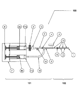
FIG. 7 is a drawing of the interior components, container assembly, of an
exemplary device of the
present invention.
FIG. 8 is a drawing of an exemplary device of the present invention.
FIG. 9 is a drawing of an exemplary device of the present invention.
=
4b
CA 2817296 2020-10-28
CA 028172962013-05-08
WO 2012/064866 PCT/US2011/060013
Fig. 10 is a drawing of an exemplary device of the present invention.
Fig. 11 is a graph of the pressure readings from experiments with an exemplary
device
comprising a pressure relief mechanism.
DETAILED DESCRIPTION
The present invention comprises methods and devices for making and using
contrast
agents for ultrasound or sonography visualization of structures. Such
structures may be
present in the bodies of humans or animals, or may be inanimate structures. As
discussed
herein, the methods and devices are used for ultrasound visualization of a
uterus and one or
more fallopian tubes of a mammal. It is to be understood that the methods and
devices are
not limited to this application, but can be used in visualization of ducts or
structures, whether
in living beings or inanimate structures.
The present invention comprises devices for making a contrast medium
composition.
As used herein, contrast agent and contrast medium mean a composition that is
visible or
visualizable by methods known to those skilled in the art, including but not
limited to
ultrasound, fluorography, radiography, or other detection methods, and the
terms may be used
interchangeably. A method of the present invention comprises use of a contrast
medium
device for generating and deliverying a contrast agent that is useful for
sonographically
observing organs or bodily structures, for example, the uterus and fallopian
tubes. A method
of the present invention comprises use of a contrast medium device for
generating a contrast
agent that is useful for sonographically observing organs or bodily
structures, for example, at
least one fallopian tube.
A contrast medium device comprises a container assembly and optionally, a
catheter
assembly fluidly coupled to the container assembly. Exemplary embodiments of a
container
assembly of the present invention are illustrated in Figs. 1-4 and Figs. 7-10.
A container
assembly may be provided with as casing (not shown in the Figs.) to enclose at
least a portion
of the container assembly. For example, a casing may enclose the components of
a container
assembly, and optionally an exit port, an actuator, and/or both plunger ends
may be found on
the exterior of the casing.
A contrast medium device comprises a container assembly and optionally, a
catheter
assembly fluidly coupled to the container assembly, and optionally, pressure
control
elements. A container assembly may comprise at least one container for a
fluid. A fluid may
be a liquid or a gas. A container assembly may comprise a first container for
a liquid, such as
5
CA 028172962013-05-08
WO 2012/064866 PCT/US2011/060013
saline, and a second container for a gas, such as air, and elements for
creating an alternating
pattern of gas and fluid. A container assembly may comprise connection
elements, such as
tubing or fluid conduits, for providing the contained fluid from a container
to a contrast
pattern generating chamber and to the catheter assembly, or from the exterior
of the container
assembly to a contrast pattern generating chamber and to a container. The
connection
elements may be used for providing fluids from the exterior of the device to
the containers.
A container may comprise one or more outlets through which the fluid, such as
gas or liquid,
exits the container, or the outlet may be used to provide a fluid, either
liquid or gas into the
container. A container assembly may comprise a component for providing force
upon the
fluid contained within the container to move fluid into, or out of, the
container. For example,
a container may be a syringe body or barrel, and the component for providing
force upon the
fluid is a syringe plunger. The container assembly may comprise a component
for activating
the component for providing force. For example, the container may be a syringe
body or
barrel, the component for providing force upon the contained fluid is a
syringe plunger, and
the component for activating the plunger may be a pump, or the hand of an
operator. An
aspect of the invention comprises an embodiment where the contrast medium
device
comprises two containers, such as two syringe bodies, and the syringe plungers
arc moved in
concert because the two plunger ends are held together by a component, such as
an actuator,
such that the syringe plungers move through the interior of the barrel of the
syringes at the
same rate, speed and distance through the interior. The syringe plungers move
at the same
rate, speed and distance because the proximal ends of each plunger are linked
together, such
as by an element, an actuator.
The container assembly may further comprise fluid connections, which are fluid
connecting elements between elements that are in fluid connection with one
another, such as
the one or more containers and a contrast pattern generating chamber. Such
fluid connections
include, but are not limited to, conduits, tubing or needles. The container
assembly may
comprise a contrast pattern generating chamber wherein a gas phase anda liquid
phase are
admixed and the composition exiting the contrast pattern generating chamber,
the contrast
medium composition, is characterized by alternating phases of gas and liquid
which form the
pattern of the contrast medium composition. The container assembly may
comprise fluid
connections which provide the contrast medium composition to a catheter
assembly or
directly to a structure to be visualized.
6
CA 028172962013-05-08
WO 2012/064866 PCT/US2011/060013
In an embodiment, a contrast medium device may comprise a container that may
function as a contrast pattern generating chamber, wherein the contrast medium
is made
within the container, no contrast pattern generating chamber is present, and
the contrast
medium composition, for example comprising gas and liquid phases, is provided
to the
exterior of the contrast medium device.
The container assembly may be in fluid connection with a catheter assembly.
The
catheter assembly may comprise a single or double lumen catheter. The catheter
may
comprise end structures, such as a balloon on the delivery end of the
catheter, wherein the
delivery end is distal from the contrast medium device and the attachment end
is proximal to
the contrast medium device. The opposite end of the catheter, the attachment
end, may have
attachment elements for attaching the catheter to for example, the contrast
medium device.
Attachment elementssuch as a luer lock, may be used to attach the catheter to
a contrast
medium device, and attachment elements are known. The catheter may comprise
other
components such as a wire, sensors, cutting elements, retrieval elements such
as clamps or
pincers. Such catheters are known in the art and one skilled in the art can
select an
appropriate catheter for the intended procedure.
The present invention comprises devices for delivery of a contrast medium to a
structure. It is contemplated by an embodiment of the present invention that
the contrast
medium is provided by the catheter assembly substantially directly to a
structure to be
visualized. In an aspect of the invention, for example, in direct delivery to
a fallopian tube,
the amount of contrast medium used per each fallopian tube evaluation may be
small, such as
less than 20 mL, less than 15 mL, less than 10 mL, less than 8 mL, less than 5
mL, less than 4
mL, less than 3 mL, less than 2 mL, less than 1 mL, less than 0.5 mL. The
amount of
contrast fluid used may be any amount that is sufficient to provide an
accurate visualization
of the structure. The contrast fluid may substantially fill the structure
visualized, or may only
be present in particular locations within the structure.
The present invention comprises contrast medium devices for delivery of a
contrast
medium to one or more structures, such as to multiple cavities, organs or
conduits that are in
fluid connection with one another. It is contemplated by an embodiment of the
present
invention that the contrast medium is provided by a catheter assembly to at
least one structure
to be visualized, and optionally, by providing contrast medium to one
structure, the contrast
medium may also flow into a second, third or further structure to be
visualized. In an aspect
of the invention, for example, visualization of a fallopian tube may first
involve providing a
7
CA 028172962013-05-08
WO 2012/064866 PCT/US2011/060013
sufficient amount of contrast medium to the uterus so that the fluid fills the
uterus to a certain
extent and then the fluid is moved into one or more fallopian tubes in fluid
connection with
the uterus. The fluid may further move into and through the fallopian tubes to
enter the
abdominal cavity. The amount of contrast medium used in a procedure to view
the uterus
and at least one fallopian tube may be from about 5 mL to about 100 mL, from
about 10 mL
to about 100 mL, from about 15 mL to about 90 mL, from about 10 mL to about 80
mL, from
about 20 mL to about 70 mL, from about 30 mL to about 60 mL. The amount of
contrast
medium generated and delivered to the patient may be about 5 mL, about 10 mL,
about 20
mL, about 30 mL, about 40 mL, about 50 mL, about 60 mL, about 70 mL, about 80
mL,
about 90 mL, or about 100 mL, or greater than 100 mL if needed for
visualization of a uterus
and fallopian tube, or for multiple visualizations. For example, a large
cavity, or a cavity
connected to several conduits may require more than 100mL for visualization of
the entire
cavity, and/or the conduits. The amount of contrast fluid used may be any
amount that is
sufficient to provide an accurate visualization of the structure to be
examined. The contrast
fluid may substantially fill the structure visualized, or may only be present
in particular
locations within the structure.
For example, a contrast medium device capable of generating a contrast medium
composition of about 20 mL, using two containers, syringe bodies, of 10 mL
each, and
transfer some or all of the contrast medium to a catheter system wherein the
deliver end is
positioned within the uterus. The contrast medium may enter the uterus and
flow directly to
the fallopian tubes where the contrast medium is visualized, for example, by
sonography.
Five to ten mL of contrast medium may be used for such a visualization of both
fallopian
tubes. The flow of contrast medium may be paused or ceased at this point. If a
second
visualization is desired, the flow of contrast medium may be resumed, and
visualization of
body structures by the presence of contrast medium may be performed.
An advantage of the present invention is that contrast medium flow is
controlled so
that some or all of the contrast medium composition may be provided to a body
structure.
The flow of the contrast medium out of the device and/or out of a catheter,
and to a body
structure may be controlled so that providing the contrast medium may be in a
continuous
flow or intermittent flow, such as providing some contrast medium, stopping
the flow,
providing contrast medium, stopping the flow, and so on. The container(s) of a
contrast
medium device may be refilled one or more times during a procedure. The flow
of contrast
medium to the structure may be controlled automatically or by an operator. The
rate of
8
delivery of contrast medium may be controlled. The rate of delivery may be in
a range
from fast to slow, and is primarily controlled by the rate of force applied to
the
component(s) for providing force upon the fluid contained within the
container(s). In
an embodiment wherein the contrast medium device comprises two containers,
such as
two syringe bodies, and the component for providing force upon the fluid
contained
within each container is a plunger, the rate of force applied to the fluid in
each
container is identical when the plungers are activated simultaneously with the
same
force applied, to provide the same rate of delivery of contrast medium from
the device.
An embodiment of the present invention comprises visualization of at least one
fallopian tube using a catheter placed at or near the opening of the fallopian
tube. Any
device that provides a catheter to the fallopian tube, may be used. A catheter
may be
connected to the contrast medium device comprising a container assembly
described
herein. A particular device for providing a catheter to a body structure, such
as
fallopian tube, and that may be useful in methods of visualizing a fallopian
tube is the
device taught in U.S. Patent 8,048,086 issued November 1, 2011; U.S. Patent
8,048,101
issued November 1, 2011; and U.S. Patent 8,052,669 issued November 8, 2009,
each
of which may be referred to in their entirety for further details. In general
these
applications disclose a device comprising a housing and an introducer shaft
that is used
to enter and traverse the uterus until the tip of the shaft approaches or
touches the
fundus of a uterus. Once the tip of the introducer shaft is at the fundus of
the uterus,
the device may be stabilized. One or more catheters, such as two, are fed
through the
introducer shaft and out into the uterine cavity. The placement of the
introducer shaft
allows for the three dimensional alignment of the catheter(s) with the comua
of the
uterus. The catheter(s) is advanced until the delivery ends(s) of the
catheter(s) are in
place in the comua. An end structure, such as a balloon, is inflated or
engaged, to
stabilize the catheter(s) in place, and the end structure may prevent or
minimize
backflow of materials exiting the catheter delivery end. Once the end
structure is
engaged, the catheter(s) is ready for delivery of materials or other
activities.
In a method of the present invention, the catheter placed by the introducer
shaft
comprises a catheter assembly. The end of the catheter opposite the delivery
end,
referred to herein as the proximal end or the attachment end, is attached to a
contrast medium device of the present invention. The contrast medium is
generated
by the actions of the container assembly and the contrast medium is provided
into and through the catheter(s) and out into
9
CA 2817296 2018-02-08
CA 028172962013-05-08
WO 2012/064866 PCT/US2011/060013
the cornua of the uterus and into a fallopian tube(s). Visualization
techniques are initiated as
the contrast medium enters the fallopian tube(s) and if possible, flows
through the tube(s) and
out into the peritoneal cavity. If a tube is blocked, the medium will not flow
in that tube but
may flow to a second, the contralateral, tube if that second tube is not
blocked. The pressure
built up by the blockage may or may not unseat an end structure on the
catheter, such as a
balloon, in an effort to relieve pressure, but if the end structure of the
catheter is moved, the
flow would then be directed into the uterus or the unblocked tube.
If the device providing the catheter uses only one catheter, then
visualization of one
fallopian tube occurs, followed by readjustment of the device, such as
rotation of the
introducer shaft, as taught in the cited patent applications, and the steps
are repeated to
provide a contrast medium to the other fallopian tube. The contrast medium
provided may be
any currently known contrast medium that may be provided through a catheter to
a location.
An embodiment of the present invention comprises using a contrast medium
device
and catheter to provide contrast medium to the uterus for visualizing at least
one fallopian
tube, or visualization of at least a portion of the uterus and at least one
fallopian tube. When
the structure to be visualized is a at least one fallopian tube, or a uterus
and/or at least one
fallopian tube, a contrast media device of the present invention, in
combination with a
catheter may be used. A catheter having elements for preventing retrograde
flow of fluid
from the uterus may be connected to a contrast medium device comprising a
container
assembly as described herein. Catheters with element(s) that prevent
retrograde flow are
known in the art, and it is within the skill of those in the art for selecting
a catheter to attach
to a contrast media device of the present invention to use in methods taught
herein.
In a method of the present invention, a catheter, such as a balloon catheter,
is a
catheter assembly. In the method, the delivery end of a catheter is placed in
the uterus, and
optionally, the structure to prevent retrograde flow into the cervix is
employed, for example,
the balloon of a balloon catheter is expanded. The end of the catheter
opposite the delivery
end, referred to herein as the proximal end or the attachment end, is attached
to a contrast
medium device of the present invention. The contrast medium is generated by
the actions of
the device and the contrast medium is provided into and through the
catheter(s) and out into
the uterus. The desired amount of contrast medium is provided and
visualization techniques
are initiated, and can be used to visualize the movement of the contrast
medium into the
uterus, to visualize at least a portion of the structure of the uterus, for
example, by providing
the contrast medium, and/or to visualize entry, transit and/or exit of the
contrast medium in at
CA 028172962013-05-08
WO 2012/064866 PCT/US2011/060013
least one fallopian tube. If a fallopian tube is blocked, the contrast medium
will not flow past
the blockage but may flow to the contralateral tube if not blocked. The
pressure built up by
the blockage may or may not be detected by an element of the contrast medium
device that is
designed to detect the fluid back pressure created by the lack of continued
flow of the fluid
through the structure and/or conduits. Should the desired pressure be reached,
the contrast
medium flow may be halted, such as by a medical professional providing
contrast medium
may stop applying pressure to the contrast medium device, and cease providing
fluid through
the catheter to the uterus. The contrast medium provided may be any currently
known
contrast medium that may be provided through a catheter to a location, or may
be the
liquid/air contrast medium disclosed herein.
A contrast medium device of the present invention may be provided with
containers
filled with fluid(s) or may be provided with empty containers that must be
first filled with
fluid(s) prior to generating and delivering contrast medium. If all of the
fluid in the contrast
medium device is used in a procedure and more contrast medium is desired, the
contrast
medium device of the present invention may be refilled, as in the containers
of the device are
filled with the respective fluid(s). In using prefilled containers, the
original containers may
be removed and new containers inserted into the device. In using refillable
containers, the
containers may be refilled without removing the delivery end of the catheter
from the patient.
The contrast medium device may be unattached from the proximal end of the
catheter and the
containers refilled with more of the same type of fluid, or a different fluid
if desired, as was
used in the first delivery of contrast medium.
In using a contrast medium device of the present invention, once an amount of
contrast medium is provided and the containers are depleted of contrast
medium, the
containers may be refilled one or multiple times so as to provide an effective
amount of
contrast medium to the structure and/or conduits of the body. For example, in
an
embodiment where the contrast medium comprises air and saline segments in a
pattern ,
which may be produced by a contrast medium device of the present invention
having two
containers, wherein one container provides saline and a second container
provides air. For
example, the containers may be 10mL syringes, wherein a first container
contains 10 mL of
saline and a second container contains 10 mL of air or a gas. The contrast
medium is
generated or produced by simultaneously moving the saline and air from each
container, such
as by applying pressure to a plunger in each container, or by a pump or other
means for
moving a fluid from a syringe or similar container. The air and saline are
moved into a
11
CA 028172962013-05-08
WO 2012/064866 PCT/US2011/060013
contrast pattern generating chamber. The contrast pattern generating chamber
may comprise
a static mixer or similar structure to mix the air and liquid fluids, creating
interspersed
segments of saline and segments of air, thus generating a contrast medium
comprising saline
with air bubbles or air segments contained therein. The static mixer may
comprise elements,
such as helical elements that simultaneously produce patterns of flow division
and radial
mixing of the air and saline fluids. Static mixers are known in the art and
generally comprise
mixer elements contained within a tube or housing, for example a cylindrical
tube. Static
mixer elements may comprise a series of baffles that are made from metal or a
variety of
plastics. The static mixer works by continuously blending two fluids as the
streams of fluids
move through the static mixer. Other mixing elements may be used in the
present invention,
and such elements are known to those skilled in the art. Alternatively, a
contrast medium
generating chamber may comprise only a conduit into which both a gas conduit
from the gas
container and a liquid conduit from the liquid container enter, and does not
comprise a static
mixer or other mixing element. As the gas and liquid are simultaneously
provided to the
contrast medium generating chamber conduit, gas segments are created within
the fluid to
generate a contrast medium composition.
When the containers of the contrast medium device are empty, for example,
before
initiation of a procedure,or substantially all of the fluids are no longer
present in the
containers, such as during a procedure, in an embodiment of the present
invention, the
containers may be filled or refilled simultaneously without disassembly of the
contrast
medium device or removal of the containers from the device housing. For
example, when a
contrast medium device comprises two containers, wherein a first container
contains saline
and a second container contains air, the first and second containers may be
filled or refilled
with saline and air, respectively, without disassembling the contrast medium
device, or
removing the containers. If a catheter is attached to the exit port of the
contrast medium
device, it may optionally be removed when filling the containers, and left in
place in the
patient. The exit port end of the contrast medium device, previously attached
to the catheter
or prior to attachment to the catheter, may be immersed in a saline solution.
If plungers are
present in the containers, such as syringe plungers or similarly acting
movable seals within
the container body, the plungers arc withdrawn up the interior of the syringe
cylinder in a
proximal direction and away from the exit port, creating a lowered pressure
within the
syringe cylinder, which causes the saline and air to enter the respective
containers (syringes).
Typically, the plungers are controlled simultaneously so that both plungers
are drawn up the
12
CA 028172962013-05-08
WO 2012/064866 PCT/US2011/060013
interior of the syringe cylinder at the same rate so that both syringes are
refilled
simultaneously with air in the air cylinder and saline in the saline cylinder.
The air cylinder
is in fluid connection with at least one in-line check valve attached to the
air syringe barrel,
and optionally with an in-line air filter, which allows for one direction
filling of the air
syringe barrel.
The path of air or gas during the filling of a container placed within a
contrast
medium device is as follows. As the air syringe barrel is withdrawn up the
interior of the
syringe in a proximal direction, away from the exit port, while the exit port
of the device is
immersed in a saline solution, air flows in through the optional air filter,
through fluid
connections to at least one one-way check valve, through the at least one
check valve,
through connectors connecting the at least one check valve and the syringe
(container) exit,
and into the air syringe barrel. When the plunger reaches the proximal end of
the syringe
cylinder, and/or is withdrawn to the desired extent, for example, the entire
length of the
syringe barrel, the syringe is filled, for example, with 10mL of air.
At the same time and rate as the air syringe plunger is being withdrawn
through the
air syringe, the fluid syringe plunger is being withdrawn at the same rate and
distance through
the fluid syringe barrel. As the exit port of the contrast medium device is
immersed in saline,
the movement of the fluid syringe plunger in a proximal direction, away from
the exit port,
causes saline to enter the exit port of the device and move through the fluid
connections to
the fluid syringe container. When the plunger reaches the proximal end of the
fluid syringe
cylinder, and/or is withdrawn to the desired extent, for example, the entire
length of the
syringe barrel, the syringe is filled with, for example, 10mL of fluid, for
example saline.
When the plungers have ceased moving, for example when each of the plungers is
at the
desired location within the syringe barrel, the air container and the fluid
container contain
substantially the same amount of air and saline in the respective containers.
The contrast
medium device is now filled or refilled. In continuing the visualization
procedure, the
catheter may be rejoined to the attachment element(s) of the exit port of the
device and with
opposite action by the plungers, moving down the syringe barrel toward to exit
port, fluid
(saline) and air are forced from the respective fluid and air containers. The
contrast medium
is generated in the contrast medium generating chamber, for example, by the
fluid/air streams
flowing through a mixer, such as a static mixer, or the fluids mixing in a
mixing chamber
lacking a mixing element, and the contrast medium composition comprising air
and liquid
segments exits the contrast medium device, for example, into and through the
attached
13
CA 028172962013-05-08
WO 2012/064866 PCT/US2011/060013
catheter. The contrast medium may enter the cavity, such as the uterus, and at
least one
fallopian tube, wherein visualization of the contrast medium within the
structure is
conducted, such as by sonographic methods, and the body structures are
examined.
The disclosure herein refers to fluids such as air or saline, but it is
contemplated that
.. the present invention is not limited to air and/or saline, and that one of
skill in the art can
substitute air and/or saline for other appropriate fluids, such as other
liquids, other gases or
known contrast medium compositions. Methods of the present invention comprise
making or
generating a contrast medium, and delivering a contrast medium to a body
structure. A
contrast medium device of the present invention is used to make a contrast
medium. For
example, an embodiment of a contrast medium device comprising one container
for fluid may
comprise a container comprising a flexible porous material contained within
the container.
An example wherein the container is a syringe body is described, such as one
shown in Fig.
4. The present invention is not limited to this design, but contemplates other
containers that
would function in a similar manner. The syringe is substantially filled with a
flexible porous
material. The flexible porous material includes, but is not limited to, strips
or pieces of
woven or nonwoven material, an open-celled material, such as a sponge, or
fragments of a
sponge, or any material that would contain a gas and release the gas when
acted upon, such as
by compression forces. For example, the flexible, porous material is an open-
celled sponge.
The sponge is placed in the container and a liquid is added, but the liquid
does not displace all
of the air in the sponge. The syringe plunger is applied to the large open end
of the syringe
and the other end of the syringe is in fluid connection with the catheter
assembly. As the
plunger is depressed into the syringe, the sponge is compressed and the air is
forced out into
the liquid, creating bubbles or air segments. The bubbles and fluid, the air
and fluid
segments, enter the catheter and transit the catheter to the structure.
Visualization of the
structure is then possible. See Fig. 5 for an illustration of visualization of
a fallopian tube.
The present invention comprises contrast medium devices comprising more than
one
container. For example, the contrast medium device may comprise two
containers, such as
one shown in Fig. 1 and Figs. 7-10, an example wherein the containers comprise
a syringe
body, also herein referred to as a syringe ban-el or syringe container.
Optionally, the interior
of the syringe barrel may be traversed by a plunger element. The plunger
element may be
moved, such as by an operator, through the interior of the syringe container
from a proximal
location in the barrel, wherein proximal refers to the end of the device
closest to the operator
and away from the exit port of the device, to a distal location, wherein
distal refers to the end
14
CA 028172962013-05-08
WO 2012/064866 PCT/US2011/060013
of the device closest to the patient and nearer to the exit port, and from a
distal location in the
barrel to a proximal location in the barrel. A plunger element may be
comprised of a piston
and a fluid seal having two surfaces, wherein the piston is attached to one
surface of the fluid
seal, the proximal surface, and the other surface, the distal surface, faces
and contacts the
deliverable fluid. A standard syringe plunger is a plunger element. The fluid
seal, having
two surfaces, forms a fluid seal within the container, so that a deliverable
fluid is maintained
or contained on the distal surface of the plunger surface (a first surface)
and no deliverable
fluid is present on proximal surface (a second surface). A deliverable fluid
is the fluid
contained within the container and which is intended to be provided to the
structure to be
visualized and/or examined. As a plunger, comprising a piston and fluid seal,
is moved
through a syringe, there may be air or a slight vacuum created on the proximal
side of the
plunger, but there is no intention to provide the air on the proximal side of
the seal, therefore
this air is not a deliverable fluid. The present invention is not limited to
this design, but
contemplates other containers that would function in a similar manner. One of
the containers,
which may be a pre-filled syringe, contains a liquid. The liquid may be any of
those
disclosed herein, such as saline or water, or known contrast agent fluids. A
second container,
which may be a pre-filed syringe, contains a gas. The gas may be any of those
disclosed
herein, such as air, carbon dioxide, oxygen, nitrogen or halocarbon compound
gases, other
gases, or known contrast agent gases. The plungers of the two syringes are
depressed
simultaneously, either manually or mechanically, and the mixture of the gas
and liquid form
an alternating pattern of gas phase and liquid phase, which is a contrast
medium composition.
The contrast medium composition then enters and transits an attached catheter
and exits into
the structure, such as the fallopian tube. Visualization of the structure is
possible by
ultrasound techniques.
Alternatively, a device of the present invention may comprise two containers,
such as
two syringes, that are provided with no fluid. In use of such a device, each
plunger is
depressed to a position in the distal end of each container, such as a
syringe, and the exit port
of the device is placed in saline. As each plunger is simultaneously moved to
a desired
proximal location within the container, air is drawn into a first container
and fluid is drawn
into a second container. Substantially simultaneous filling of a dual
container device is
disclosed herein. Once the containers are filled, the plungers may be
depressed, moving the
surface of the fluid seal to a more distal location and dispensing the fluids
from the
containers. The fluids are mixed, gas and liquid are admixed to form a
contrast medium
CA 028172962013-05-08
WO 2012/064866 PCT/US2011/060013
composition comprising air and liquid segments, and the contrast medium
composition flows
out the exit port of the device, and optionally into a catheter placed within
the structure to be
examined. An embodiment of the present invention may comprise movement of one
plunger,
and filling with a fluid into, or providing a fluid from one container.
Compositions of the present invention comprise a contrast medium made using
the
methods taught herein. A contrast medium of the present invention comprises a
gas phase
within a liquid carrier. The gas phase may be a bubble or may be a liquid-
free, gas-filled area
adjacent to a liquid phase area, and the alternating gas-filled area and
liquid area may repeat
multiple times. The sizes of the gas-filled areas or the liquid filled areas
may be uniform in
size or not. The present invention contemplates an aspect in which providing a
contrast
medium in reduced volumes is used, compared to amounts currently used which
may be 20
mL or more, and providing the contrast medium substantially in or very near
the structure to
be visualized (i.e. fallopian tube).
The present invention contemplates providing an amount of contrast medium that
is
effective to view a structure. For example, an effective amount of a contrast
medium may
comprise 5 mL to 200 mL, depending on the volume of the structure and the
number of
structures to be examined. For example, if a device of the present invention
is used to
provide contrast medium to the uterus and fallopian tubes, an effective amount
of contrast
medium to be provided to those structures may be greater than the amount used
to provide a
contrast medium directly to the fallopian tubes. The present invention
controls the amount of
gas and liquid used in combination to form the mixed gas/liquid composition,
which enters
the structure. The pattern of the contrast medium composition can range from
predominantly
a gas (air or other gas) phase to predominantly a liquid (saline or other
liquid) phase and can
be provided in a regular pattern or in an irregular pattern. The ratios of the
gas to liquid may
be determined by the size of the respective syringe. The larger the air
syringe the greater the
air segment in the pattern of the composition. The use of a porous structure
may create a
more random or irregular pattern. The amount of contrast medium delivered may
be
controlled by the amount of syringe plunger displacement or by refilling the
containers one or
more times.
A contrast medium device of the present invention generates and delivers a
reproducible and reliable pattern of alternating air and fluid that is visible
by detection
methods such as sonography. The air/liquid pattern produced by a composition
generated by
a device of the present invention is reproducible in that a substantially
regular repeating
16
CA 028172962013-05-08
WO 2012/064866 PCT/US2011/060013
pattern of alternating air and liquid is generated as a contrast medium
composition by the
device, is provided to a body structure, and for example, is visible with a
fallopian tube. The
pattern is consistently produced by a device, as in a device of the present
invention generates
a contrast medium composition wherein the distance between the air/saline
interfaces is short
enough and repeats regularly enough that movement of the composition through a
structure is
visible, such as by sonography. The consistent pattern may be viewed in the
uterus and in
open, not blocked, fallopian tubes by detection methods, such as sonography.
It is
contemplated by the present invention that the distance between interfaces of
a contrast
medium of the present invention is not necessarily identical for every pair of
interfaces but
that the distances are sufficiently similar in size so as to form the
perception of a repeating
pattern of light and dark by detection means such as sonography, and that the
structure of a
body structure, such as fallopian tube, can be viewed by the movement of the
regular pattern
of light and dark produced by the interfaces under detection means such as
sonography.
For example, in order to evaluate the fallopian tubes for patency, to
determine
whether the tube is open and lacking obstructions or blockages, it is desired
that the saline
and air composition delivered have air/saline interfaces that are in a
frequent regular
alternating pattern of varying intervals. Saline alone appears black when
viewed by
sonography as saline reflects less sound echoes to the probe, therefore, long
intervals of
saline may present a problem for the user to visualize a body structure, such
as fallopian tube.
Air appears white, as does bone, as air reflects more sound echoes to the
probe, therefore,
long intervals of air may be misinterpreted and easily confused with other
body tissues,
leading to uncertainty in the diagnosis of fallopian tube patency. Movement of
the saline and
air interfaces with a repeating pattern, as described by the current
invention, that is frequent,
regular, and alternating allows for an effect called reverberation that is
caused by sound
interfacing with two structures of sufficiently different reflective
properties. The described
invention also allows for a comet-tail effect, a type of reverberation caused
by a number of
small, highly reflective interfaces, such as air bubbles in a fluid. If the
pattern is too erratic,
as is seen in literature of historical and previously used methods, a
confident and accurate
diagnosis of the structure or patency of a fallopian tube cannot be assessed,
and certainly not
easily and reliably. With an erratic and not regular pattern, the complexity
of the Sono
Hysterography procedure increases dramatically, making the reliability of the
procedure
questionable and the learning curve, for a medical professional to learn and
perform the
procedure, very steep. Specifically, erratic patterns consisting of small
pattern frequencies
17
CA 028172962013-05-08
WO 2012/064866 PCT/US2011/060013
creating a long segment of either air or saline can lead to misinterpretations
as there will not
appear to be movement of the contrast medium, and it is the perception of the
movement of
the contrast medium in and through a structure that is necessary for the
medical professional
to make an evaluation of the fallopian tubes.
Additionally, in sonographic procedures examining the uterus and fallopian
tubes, it
can be challenging to obtain the optimal sonographic probe position/location
in the correct
plane due to varying patient anatomical positions of the uterus and fallopian
tubes.
Therefore, the consistent movement of a frequent, regular alternating pattern
of the contrast
medium, such as that generated by a device of the present invention, will
increase the
likelihood of the body structure(s) being viewed by the physician (medical
professional) to
make the intended evaluation. During the procedure, the physician may have
difficulty
viewing one of the fallopian tubes, such as where flow is easily viewed in one
fallopian tube
and the other tube is either blocked, difficult to locate or experiencing
tubal spasm. In such
situations and others like it, the user may need to deliver additional saline
and air to the
patient to rule out or confirm blockage in the unviewed tube, hence the aspect
of the
invention enabling quick and easy refilling of the device with fluids, such as
air and saline, is
advantageous for the ease and convenience of the procedure. Patient discomfort
or a decision
to delay the procedure a few minutes to allow the tubes to relax to challenge
a difficult tube
evaluation may also necessitate delay in delivery of the saline and air,
extending the overall
procedure time. Thus, providing a contrast medium composition that provides a
regularly
repeating pattern of interfaces of air segments and liquid segments, such as
after a pause in
the procedure or after a refilling of the device containers, is an advantage
of the present
invention in view of prior devices.
A composition of the present invention may comprise a liquid and a gas, and
optionally, surfactants, emulsifiers, or other stabilizing agents. The liquid,
which may be
seen as a carrier of the gas phase, may be any liquid that is substantially
free of solids and
flows at normal or bodily temperatures. For example, the liquid may be water
or
physiologically acceptable aqueous solutions including, but not limited to,
physiological
electrolyte solutions, physiological saline solutions, Ringer's solution or
aqueous solutions of
sodium chloride, calcium chloride, sodium bicarbonate, sodium citrate, sodium
acetate, or
sodium tartrate, glucose solutions, or solutions of mono- or polyhydric
alcohol, e.g., ethanol,
n-butanol, ethylene glycol, polyvinylpyrrolidone, or mixtures or combinations
of these.
Further, the liquid canier may comprise physiologically acceptable non-aqueous
solutions,
18
CA 028172962013-05-08
WO 2012/064866 PCT/US2011/060013
including, but not limited to, anhydrous or substantially anhydrous carrier
liquids, alcohols,
glycols, polyglycols, synthetic perfluoranated hydrocarbons, or in mixtures or
combination
with other non-aqueous or aqueous liquids.
Contrast media compositions of the present invention may comprise surfactants
or
compounds that stabilize the gas-liquid interface. Surfactants may be provided
in the liquid
phase of the contrast medium. For example, if a contrast medium composition
comprises air
and a liquid, such as saline, one or more surfactants may be added to the
saline. Surfactant
compositions may be useful when the contrast medium is provided to a structure
that is larger
than the catheter size used to transmit the contrast medium. Surfactants
include tensides,
such as lecithins; esters and ethers of fatty acids and fatty alcohols with
polyoxyethylene and
polyoxyethylated polyols like sorbitol, glycols and glycerol, cholesterol; and
polyoxy-
ethylene-polyoxypropylene polymers, viscosity raising and stabilizing
compounds, mono-
and polysaccharides (glucose, lactose, sucrose, dextran, sorbitol); polyols,
e.g., glycerol,
polyglyeols; and polypeptides like proteins, gelatin, oxypolygelatin, plasma
protein,
.. amphipathic compounds capable of forming stable films in the presence of
water and gases,
such as the lecithins (phosphatidyl-choline) and other phospliolipids, inter
alia phosphatidic
acid (PA), phosphatidylinositol, phosphatidylethanolamine (PE),
phosphatidylserine (PS),
phosphatidylglycerol (PG), cardiolipin (CL), sphingomyelins, the plasmogens,
the
cerebrosides, natural lecithins, such as egg lecithin or soya bean lecithin,
or synthetic
lecithins such as saturated synthetic lecithins, for example,
dimyristoylphosphatidylcholine,
dipalmitoylphosphatidylcholine or distearoylphosphatidylcholine or unsaturated
synthetic
lecithins, such as dioleylphosphatidylcholine or
dilinoleylphosphatidylcholine, free fatty
acids, esters of fatty acids with polyoxyalkylene compounds like
polyoxypropylene glycol
and polyoxyalkylene glycol; ethers of fatty alcohols with polyoxyalkylene
glycols; esters of
fatty acids with polyoxyalkylated sorbitan; soaps; glycerol-polyalkylene
stearate; glycerol-
polyoxyethylene ricinoleate; homo- and copolymers of polyalkylene glycols;
polyethoxylated
soya-oil and castor oil as well as hydrogenated derivatives; ethers and esters
of sucrose or
other carbohydrates with fatty acids, fatty alcohols, these being optionally
polyoxyalkylated;
mono- di- and triglycerides of saturated or unsaturated fatty acids;
glycerides of soya-oil and
sucrose, block copolymers of polyoxypropylene and polyoxyethylene (poloxamers)
polyoxyethylenesorbitans, sorbitol, glycerol-polyalkylene stearate,
glycerolpolyoxyethylene
ricinoleate, homo- and copolymers of polyalkylene glycols, soybean-oil as well
as
hydrogenated derivatives, ethers and esters of sucrose or other carbohydrates
with fatty acids,
19
CA 028172962013-05-08
WO 2012/064866 PCT/US2011/060013
fatty alcohols, glycerides of soya-oil, dextran, sucrose and carbohydrates.
Surfactants may be
film forming and non-film forming and may include polymerizable amphiphilic
compounds
of the type of linoleyl-lecithins or polyethylene dodecanoate, phosphatidic
acid,
phosphatidylcholine, phosphatidylethanolamine, phosphatidylserine,
phosphatidylglycerol,
phosphatidylinositol, cardiolipin, sphingomyelin and biocompatible and
amphipathic
compound capable of forming stable films in the presence of an aqueous phase
and a gas,
phospholipids including phosphatidylcholine (PC) with both saturated and
unsaturated lipids;
including phosphatidylcholine such as
dioleylphosphatidylcholine;
dimyristoylphosphatidylcholine (DMPC),
dipentadecanoylphosphatidylcholine-,
dilauroylphosphatidylcholine (DLPC), dipalmitoylphosphatidylcholine (DPPC);
disteraoylphosphatidylcholine (DSPC); and diarachidonylphosphatid-ylcholine
(DAPC);
ph osphati dyl eth an ol am i nes (PE), such as di
oleylphosph ati dyleth anolam in e,
dipaimitoylphosphatidylethanolamine (DPPE) and
distearoylphosphatidylethanolamine
(DSPE); phosphatidylserine (PS) such as dipalmitoyl phosphatidylserine (DPPS),
disteraoylphosphatidylserine (D SP S); phosphatidylglycerols (PG),
such as
dip alm itoylpho sphati dylglycerol (DPP G), di stearoylpho sphatidyl glyc
erol (D SP G); and
phosphatidylinositol. Surfactants, emulsifiers, or other stabilizing agents
may aerosolized
within the gas phase.
Contrast medium compositions may comprise gases, and any physiologically
acceptable gas rnay be present in the compositions of the present invention.
The term "gas" as
used herein includes any substances (including mixtures) substantially in
gaseous form at the
normal human body (37 C). Close to 200 different gases have been identified
as potentially
useful for making ultrasound contrast agents, and include oxygen, air,
nitrogen, carbon
dioxide or mixtures thereof, helium, argon, xenon, krypton, CHC1F2 or nitrous
oxide, sulfur
hexafluoride, tetrafluoromethane, chlorotrifluoromethane,
dichlorodifluoromethane,
bromotrifluoromethane, bromochlorodifluoromethane,
dibromodifluoromethane
dichlorotetrafluoroethane, chloropentafluoroethane, hexafluoroethane,
hexafluoropropylene,
octafluoropropane, hexafluoro-butadiene, octafluoro-2-butene,
octafluorocyclobutane,
decafluorobutane, perfluorocyclopentane, dodecafluoropentane, fluorinated
gases including
materials which contain at least one fluorine atom such as SF6, freons
(organic compounds
containing one or more carbon atoms and fluorine, i.e. CF4, C2F6, C3F.8, C4F8,
C4F 10, CBrF3,
CC12F2, C2C1F5 and CBrC1F2 and perfluorocarbons. The term perfluorocarbon
refers to
compounds containing only carbon and fluorine atoms and includes saturated,
unsaturated,
CA 028172962013-05-08
WO 2012/064866 PCT/US2011/060013
and cyclic perfluorocarbons such as perfluoroalkanes such as perfluoromethane,
perfluoroethane, perfluoropropanes, perfluorobutanes (e.g. perfluoro-n-butane,
optionally in
admixture with other isomers such as perfluoro-isobutane), perfluoropentanes,
perfluorohexanes and perfluoroheptanes; perfluoroalkenes such as
perfluoropropene,
perfluorobutenes (e.g. perfluorobut-2ene) and perfluorobutadiene;
perfluoroalkynes such as
perfluorobut-2-yne, and perfluorocycloalkanes such as perfluorocyclobutane,
perflu oromethylcyclobutane, peril
uoro dimethy lcyc lob utanes ,
perfluorotrimethylcyclobutanes,
perfluorocyclopentane, perfluoromethycylopentane,
perfluorodimethylcyclopentanes, perfluorocyclohexane,
perfluoromethylcyclohexane and
.. perfluorocycloheptane.). The saturated perfluorocarbons, which are usually
preferred, have
the formula C11Fn+2, where n is from 1 to 12, preferably from 2 to 10, most
preferably from 3
to 8 and even more preferably from 3 to 6. Suitable perfluorocarbons include,
for example,
CF4, C2F6, C3F8, C4F8, C4F10, C5F12, C6F12, C7F14, C8F 18, and C9F20.
The present invention comprises embodiments of contrast medium devices and
.. systems. A system of the present invention may comprise separable
components; a contrast
medium device comprising a container assembly, and a catheter assembly that
provides the
contrast medium composition, the fluid output of device, in, near to or in the
targeted duct or
cavity. Alternatively, a system of the present invention may be a single, one-
piece
construction with a contrast medium device comprising a container assembly
adjoined to a
catheter assembly. A contrast medium device may comprise a container assembly
that
provides a contrast medium comprising a gas phase and a liquid phase. A
contrast medium
device may comprise a container assembly comprising a modified conventional
multiple
syringe pump, either a mechanical or a manual handheld device capable of
accepting
variously sized syringes. The syringe outputs are directed into a mixing
chamber or conduit
.. where the appropriately created train of gas (i.e. air) and liquid (i.e.
saline) are then driven
into the input of a catheter assembly. For example, directed delivery of
contrast medium in
the proximity or within the duct (i.e. fallopian tube) will allow for
sonography of the
structure. A contrast medium composition may be provided directly to the
fallopian tube, by
which is meant that the contrast medium composition is delivered only to the
fallopian tube,
or only to the fallopian tube first, and not by a filling of the uterus with a
fluid and having
that fluid overflow into the fallopian tubes. A contrast medium composition
may be provided
to the uterus directly, which may allow for visualization of the uterus, and
by providing a
sufficient amount of contrast medium, the contrast medium may flow to one or
more
21
CA 028172962013-05-08
WO 2012/064866 PCT/US2011/060013
fallopian tubes. The contrast medium may or may not enter and flow through the
fallopian
tube, depending on the patency of the fallopian tube. Providing a composition
directly to a
structure herein means that the composition is provided at or near an opening
of the structure
to be assessed, so that the composition enters the structure and does not flow
into the
.. structure from a remote site of delivery of a composition.
An aspect of the invention comprises use of a device of the present invention
with known hysterosalpingography procedures. For example, such procedures may
be
performed prior to or after use of a device of the present invention. A
procedure may
comprise providing saline only to the uterine cavity to at least in part fill
the uterus or to
distend the uterus. The uterus may be visualized by detection methods, such as
sonography.
The saline is then released from the uterus, such as by releasing a balloon
used to seal the
uterus closed from the cervix, or by withdrawing the catheter that provided
the saline to the
uterus. Alternatively, the saline may flow out of the fallopian tubes. After
such a pre-
treatment procedure, a contrast medium device of the present invention may be
used by
.. attaching the device to a catheter having its delivery end within the
uterus, generating a
contrast medium composition and providing the contrast medium composition to
the uterus
and at least one fallopian tubes. Post-treatments may also be provided to the
uterus or
fallopian tubes after providing the contrast medium composition. For example,
a therapeutic
composition or an embryo composition may then be provided to the uterus or
fallopian tube.
Though not wishing to be bound by any particular theory, it is theorized that
providing a contrast medium composition of the present invention, using a
device of the
present invention aids in the fertility of a patient who has undergone the
methods described
herein. It is thought that there is a higher incidence of becoming pregnant
found in women
who have undergone a procedure comprising using a contrast medium device and
air/saline
contrast medium composition of the present invention. The present invention
comprises a
method for enhancing pregnancy in a female, aiding in or obtaining a pregnant
condition in a
female, or increasing the fertility of a female, comprising, providing a
contrast medium
generating and delivery device comprising a container assembly comprising at
least one
container for containing a fluid, a component for moving a fluid from the
container, and
connections for fluid connection of at least one container to a contrast
medium generating
chamber, such as a device disclosed herein, filling at least one container
with a fluid; moving
the fluid from at least one container to a contrast medium generating
container to generate a
contrast medium composition; and delivering the contrast medium composition to
a body
22
CA 028172962013-05-08
WO 2012/064866 PCT/US2011/060013
structure of the female. Visualization by detection methods such as sonography
may or may
not be performed.
In methods of the present invention, one or both fallopian tubes may be viewed
simultaneously, sequentially or in separate procedures. In some instances, it
may not be
possible to view both fallopian tubes in the same plane of sonographic
imaging. One or both
fallopian tubes may not fill simultaneously, for example, should a spasm
constrict the
opening or a portion of a fallopian tube.
An aspect of the present invention comprises a contrast medium device
comprising a
container assembly comprising a contrast pattern generating chamber having a
diameter in a
range of 0.3 to 1.8 ratio to the diameter of the structure to be visualized.
The diameter of the
contrast pattern generating chamber may be in a ratio of 0.1 to 100 the
diameter of the
structure to be visualized. The contrast pattern generating chamber may have a
diameter ratio
of 0.5 to 1 of the structure to be visualized, a diameter ratio of 1 to 1 of
the structure to be
visualized, a diameter ratio of 1 to 1.5 of the structure to be visualized, a
diameter ratio of 1
to 2 of the structure to be visualized. An aspect of a contrast medium device
comprises a
container assembly comprising a contrast pattern generating chamber that has a
diameter
substantially equal to the diameter of the structure to be visualized, wherein
the ratio of the
diameters is 1.
An aspect of the present invention comprises a contrast medium device
comprising a
container assembly comprising a contrast pattern generating chamber comprising
a static
mixer used to mix two or more fluids provided from the container(s). As two or
more fluids
enter the static mixer, placed in line and in fluid connection with two or
more containers and
before the exit port, the static mixer mixes the two or more fluids. For
example, in an
embodiment of a contrast medium device of the present invention, the fluid of
one container
is saline and the fluid of a second container is air. As the saline and air
are moved from the
respective containers into the static mixer, bubbles of air within the saline
or segments of air
and saline are created by the static mixer. Upon leaving the static mixer and
exiting the
contrast medium device through the exit port, and optionally into a catheter,
the mixture of
air and saline, primarily viewed as bubbles of air within the saline in an
open space like the
uterus or a patterned sequence of air and saline segments when entered into a
tube, is a
contrast medium that can be used to visualize a structure.
The interfaces of the alternating gas and liquid phases must be present in
sufficient
numbers if a duct, tube or structure is to be visualized by this contrast
medium, and both
23
CA 028172962013-05-08
WO 2012/064866 PCT/US2011/060013
phases must be present in the viewing region during the time of viewing. It is
the presence of
both phases traversing the viewing region that provide the visualization
contrast. For
example, if only one phase (either liquid or gas) is visible in the viewing
region at a given
time, assessment is difficult or impossible. By the creation of multiple
interfaces between the
two phases in the contrast medium, observation of structure is possible due to
the flow of the
contrast medium comprising the interfaces of the phases.
An aspect of the present invention comprises contrast medium devices
comprising
contrast pattern generating chambers having diameters similar in diameter to
the structure
being observed. For example, if gas phase is created that is smaller than the
diameter of the
structure to be observed, the gas will rise to the upper portion of the duct
and coalesce with
another gas phase and fill the diameter of the structure. An aspect of the
present invention
comprises contrast medium devices comprising contrast pattern generating
chambers having
diameters that are either larger or smaller in diameter to the structure being
observed. For
example, if very small gas phases are created in the contrast pattern
generating chamber, the
small gas phases can be maintained in a larger diameter structure using
dispersing agents,
surfactants, or other similar acting components in the liquid or gas phase.
Such small gas
phases may be achieved by vibratory manipulation of the container assembly.
The higher the
frequency of the oscillations, the smaller the released gas phase bubbles.
An aspect of the present invention comprises contrast medium devices
comprising
contrast pattern generating chambers comprising structures that can mix two or
more fluids.
For example, a contrast pattern generating chamber may comprise a static
mixer. Static
mixers and similar elements that can mix two or more fluids are known to those
skilled in the
art, and the present invention is not limited to the illustrations used
herein. By the term
mixing, it is to be understood that two or more fluids are mixed, but that
interfaces between
the two or more fluids is maintained in the mixed composition. It is the
interfaces between
the fluids that provide the contrast for visualization of structure. The
interfaces may be
maintained by using dispersing agents, surfactants, or other similar acting
agents in the liquid
or gas phase. A contrast medium of the present invention comprises interfaces
between air
and a fluid, such as saline, wherein the interfaces are provided by bubbles of
air in saline or
segments of air and saline, and the interfaces are maintained for a time
sufficient to determine
physical aspects of structures such as a uterus and/or at least one fallopian
tube, or two
fallopian tubes. For example, physical aspects may comprise the shape of a
cavity, polyps
within a cavity, the patency of conduits, and/or the blockage of conduits.
Cavities herein may
24
CA 028172962013-05-08
WO 2012/064866 PCT/US2011/060013
comprise any cavity of the body, for example, the uterus. Conduits herein may
comprise any
conduit of the body, for example, a fallopian tube.
A manual means of creating a contrast medium can be achieved by the use of a
contrast medium device comprising a container assembly comprising a single
syringe and a
porous substance, such as open cell foams, sponges, or woven or non-woven
fabrics or fibers
or combinations thereof. The syringe is charged with one or more of these
substances in a
loosely fitted fashion and the plunger is then replaced in the fully retracted
position. The
contrast medium is then injected or otherwise fed or drawn into the syringe
chamber
containing the porous substance(s). Upon controlled depression of the syringe
plunger, the
fluid and air or other gas egresses in a manner similar to the dual syringe
system described
above. The catheter assembly delivers the contrast medium into the structure
being assessed.
A use of the devices disclosed herein is to deliver contrast medium
compositions to a
structure to be visualized. Diagnostic or therapeutic treatments may be
provided to humans
or animals by delivering compositions, such as contrast medium compositions,
or
compositions comprising therapeutic agents to a structure by using the
contrast medium
device and a catheter assembly as described herein. For example, therapeutic
agents may be
provided to a fallopian tube in combination with alternating phase interfaces
provided by the
introduction of a gas to a composition comprising therapeutic agents, and for
treatment of the
fallopian tube such agents include, but are not limited to, methotrexate,
hormones, fertility
enhancing compounds, fertility interfering compounds, motility enhancing
compounds,
motility interfering compounds, compounds affecting the ciliaideciliation
cycle, cilia growth
enhancing or interfering compounds, ovarian follicle treatment compounds,
antibacterial,
antimicrobial, antifungal, antiviral, antimycoplasmal, or antiparisital
compounds, compounds
that reduce inflammation or scar tissue formation, composition comprising one
or more
antibiotics, antimycoplasma agents, or antiviral compounds; compositions
comprising
mucoproteins, electrolytes or enzymes to enhance or inhibit fertility,
progesterone, estrogen,
adrenergic active compounds, noradrenergic active compounds, nonsteroidal anti-
inflammatory drug, prostaglandins, other compounds that may treat or prevent
conditions
related to the fallopian tube, uterus, ovaries, or other organs or coverings
reached by a
composition flowing from the cornua or ostia of a fallopian tube or
combinations thereof.
Therapeutic compositions comprise hormones for fertility, fertility enhancing
compounds,
gametes, sperm, ova, combinations of sperm and ova, one or more zygotes, or
one or more
embryos, or combinations thereof. In methods where delivery of such diagnostic
or
CA 028172962013-05-08
WO 2012/064866 PCT/US2011/060013
therapeutic agent compositions are provided by directly providing such
compositions to
structures, the compositions may further comprise the intermingling of a gas
with the
compositions comprising diagnostic or therapeutic agents, and the delivery of
the
compositions may be monitored by techniques such as ultrasound. A composition
comprising therapeutic agents combined with the interfaces created by
combining a gas with
the therapeutic composition using a contrast medium device of the present
invention may
provide both treatment and diagnosis of the condition of a structure in one
step of delivering
the composition. Alternatively, a combined therapeutic agent composition with
interfaces
from gas/liquid phases may be employed to limit or locate the medicament in
the targeted
structure with the support of sonographic imaging allowing for diagnosis and
treatment to
occur simultaneously or in sequence.
FIG. 1 presents a schematic of an embodiment of contrast medium device 100
comprising container assembly 101, and shows a portion of a catheter assembly
102 in fluid
connection with container assembly 101, for creating alternating and
repetitive interfaces of
gas and liquid phases. The container assembly 100 may be coupled to a catheter
assembly
comprising a catheter I. The dimensions of a contrast pattern generating
chamber 3 and/or a
catheter may have diameters so as to maintain the distinct gas/liquid phases
and thereby
minimize coalescing of like phases. In some embodiments, diameters of the
contrast
generating chamber and the catheter may range from about 0.5 mm to about 5.0
mm. A
pressure relief valve 2 may minimize undue pressure build up in a structure,
such as in a
fallopian tube, if the structure is blocked, such as if a fallopian tube is
not patent. Such valves
may be used in line in other locations in the device (not shown) or
embodiments may have no
valves. It may also function as a secondary relief to an end structure on a
catheter, such as a
balloon, when the catheter is positioned in the entryway to the fallopian
tube, the cornua, and
the end structure acts to hold the catheter in place.
The contrast pattern generating chamber 3 creates the phases with interfaces
between
a liquid (e.g., saline) phase 14 and the gas (e.g., air) phase 13. Formation
of interfaces
between gas and liquid phases occurs as the two media enter the contrast
pattern generating
chamber upon being advanced by dual syringe pump 7. A rubber septum 4 permits
a needle
9 to be inserted into contrast pattern generating chamber 3 with air tight
sealing. A liquid
phase is introduced into contrast pattern generating chamber 3 through a
connection 10,
which may be tubing. The gas or liquid may be provided from either container.
Valves may
be added in line, such as in order to prevent possible flow along the path of
least resistance, a
26
CA 028172962013-05-08
WO 2012/064866 PCT/US2011/060013
one-way check valve 12 may be positioned posterior to needle 9. Preceding the
check valve
is an in-line aseptic filtration device 5 of 0.2 or so micron porosity, such
filters may be used
in line for either or both media. Embodiments of the present invention may
comprise devices
that do not include such valves or filters. Syringe 1 la as well as syringe 1
lb may be pre-
loaded with their respective medium, either liquid or gas, and placed and
locked into dual
syringe pump 7. The syringe pump drive block 8 advances the respective gas and
liquid
syringe plungers 6a and 6b in a simultaneous fashion. Junction 15 is formed
between the
contrast generating chamber and a catheter. Vibrator 16 is an optional element
that is used to
create vibrations through needle 9 to create smaller phases, such as bubbles,
of the phase
exiting needle 9, either gas or liquid.
An alternative embodiment comprises a dual pump where the drive block
comprises
two separate drivers for the two individual syringes. This permits the
modification of the
interface pattern, or the gas/liquid phases, to provide one phase in shorter
or longer segments
over the other. This could be accomplished by a slower (or faster) rate of
delivery by one
plunger over the other.
Needle 9 diameter may be somewhat smaller or slightly smaller than the
diameter of
the contrast pattern generating chamber to allow the phase delivered through
needle 9 to be
affected by the other phase in the contrast pattern generating chamber 3, so
that the phase
delivered by needle 9 is dispersed in discrete amounts. For example, surface
tension of a
liquid, delivered through needle 9, may cause a definite amount of liquid to
detach from the
needle end and form a liquid phase within the gas in the contrast pattern
generating chamber.
For example, the needle gauge can range from about 10 to 30.
Figure 2 is similar to Figure 1 except that contrast medium device 200 has a
contrast
pattern generating chamber having a diameter larger than a delivery catheter.
Figure 2 is
numbered similarly to Figure 1, showing container assembly 201 in fluid
connection with
catheter assembly 202.
Figure 3 is similar to Figure 1 except that contrast medium device 300 has a
contrast
pattern generating chamber having a diameter larger than a delivery catheter,
and no needle 9
is present. Figure 3 is numbered similarly to Figure 1, showing container
assembly 301 in
fluid connection with catheter assembly 302.
Fig. 4 is a schematic of an embodiment of a container assembly 200 used for
creating
and delivering an alternating gas/liquid contrast medium to a catheter
assembly or similar
device. The syringe 10 is packed with a porous substance 20. The porous
substance 20 is
27
CA 028172962013-05-08
WO 2012/064866 PCT/US2011/060013
partially saturated with a liquid 30. This may be achieved by withdrawal of
the plunger 40,
and submersing the syringe in a liquid, injection of liquid via the syringe
opening 50 or other
suitable means of placement of the liquid in the interstices of the porous
substance 20. For
example, the porous substance may be provided in a wetted state, with the
liquid already
associated with the porous substance, prior to placement in the container. The
syringe
opening 50 is properly coupled with or without an aseptic filtration
component, to a catheter
assembly or similar delivery component to transfer the contrast medium to the
desired site.
The plunger 40 is gradually advanced so that the liquid and gas phases
alternatively exit the
syringe opening 50 to the catheter assembly and is delivered to the intended
site to be
imaged.
Fig. 5 is a schematic of visualization of a fallopian tube using a contrast
medium
composition of the present invention, and a delivery device of U.S. Patent
Application Serial
Nos. 12/240,738 and 12/240,791. Introducer shaft 60 is shown positioned in the
uterus 120.
The catheter assembly 70 is extended from introducer shaft 60 and delivery end
of the
catheter 80 is in place in the cornua of the uterus. Contrast medium 130 is
present in
fallopian tube 90, and comprises contrast medium 130 with a fluid phase 100
and a gas phase
110.
Fig. 6 illustrates the delivery of an air/saline contrast medium composition
made by a
device of the present invention wherein the air/saline composition is
delivered directly to the
uterus of a female mammal. The delivery end of a catheter 1 with a balloon
element to block
retrograde flow is placed within the uterus 2 of a mammal. A gas/liquid
contrast medium 3 is
generated and delivered through catheter 1 and out into uterus. The contrast
medium flows
through the uterus and into the fallopion tubes 4 which are visible because of
the alternating
pattern created by the gas/liquid segments of the contrast medium.
Fig. 7 illustrates the container assembly of a contrast medium device 20
comprising a
dual container embodiment of the present invention. Not shown in the figures
is a casing
which may enclose the components shown in the figures, but optionally, may not
encase the
air and exit port 6, and/or the plunger ends. From the proximal end of the
figure, elements of
the device are described. Plunger ends 7a and 7b, are attached to syringe
plungers (not
shown) maintained within the syringe bodies 4a and 4b. Not shown is an element
connecting
the two plunger ends so that plunger ends 7a and 7b may be actuated
simultaneously. See
Fig. 8 for actuator 35, as an example of an element connecting the two plunger
ends. Also
not shown is the entire length of each plunger, wherein each plunger end may
be connected to
28
CA 028172962013-05-08
WO 2012/064866 PCT/US2011/060013
a piston and a fluid seal displaced within the syringe body. Syringe body
(Container) 4b is
hollow and can contain a liquid, such as saline, and is in fluid connection
with a conduit for
fluid, connection 3b. Connection 3b connects to contrast medium generating
chamber 8,
which comprises a conduit in fluid connection respectively with each container
(connections
ha and 11b) and a mixing chamber 1, and static mixer 12. Syringe body
(Container) 4a is
hollow and can contain a gas, and is in line and in gas connection with a
check valve 2a that
is line with connection 10 in the air path from the air port opening 5.
Connection 10 is in line
and in fluid connection with an air filter 9 which is in line and in fluid
connection with an air
port opening 5. For filling container 4a, air can be drawn in through air port
opening 5, into
and through filter 9, through connection 10, through check valve 2a, through
connection 3a,
through container exit port 14a and into container 4a. For providing air to
contrast medium
generating chamber 8, air is moved from container 4a through container exit
port 14a and into
connection 3a by applying pressure to plunger end 7a, which moves the plunger
piston and
fluid seal through the interior body of container 4a. Check valve 2b is in
fluid (gas)
communication with contrast generating chamber 8 so that gas from container 4a
is moved
from container 4a, through container exit port 14a, through connection 3a,
through check
valve 2b, to the proximal end of contrast medium generating chamber 8 at
connection 11 a in
contrast generating chamber 8 and to the mixing chamber 1, which may comprise
a static
mixer 12. Contrast medium generating chamber 8 is in fluid connection with
exit port 6. A
connector, connector 7 is shown, and may be used for connecting a catheter to
a contrast
medium device as shown. Connecting elements, such as connector 22, such as
luer locks or
other tubing or conduit connectors, may be used to attach separate elements of
the device.
In providing saline or any other fluid to contrast medium generating chamber
8, saline
(or other fluid) is moved from container 4b through container exit port 14b
and into
connection 3b by applying pressure to plunger end 7b, which moves the plunger
piston and
fluid seal through the interior body of container 4b. From connection 3b,
saline enters the
proximal end of contrast medium generating chamber 8, comprising connection
lib, which is
in line and in fluid connection with mixing chamber 1. The distal end of
contrast medium
generating chamber 8 is in fluid connection with static mixer 12, and exit
port 6.
In filling the device of Fig. 7 with a liquid, such as saline, exit port 6 is
immersed in
the fluid, such as saline, found in a container such as a bowl or other
container of fluid. The
piston and fluid seal end of plunger 7b are located in a more distal position
within container
4b, and a force is applied to move the plunger end, and the piston and fluid
seal, away from
29
CA 028172962013-05-08
WO 2012/064866 PCT/US2011/060013
exit port 6and towards the proximal end of container 4b. As the fluid seal
moves through the
container in a proximal direction, saline is drawn into and through exit port
6, through the
contrast medium generating chamber 8 and connections 11 a and Jib, where
saline is
prevented from flowing any further than 11 a connection by check valve 2b (a
one way valve),
and saline continues to flow through connection 3b, container exit 14b and
into container 4b.
Fig, 8 illustrates an alternative embodiment of a contrast medium device
wherein the
static mixer 12 (shown in Fig. 7) is shortened or not present, and exit port 6
is connected to
mixing chamber 1 of contrast medium generating chamber 8. The other elements
are similar
to Fig. 7.
Figure 9 is in illustration of the interior components (container assembly) of
a contrast
medium device of the present invention with pressure management elements
present. The
structures are numbered as in Fig. 7. The distal end of connector 7 is in line
and in fluid
connection with a channel 21a of the pressure relief mechanism, comprising at
least elements
21a, 21b, 22, 16 and 17. Channel 21a of the pressure relief mechanism is in
fluid connection
with connection 19, optionally comprising a stopcock valve, which can be in an
open or
closed position by use of the stopcock handle 15, and in line and in fluid
connection with
connector 18 and exit port 20. . Relief valve 21b of the pressure relief
mechanism is in a
closed position and is opened when the fluid pressure at the valve exceeds the
allowed
pressure. When the allowed pressure is exceeded, relief valve 21b opens, and
fluid flows
from channel 21a (or from connection 19 to channel 21a) through relieve valve
21b and into
and through connector 16, which is connected to the relief valve 21b by way of
tubing
connection 22. Connector 16 is an attachment element for attaching a
container, bag or
collection device (not shown) for the fluid flowing through exit port 17. If
relief valve 21b
opens, stopcock 15 may be turned to close connection 19 so that fluid ceases
to flow to relief
valve 21b.
Fig. 10 is a illustration of the interior elements of a device of the present
invention,
and shows actuator 35 which when moved toward or away from the exit port 20,
moves both
plunger ends 7a and 7b (partially obscured in the drawing by actuator 35)
simultaneously.
The actuator may be external to a casing enclosing the interior elements of
the device.
Methods of the present invention comprise using a contrast medium to observe
structures via ultrasound techniques. The present invention comprises making a
contrast
medium comprising liquid and gas phases in a pattern using a contrast medium
device as
described herein. The contrast medium is delivered directly to or in the
structure to be
CA 028172962013-05-08
WO 2012/064866 PCT/US2011/060013
visualized by sonography. For example, if fallopian tubes are to be examined,
the contrast
medium may be delivered to the uterine comua or at the opening of the
fallopian tube by a
catheter. Alternatively, a sufficient amount of contrast medium may be
provided to the uterus
and fallopian tubes so that the entire organ system, uterus and fallopian
tubes, may be
assessed by visualization techniques. Complete uterine cavity dissention may
not be required
to assess one or both fallopian tubes. In contrast, other known systems
require filling the
entire uterus with a liquid, such as saline, and then adding mixed gas/liquid
composition to
the saline-filled uterus and waiting until the gas/liquid mixture reaches the
fallopian tubes.
Procedural limitations exist with such a method in that it requires charging
the uterus with
enough saline for distension before the introduction of the air and saline to
visualize the
fallopian passages, the air present in the uterus or tubes may create air
pockets that change
fluid flow, and the patient may need to be maneuvered to odd positions for gas
flow in a
useful direction. The physician must perform multiple switching steps of a
complex nature.
The present invention may comprise a single step process which uses a simple
automated
contrast medium device or a handheld contrast medium devices.
A method of the present invention comprises providing a contrast medium device
comprising a first and a second container, for example, each capable of
containing 10 mL of
fluid and each container is fitted with a plunger, wherein the containers are
in fluid
connection with an exit port and the second container is connected through at
least one check
valve with an air port; filling the two containers simultaneously by placing
the exit port
within a container of saline and withdrawing the two plungers through the two
containers and
away from the exit port. One container fills with saline, whereas the second
container fills
with air. When the plungers are withdrawn to a determined location, the first
container
contains saline and the second container contains air. In
depressing the plungers
simultaneously, so that each plunger moves through its container toward the
exit port, the
saline and air are moved from the respective container and into and through
separate fluid
connections to a contrast medium generating chamber comprising a mixing
chamber
comprising a static mixer, or alternatively, to a contrast medium generating
chamber
comprising a mixing chamber that is in fluid connection with the two separate
connections
from each container, and functions as a container space, a conduit, where the
two separate
connections from the containers are joined (a conduit mixing chamber), and a
static mixer is
not present. A contrast medium composition is generated and moves from the
contrast
medium generating chamber to enter a catheter and into a body structure. Tin a
static mixer,
31
CA 028172962013-05-08
WO 2012/064866 PCT/US2011/060013
the air and saline are mixed to form a contrast medium composition comprising
air/saline
interfaces. Alternatively, in an embodiment where the contrast medium
generating chamber
does not comprise a static mixer, but comprises a conduit mixing chamber, the
air and saline
exit their respective channels and become interspersed and mixed to form air
segments
.. (bubbles) within the fluid upon the admixing of the fluid streams, carried
by the separate
connections, within the mixing chamber. Such air/saline interfaces may be the
result of
bubbles of air surrounded by and within the saline or air and saline segments.
The contrast
medium composition exiting the static mixer or the conduit mixing chamber
flows, optionally
through an exit conduit, and out an exit port. A catheter may be connected to
the conduit and
the contrast medium composition flows into the catheter. After dispersing the
fluids of the
containers, the containers may be refilled one or more times so that a
sufficient amount of
contrast medium is made and delivered. Alternatively, containers may be
provided that are
prefilled with fluids, such as air and saline, to initially provide contrast
medium and either the
first prefilled containers are replaced by new prefilled containers containing
fluids, such as air
and saline, or the first prefilled containers, now empty, may be refilled by
steps described
above.
With the present invention, delivery of the contrast medium comprising a
gas/liquid
interface pattern from the contrast medium device to the fallopian tubes may
confirm patency
of the tubes by the unobstructed flow during visualization and may not result
in an
unnecessary buildup of material in the cul-de-sac. The delivery volume may be
confined to
the potential volume of the fallopian duct, approximately about 2 milliliters,
for a single
evaluation and may comprise a greater amount to confirm the initial
observations. Imaging a
fallopian tube may comprise use of a combined fluid/gas phase composition of
from about
0.5 mL to about 20 mL, from about 1 mL to about 15 mL, from about 1 mL to
about 5 mL,
.. from about 1 mL to about 10 mL, from about 10 mL to about 20 mL, from about
1 mL to
about 3 mL, from about 15 mL to about 20 mL. Imaging a uterus and at least one
fallopian
tube may comprise use of a combined fluid/gas phase contrast medium
composition of from
about 10 mL to about 150 mL, from about 10 mL to about 100 naL, from about 10
mL to
about 50 mL, from about 20 mL to about 100 mL, from about 10 mL to about 80
mL, from
about 10 mL to about 90 mL, from about 20 mL to about 90 mL. Tubal blockage
may be
evident by the lack of contrast medium mobility along the fallopian tube into
the peritoneal
cavity. Ensuing pressure relief may be provided by a relief valve in the
device or by
movement of an end structure on the delivery catheter from its position in the
cornua. A
32
CA 028172962013-05-08
WO 2012/064866 PCT/US2011/060013
device of the present invention lends itself to being automated once the
syringes have been
inserted into a pumping system or activation by manual delivery once syringes
are inserted
into or attached to a handheld device.
An embodiment of the present invention contemplates a contrast medium delivery
device that does not require supplemental systems, such as a liquid
reservoir(s) or valve
control of the fluid flow from such liquid reservoirs attached to the device.
Simplified
devices and methods, such as those of the present invention comprising
refillable containers,
lead to a higher likelihood of a successful procedure and outcome. Further,
the present
device is able to maintain the pattern of alternating phases for periods of
time that are useful
for sonography. This permits the user the freedom to properly locate
structures and
reposition the patient or structure, or catheter during the procedure.
Generally, there is no
coalescing of individual phases. The pattern of gas/liquid phases or
interfaces created by the
contrast medium device is visually observed at the onset and each segment of
media and rate
of delivery can be controlled to suit the needs of the user.
The mixing of the fluids (air and saline) leads to the consistency of the
alternating
pattern of interfaces of air/liquid and is desired for optimally viewing the
travel of the
imaging agent in the fallopian tubes. Due to the size of the tubes, the
consistency of the
alternating pattern is needed to allow for visualization of flow with
ultrasound. Other devices
that have attempted to deliver saline and air did so inconsistently and the
procedure was
considered too difficult to make an assessment of the fallopian tubes and/or
uterus by medical
personnel. With previous devices, the procedures took much practice and
learning of skills to
use the devices and obtain visualization that was sufficient for diagnosis,
whereas with the
consistent alternating pattern generated by the present invention, most
physicians can become
proficient with the device and acquire sufficient good data routinely, after
only a few uses
and patients. An example of a contrast generating chamber, can be purchased
commercially
from Micromedics (Part: Blending Connector with static mixers) and is an
example of a
component that can bring the liquid and gas together consistently to create an
alternating
pattern that is easily visualized. The Micromedics part has a static mixer
made of Keptal F30
that is in a configuration that creates turbulence of the liquid and gas.
Other materials, such
as foam or any porous material would serve a similar purpose to create
turbulence and mix
the liquid and gas. Alternatively,wherein the contrast medium generating
chamber comprises
a mixing chamber without a static mixer, such as one where the mixing chamber
is a
contained space at the juncture of two individual connections(a conduit mixing
container) the
33
CA 028172962013-05-08
WO 2012/064866 PCT/US2011/060013
alternating pattern may or may not be as consistent as a contrast medium
generating chamber
with a static mixer. The diameter of the separate connections, the contrast
medium
generating chamber, the mixing chamber and the exit port leading to a catheter
influence the
interval of saline and air created and may be optimized to ensure a consistent
alternating
pattern The liquid appears black under ultrasound while the gas appears white.
The
alternating pattern of black and white, that move through the cavity or
conduits, allows for
assessment of a small diameter tube, such as the fallopian tube or a duct. In
some
passageways of structures where the volumes and diameters are larger, the two
phases of
fluids may be maintained by additives or surfactants, such as those disclosed
herein. The
contrast medium device may comprise containers larger than the syringes shown
herein. For
example, one container may be used, and the container may contain a liquid
that is foamed.
The foam may be created by shaking, adding foaming agents, by sonication or
stirring. The
foam may be transferred to the cavity to be imaged by transporting the foam
from the
container assembly through a catheter assembly to the structure to be
assessed. It is apparent
that other methods of creating the dispersion are possible and can include
mechanized means
to do so. The phase created by these methods permits one to regulate the sizes
of the
resultant foam by control of shaking or agitation as well as the types and
concentrations of
the dispersants.
The methods of the present invention allow for assessment of passageways, such
as
the fallopian tube and uterine cavity, by ultrasound and provide a simple,
safe and
inexpensive outpatient method. Methods of the present invention comprise
sonigraphically
observing a location of a body, such as a uterus and its associated fallopian
tubes, using the
devices and compositions disclosed herein.
In general, the present invention comprises methods and devices for
visualizing
structures, by providing contrast medium compositions to the structure and
visualization
techniques such as ultrasound. Visualization of the contrast medium in or
around the
structure provides information to the viewer and such methods and devices can
be used for
diagnosis and treatment of conditions related to the structure viewed. The
methods and
devices of the present invention are useful for diagnosis and treatment of
conditions related to
uteri and/or fallopian tubes of humans and animals.
A contrast medium device of the present invention comprises a container
assembly
comprising at least one container for containing a fluid, a component for
moving a fluid from
the container, components for connecting the at least one container to the
container assembly,
34
CA 028172962013-05-08
WO 2012/064866 PCT/US2011/060013
and optionally comprising a catheter assembly in fluid connection with the
container
assembly. In embodiments of the invention, the container is a syringe and the
component for
moving a fluid from the container is a syringe plunger. Embodiments may
further comprise a
component for activating the syringe plunger, and the component is a
mechanical pump or
hand action. The devices may further comprise connecting elements to fluidly
connect parts
of the devices, valves, needles, filters, vibrators, pumps and other
components.
Embodiments comprise devices where at least one container further comprises a
porous substance and a gas. A porous substance may be any substance that can
contain gas
and liquid and release the gas and liquid easily upon compression or physical
force upon the
porous substance. For example, a porous substance may be a sponge, such as
open cell
polyurethane sponge, that may be compressible. For example, a porous substance
may be
material that contains a gas and a liquid is rigid, but collapses upon
compression, to release
the gas and liquid. A porous substance may be provided to a container in a dry
state, wherein
the porous substance contains gas, and a liquid may be added to the container
so that the
porous substance contains both a gas and a liquid. Alternatively, the porous
substance may
be wet, containing both liquid and gas, and thus be provided to a container.
More liquid may
be added to the container or not after insertion of the wetted porous
substance. It is theorized
that the porous substance comprises a gas within its pores and a liquid
associated therewith
the porous substance. The liquid and gas may be found within the pores or
associated with
the porous material in an easily releasable fashion, such as by surface
tension, hydrogen
bonding or other weak bonding associations.
The liquids provided to containers or porous substances may further comprise a
surfactant, emulsifier, other stabilizing agents, or other dispersing agents.
The liquids
provided to containers or porous substances may further comprise liquids that
are foamed.
Liquids may be foamed by methods known in the art.
Embodiments of the present invention comprise contrast medium devices comprise
a
container assembly comprising two containers and a pattern contrast generating
chamber in
fluid connection with the containers. For example, the containers may be
syringes, each
comprising a component for moving fluid from the container that is a syringe
plunger. Such
embodiments may further comprise a component for activating the syringe
plungers and the
component is a mechanical pump or hand action. In two container devices, one
container
contains a gas and the other container contains a liquid. For example, where
the containers
are syringes, one contains a gas and the other syringe contains a liquid. Tn
the present
CA 028172962013-05-08
WO 2012/064866 PCT/US2011/060013
invention, where two or more containers are used in a device, the containers
may be of the
same or different size, volume, diameter, length or made from the same or
different materials.
A method of the present invention comprises viewing structures using
ultrasound
techniques known to those skilled in the art. A method of sonographic
visualization of a
structure comprises, creating a contrast medium comprising alternating phases
of a gas and a
liquid in a contrast medium device comprising at least one container;
providing the contrast
medium to a catheter assembly, wherein the catheter assembly comprises a
catheter delivery
end positioned at or near a structure to be visualized; delivering the
contrast medium directly
to the structure to be visualized; and viewing the contrast medium in the
structure by
ultrasound. A method of sonographic visualization of a structure comprises
observing a
structure having a contrast medium of the present invention contained within
it, or flowing
through the structure. Methods of the present invention comprise making a
contrast medium
comprising admixing a gas and liquid in a contrast medium device such that
alternating
phases of gas and liquid, with visible interfaces between the phases that form
a visible pattern
by ultrasound, are created to form a contrast medium composition.
The present invention comprises ultrasound to visualize the contrast medium
within a
structure. The procedures performed with ultrasound, generally use a
transvaginal probe
where the probe can be placed closer to the fallopian tubes. Positioning of
the probe to
achieve a sagittal view allows for visualization of the sonographic imaging
agent in the
uterine cavity with the catheter in place, verifying forward flow into the
uterine cavity absent
retrograde flow back towards the vagina. Positioning of the probe to achieve a
transverse
view allows for visualization of the sonographic imaging agent from the
uterine cavity into
the fallopian tubes, which may result in both tubes or each tube viewed in a
certain plane.
Any structure that is viewable using ultrasound may be viewed using the
contrast
medium compositions of the present invention, and contrast medium compositions
made by
the contrast medium devices taught herein. For example, a structure to be
visualized is at
least one fallopian tube of a human or animal.
The contrast medium compositions of the present invention may be made with a
liquid that is flowable and forms a discrete liquid phase when in contact with
a gas. The
contrast medium liquid may comprise visualizable liquids or not. The contrast
medium
composition may further comprise a therapeutic composition. Therapeutic
compositions
comprise therapeutic agents including, but not limited to, methotrexate,
hormones, fertility
enhancing compounds, fertility interfering compounds, motility enhancing
compounds,
36
CA 028172962013-05-08
WO 2012/064866 PCT/US2011/060013
motility interfering compounds, compounds affecting the cilia/deciliation
cycle, cilia growth
enhancing or interfering compounds, ovarian follicle treatment compounds,
antibacterial,
antimicrobial, antifungal, antiviral, antimycoplasmal, or antiparisital
compounds, compounds
that reduce inflammation or scar tissue formation, composition comprising one
or more
antibiotics, antimycoplasma agents, or antiviral compounds; compositions
comprising
mucoproteins, electrolytes or enzymes to enhance or inhibit fertility,
progesterone, estrogen,
adrenergic active compounds, noradrenergic active compounds, nonsteroidal anti-
inflammatory drug, prostaglandins, other compounds that may treat or prevent
conditions
related to the fallopian tube, uterus, ovaries, or other organs or coverings
reached by a
composition flowing from the cornua or ostia of a fallopian tube or any
combination thereof,
or combinations thereof. Treatment compositions comprise hormones for
fertility, fertility
enhancing compounds, gametes, sperm, ova, combinations of sperm and ova, one
or more
zygotes, or one or more embryos, or combinations thereof
Methods of visualization of structures may comprise use of compositions made
by a
contrast medium device of the present invention. In embodiments, the contrast
medium
device comprises a container containing a porous substance and a fluid. The
porous
substance further comprises a gas, and the liquid may comprise a surfactant,
emulsifier, other
stabilizing agents, or other dispersing agents. The liquid may be foamed.
Methods of the present invention comprise delivery of a contrast medium
composition
of the present invention directly to the structure. For example, a contrast
medium
composition may be delivered directly to a fallopian tube. The composition may
be delivered
by a catheter and the catheter may be provided to the location by devices
known in the art and
by those taught herein. For example, the catheter may be provided so that the
catheter
delivery end is positioned in the cornua of a uterus. The contrast medium
composition is
provided through the catheter and out into the opening of the fallopian tube,
and the
composition flows through the fallopian tube, if possible. The contrast medium
composition
is visible by ultrasound and the condition of the fallopian tube can be
determined by the
visualization, diagnoses may be provided or treatment to the fallopian tube or
other structures
rnay be provided. For example, the pateney or occlusion of at least one
fallopian tube is
determined when viewing the at least one fallopian tube by ultrasound. Methods
of the
present invention comprise using small amounts of contrast medium composition
to assess or
treat structures, such as a fallopian tube, and the amount of contrast medium
to be provided to
the structure is less than 20 mL for a single evaluation.
37
CA 028172962013-05-08
WO 2012/064866 PCT/US2011/060013
A contrast medium generating and delivery device of the present invention
comprises a
container assembly comprising at least one container for containing a fluid, a
component for
moving a fluid from the container, and connections for fluid connection of at
least one
container to a contrast medium generating chamber. A contrast medium
generating and
delivery device of the present invention may comprise two containers, each
with a component
for moving a fluid from a container. In an aspect, each container is a syringe
and the
component for moving a fluid from the container, is a syringe plunger. A
contrast medium
generating and delivery device of the present invention may further comprise a
component
for actuating both syringe plungers simultaneously, referred to herein as an
actuator. An
actuator joins the ends of the syringe plungers so that the actuator moves the
plungers
simultaneously in the same direction, same rate and same distance. A contrast
medium
generating and delivery device of the present invention may comprise a
contrast medium
generating chamber comprising a static mixer. A contrast medium generating and
delivery
device of the present invention may comprise a contrast medium generating
chamber
comprising a conduit mixing chamber. A contrast medium generating and delivery
device of
the present invention may comprise a container in fluid connection with an air
port to
atmosphere or other gas source. A contrast medium generating and delivery
device of the
present invention may comprise at least one check valve is in fluid connection
with the
container in fluid connection with the air port. A contrast medium generating
and delivery
device of the present invention may comprise at least two check valves are in
fluid
connection with the container in fluid connection with the air port. A
contrast medium
generating and delivery device of the present invention may comprise a
pressure relief
mechanism. A pressure relief mechanism may comprise a pressure release valve.
A contrast
medium generating and delivery device of the present invention may comprise a
contrast
medium generating chamber comprises a mixing chamber and does not include a
static mixer.
A contrast medium generating and delivery device of the present invention may
comprise an
exit port in fluid connection with the contrast medium generating chamber. A
contrast
medium generating and delivery device of the present invention may comprise a
catheter
attached to the exit port.
A method of the present invention comprises a method of sonographic
visualization of
a body structure, comprising, providing a contrast medium generating and
delivery device
comprising a container assembly comprising at least one container for
containing a fluid, a
component for moving a fluid from the container, and connections for fluid
connection of at
38
CA 028172962013-05-08
WO 2012/064866 PCT/US2011/060013
least one container to a contrast medium generating chamber; filling at least
one container
with a fluid; moving the fluid from at least one container to a contrast
medium generating
container to generate a contrast medium composition; providing the contrast
medium
composition to a body structure via a catheter comprising a catheter delivery
end positioned
in the same or a different body structure; viewing the contrast medium
composition in one or
more body structures by ultrasound. A method of the present invention may
comprise at least
one container prefilled with fluid. A method of the present invention may
comprise a
contrast medium device comprising two containers, wherein a first container is
filled with air
and a second container is filled with saline. A method of the present
invention may comprise
providing saline and air to a contrast medium generating chamber wherein the
saline and air
are mixed to form a contrast medium composition comprising air segments and
saline
segments in a pattern of regular frequency. A method of the present invention
may comprise
providing a contrast medium composition to the uterus and the fallopian tubes,
which may be
viewed using sonography.
A method of the present invention comprises diagnosing the patency of a
fallopian
tube, comprising, providing a contrast medium generating and delivery device
comprising a
container assembly comprising at least one container for containing a fluid, a
component for
moving a fluid from the container, and connections for fluid connection of at
least one
container to a contrast medium generating chamber; filling at least one
container with a fluid;
moving the fluid from at least one container to a contrast medium generating
container to
generate a contrast medium composition; providing the contrast medium
composition to a
body stmcture via a catheter comprising a catheter delivery end positioned in
the same or a
different body structure; and viewing the contrast medium composition in one
or more body
structures by ultrasound. A method of the present invention may comprise
wherein the
contrast medium generating and delivery device comprises two containers,
wherein the
containers are conjoined to effect simultaneous action, wherein a first
container is filled with
air and a second container is filled with saline, and providing saline and air
to a contrast
medium generating chamber wherein the saline and air are mixed to form a
contrast medium
composition comprising air segments and saline segments in a pattern of
regular frequency.
A method of the present invention may comprise providing a contrast medium
composition to
the uterus and at least one fallopian tubes and wherein the uterus and/or at
least one fallopian
tube is viewed using sonography.
39
It must be noted that, as used in this specification and the appended claims,
the
singular forms "a," "an," and "the" include plural referents unless the
context clearly dictates
otherwise.
It should be understood, of course, that the foregoing relates only to
preferred
embodiments of the present invention and that numerous modifications or
alterations may be
made therein without departing from the spirit and the scope of the invention
as set forth in
this disclosure.
The present invention is further illustrated by the following examples, which
are not
to be construed in any way as imposing limitations upon the scope thereof. On
the contrary,
it is to be clearly understood that resort may be had to various other
embodiments,
modifications, and equivalents thereof which, after reading the description
herein, may
suggest themselves to those skilled in the art without departing from the
spirit of the present
invention and/or the scope of the appended claims.
CA 2817296 2018-02-08
CA 028172962013-05-08
WO 2012/064866 PCT/US2011/060013
EXAMPLES
EXAMPLE 1
Preparation of contrast medium with dual syringe pump
A container assembly comprising a dual pump was made, as generally depicted in
Figure 1, with two syringes, one 6cc and the other 20cc in volume. The 6cc
syringe was
completely filled with saline and the 20cc was filled with air. Sterile 0.2
lam filters (Sartorius
Minisart or Whatman Syrfil-MF) were attached to the syringes, as sterile
technique was
desired. A 27 gauge, 3.5" length spinal needle was used to inject a gas phase
into a fluid
phase in the contrast pattern generating chamber to create the alternating air
and liquid phase
interfaces. A PICC-Nate catheter T-port extension and two lengths of extension
tubing were
utilized in the set-up.
Variations of syringe ID, pump volume, pump rate and pump delay settings were
evaluated and yielded an acceptable contrast medium, as visualized in a
catheter assembly
forward of the container assembly. The contrast medium was delivered into
clear PVC
tubing that simulated the dimensions of a fallopian tube. The user could alter
the pattern
created with the gas and liquid phases by allowing for increased volumes of
gas or liquid and
the speed by which the contrast medium was delivered by adjusting the settings
on the pump.
A fairly regular pattern of gas/liquid phase interfaces was created by the
contrast medium
device.
EXAMPLE 2
Preparation of contrast medium with handheld dual syringes
The assembly of Example 1 was followed using a housing to support the dual
syringes. A block was placed behind the plunger of the 6cc syringe containing
saline to align
with the plunger distance of the 20cc syringe containing air. The creation of
the contrast
medium and its delivery to a catheter were controlled and manipulated by hand
force on the
plungers of the dual syringes as necessary to deliver the contrast medium into
the catheter.
When the two plungers of the syringes were pushed simultaneously, the pattern
of the
contrast medium was uniform, with substantially equal amounts of air and
saline phases,
alternating in the catheter. When one plunger was pushed, followed by pushing
of the
plunger of the other syringe, the pattern was sometimes regular and sometimes
irregular,
depending on the activation of the individual plungers. Although the sizes of
the individual
41
CA 028172962013-05-08
WO 2012/064866 PCT/US2011/060013
segments of air and saline phases were not uniform, the phases of liquid/air
were repeated
sufficiently to view easily. The contrast medium was delivered into clear PVC
tubing that
simulated the dimensions of the fallopian tube.
EXAMPLE 3
Preparation of contrast medium with syringe containing porous substance
A sterile Optipore scrubbing sponge was cut lengthwise in two equal parts. The
plunger from a 60 cc syringe was removed and the sponge halves were inserted,
one behind
the other. The plunger was reinserted in the syringe and depressed to the 15
cc mark. The
syringe tip was submerged into a sterile container of saline and the plunger
was withdrawn to
the 30 cc mark. The container assembly was now assembled and loaded. The
container
assembly was attached to a catheter assembly and the plunger was depressed to
create an air
and saline composition, a contrast medium composition, for sonographic
visualization. The
contrast medium was delivered into clear PVC tubing that simulated the
dimensions of the
fallopian tube. An irregular pattern or random pattern was visualized as the
user controlled
the delivery of the contrast medium. Although the sizes of the individual
segments of air and
saline phases were not uniform, the phases of liquid/air were repeated
sufficiently to view
easily.
EXAMPLE 4
Study of contrast medium created by dual syringe pump in simulated model
A contrast medium device of Figure 1 and Example 1 was used to deliver
contrast
medium created by the device, made with saline as the liquid phase and air as
the gas phase,
to a channel sized to mimic the human fallopian tubes in an ultrasound phantom
model
(purchased from Blue Phantom, a division of Advanced Medical Technologies,
LLC,
Kirkland, Washington). The delivery end of a catheter assembly was positioned
in the
simulated fallopian tube. The contrast medium device pump was activated,
creating the
contrast medium, and the contrast medium was delivered to the model fallopian
tube and
resembled the pattern shown in Figure 5. An ultrasound machine (manufactured
by GE
Medical Systems, model: Voluson 730Pro) was used to visualize the contrast
medium
created, which traveled in real-time down the channel or simulated fallopian
tube and the
gas/liquid phase contrast was visualized with the ultrasound probe.
EXAMPLE 5
42
CA 028172962013-05-08
WO 2012/064866 PCT/US2011/060013
Study of contrast medium created by dual syringe pump in human subjects
A contrast medium device of Figure 1 and Example 1 was used to deliver
contrast
medium to human subjects' fallopian tubes. The contrast medium composition was
created
by the device using saline as the liquid phase and air as the gas phase, each
traveling through
an aseptic filter of approximately 0.2 microns in size to ensure sterility.
The catheter
assembly was provided to the human patients using a delivery system, described
in U.S.
Patent Application Serial No. 11/065,886 placed at the cornua of each subject.
The contrast
medium was delivered through the catheter of the delivery system and was
visualized using
an ultrasound instrument (manufacturer: GE Medical Systems, Model: Logic 500).
Tubal
patency was evidenced by contrast medium traversing the fallopian tubes and
exiting into the
peritoneal cavity. This evaluation was conducted in real-time with assessment
of contrast
medium flow evident upon proper positioning of the delivery system.
EXAMPLE 6
Study of contrast medium created by syringe containing porous substance in
simulated model
A contrast medium device like that shown in Figure 4 and Example 3 was
used to deliver contrast medium created by the device, wherein saline was the
liquid phase
and air was the gas phase, to a channel sized to mimic the human fallopian
tubes in an
ultrasound phantom model (purchased from Blue Phantom, a division of Advanced
Medical
Technologies, LLC, Kirkland, Washington). The porous substance used was a
highly porous
polyurethane open cell foam designed for protective packaging material. A
delivery end of a
catheter assembly was positioned in the simulated fallopian tube and the
contrast medium
device was activated by hand, creating a contrast medium that was more
irregular in pattern
than that shown in Figure 5. An ultrasound machine (manufactured by GE Medical
Systems,
model: Voluson 730Pro) was used to visualize the contrast medium created,
which traveled in
real-time down the channel or simulated fallopian tube and the gas/liquid
phase contrast
medium composition was visualized with the ultrasound probe.
EXAMPLE 7
Study of contrast medium created by syringe containing porous substance in
human subjects
A contrast medium device like that shown in Figure 4 and Example 3 was used to
deliver contrast medium created by the device wherein saline was the liquid
phase and air
was the gas phase, to human subjects' fallopian tubes by way of a catheter
assembly
43
CA 028172962013-05-08
WO 2012/064866 PCT/US2011/060013
incorporated in a delivery system as described in U.S. Patent Application
Serial No.
11/065,886. The delivery device was placed in the uterus of a human subject
and the delivery
end of one or both catheters were in place in the cornua of the uterus. A 60cc
sterile syringe
was packed with a 3X2" sterile Optipore wound cleansing sponge (manufactured
for
ConvaTec, division of E.R. Squibb & Sons, LLC, Princeton, NJ). The sponge was
constructed of polyurethane and was highly porous in nature. Saline was drawn
into the
syringe so as to fill the syringe, but not to remove the air trapped in the
sponge. The syringe
was attached to the attachment end of one or both catheters of the delivery
device. When the
plunger of the syringe was depressed, the contrast medium was formed and was
delivered
through the catheter assembly, and out into the fallopian tube(s). The
contrast medium was
visible under ultrasound (manufacturer: Philips, Model: HD3). This evaluation
was
conducted in real-time with assessment of contrast medium flow evident upon
proper
positioning of the delivery system.
EXAMPLE 8
Study of contrast medium created by four channel configurations in bench model
A contrast medium device like that shown in Fig. 7 was altered to produce the
following four configurations to evaluate the contrast media intervals:
a) Contrast medium device as depicted in figure 7 (with static mixer) with
each fluid channel
having an ID (interior diameter) of 0.022"moving to ID 0.100" in the static
mixer.
b) Contrast medium device as depicted in figure 8 (no static mixer) with each
fluid channel
having an ID of 0.022".
c) Contrast medium device like figure 8 with each fluid channel having an ID
of 0.045".
d) Contrast medium device like figure 8 with each fluid channel having an ID
of 0.017".
All contrast medium devices were connected to a 2.2mm tube, which simulated
the
diameter of the fallopian tube to evaluate the interval of fluids created. It
was found that with
these fluid channel inner diameters and with or without static mixer, the
length of the saline
compared to the length of the air intervals created were substantially the
same, with an
average length of about 5mm.
EXAMPLE 9 Saline and air analyzed for differences in frequency of pattern and
segment
length with various rates of delivery
44
CA 028172962013-05-08
WO 2012/064866 PCT/US2011/060013
A system for simulating the female reproductive system, including cervix,
uterine cavity
and fallopian tubes was created to allow for analysis of various delivery
rates of the device
described in Fig. 7 and possible effects of the frequency of air and saline
pattern and the
lengths of the respective segments. The length of air segments and rate of
delivery for
several experiments are shown in Table 1.
The testing apparatus consisted of a clear soft elastomer material sandwiched
between two
clear acrylic plates. The bottom plate had a similarly sized depression
representative of the
uterine cavity where the elastomer was placed over. The acrylic plates were
bolted together
creating a water tight seal. The top plate consisted of the following: a) one
inlet fitted with a
2mm bore elastomer tubing of short length to simulate the internal cervical os
that was
sufficient to accommodate a standard intrauterine balloon catheter and b) two
outlets fitted
with two 2mm bore elastomer tubing to simulate the inner diameter of each
fallopian tube.
Each tube length was approximately 400mm to allow for multiple data readings.
The testing
apparatus was fixtured to a standard table top model tensile testing machine
(Instron). A
bracket was fabricated to hold the device to the Instron but allowed for
removal as needed for
refilling of the saline and air. The device was connected to the testing
apparatus containing
the standard intrauterine catheter. The cross head of the Instron was placed
against the
plunger of the fully filled device and advanced forward at a set rate,
simulating delivery.
TABLE 1
Rate of Deliver Average Frequency o Average Length of Average Length o
Saline-Air Pattern Segment Saline Segment
1.46 cc/sec 10 air/ 9 saline 7.6mm 3.5 mm
0.73 cc/sec 6 air/ 6 saline 9.8 mm 7.5 mm
0.29 cc/sec 4 air/ 3 saline 17.0 mm 8.9 mm
EXAMPLE 10 Saline and Air Delivered By a Device incorporating a Pressure
Relief
mechanism
The device described in Fig. 9, included a 3.0 PSI (155 mmHg) pressure relief
valve. A
testing apparatus consisting of the device connected to a disposable pressure
transducer (Utah
Medical P/N DPT-100), which was then connected to a pressure monitor
(PendoTech
PressureMat 3Plus), which was then connected to a standard digital computer
was used for
measuring the pressure achieved when delivering saline and air. A fluid
containment bag
CA 028172962013-05-08
WO 2012/064866 PCT/US2011/060013
was attached to the relief port of the pressure relief valve to capture excess
fluid expelled
from the pressure relief valve when activated.
The device of this example was designed to limit the injection pressure of
saline and air
instilled into a closed system to a value at or below 200mm Hg. When the
inline pressure
met or exceeded the pressure rating of 3.0 PSI (155 mmHg), the valve opened,
and fluid was
expelled from the relief port of the pressure relief valve.
Fluid injection pressure measurements of the device were captured in a
simulated closed
system, which was achieved by placement of a cap over the port on the opposite
end of the
transducer. The transducer was primed with the saline, and it was visually
verified that no air
bubbles were in proximity of the sensing portion of the transducer before
pressure
measurements were obtained to ensure accurate fluid pressure readings. The
test was repeated
6 times with results depicted in the graph shown in Fig. 11.
46