Note: Descriptions are shown in the official language in which they were submitted.
CA 02859193 2014-08-12
GENETIC POLYMORPHISMS IN
AGE-RELATED MACULAR DEGENERATION
SEQUENCE LISTING
This application contains a sequence listing in electronic form in ASCII text
format.
A copy of the sequence listing is available from the Canadian Intellectual
Property Office.
The sequences in the sequence listing are reproduced in the Sequence Table
which follows.
FIELD OF THE INVENTION
This invention relates generally to treatment of human disease. More
specifically, the
invention relates to wet age-related macular degeneration (AMD).
BACKGROUND OF THE INVENTION
AMD is a leading cause of severe, irreversible vision loss among the elderly.
Bressler
(2004) JAMA 291:1900-01. It is characterized by a broad spectrum of clinical
and pathologic
findings, including pale yellow spots known as drusen, disruption of the
retinal pigment
epithelium (RPE), choroidal neovascularization (CNV), and di sciform macular
degeneration.
The disease is classified into two forms: non-exudative (dry) and exudative
(wet or
neovascular). Recently, several therapies have been developed for treatment of
wet AMD ¨
photodynamic therapy using verteporfin (Visudyneg); a VEGF-binding aptamer,
pegaptanib
(Macugen ,); and an anti-VEGF antibody fragment, ranibizumab (Lucentist).
Genetic polymorphisms occur in a population when different alleles in
particular genes
result in different phenotypes, including disease development or progression
and
responsiveness to therapeutic drugs. Multiple polymorphisms have been
identified that are
associated with development or progression of AMD (e.g., Despriet et al.
(2007) Arch.
Ophthainzol. 125:1270-71; Seddon et al. (2007)JAMA 297:1793-99, 2585; Boon et
al. (2008)
Am. J. Human Genet. 82:516-23). Previous work has shown that particular
polymorphisms at
amino acid position 402 of the complement factor I-1 (CFH) gene are associated
with response
to PDT with verteporfin or off-label bevacizumab therapy for AMD (Brantley et
al. (2008) Eye
published online 22 February, pp. 1-6; Brantley et al. (2007) Ophthalmology
114:2168-73).
Identification of additional polymorphisms associated with development of
disease and/or
1
CA 02859193 2014-08-12
WO 2011/050034 PCT/US2010/053334
predictive of the efficacy or safety of particular therapies may be used to
tailor therapies to
those patients who would best benefit from them.
SUMMARY OF THE INVENTION
The present invention is based in part on the identification of genetic
polymmphisms that
.. are predictive of AMD risk or an increased likelihood that treatment with
high-affinity anti-
VEGF antibodies will benefit patients with AMD.
In one aspect, the invention provides a method of predicting whether a wet AMD
patient has an increased likelihood of benefiting from treatment with a high-
affinity anti-
VEGF antibody, comprising screening a sample isolated from said patient for a
genomic
.. polymorphism in the matrix metalloprotease 25 gene (MMP25) allele
corresponding to
rs1064875, wherein the patient has an increased likelihood of benefiting from
said treatment if
the corresponding genotype comprises AA or AG. In some embodiments, the
genotype
comprises AA. In some embodiments, the genotype comprises AG.
In another aspect, the invention provides a method of predicting whether a wet
AMD
patient has an increased likelihood of benefiting from treatment with an anti-
VEGF antibody,
comprising screening a sample isolated from said patient for a genomic
polymorphism in the
discoidin domain receptor family member 2 gene (DDR2) allele corresponding to
rs10917583,
wherein the patient has an increased likelihood of benefiting from said
treatment if the
corresponding genotype comprises AA or AC. In some embodiments, the genotype
comprises
AA. In some embodiments, the genotype comprises AC.
In another aspect, the invention provides a method of predicting whether a wet
AMD
patient has an increased likelihood of benefiting from treatment with an anti-
VEGF antibody,
comprising screening a sample isolated from said patient for a genomic
polymorphism in the
basic leucine zipper transcription factor, ATF-like (BATF) allele
corresponding to rs175714,
wherein the patient has an increased likelihood of benefiting from said
treatment if the
corresponding genotype comprises AA or AG. In some embodiments, the genotype
comprises
AA. In some embodiments, the genotype comprises AG.
In some embodiments, the anti-VEGF antibody binds the same epitope as the
monoclonal anti-VEGF antibody A4.6.1 produced by hybridoma ATCCO HB 10709. In
some
embodiments, the anti-VEGF antibody has a heavy chain variable domain
comprising the
following heavy chain complementarity determining region (CDR) amino acid
sequences:
CDRH1 (GYDFTHYGMN; SEQ ID NO: 1), CDRH2 (WINTYTGEPTYAADFKR; SEQ ID
2
CA 02859193 2014-08-12
WO 2011/050034 PCT/US2010/05333-1
NO: 2) and CDRH3 (YPYYYGTSHWYFDV; SEQ ID NO: 3) and a light chain variable
domain comprising the following light chain CDR amino acid sequences: CDRL1
(SASQDISNYLN; SEQ ID NO: 4), CDRL2 (FTSSLHS; SEQ ID NO: 5) and CDRL3
(QQYSTVPWT; SEQ ID NO: 6). In some embodiments, the anti-VEGF antibody has the
heavy chain variable domain and light chain variable domain of Y0317. In some
embodiments, the anti-VEGF antibody is ranibizumab.
In another aspect, the invention provides a kit for predicting whether a wet
AMD
patient has an increased likelihood of benefiting from treatment with
ranibizumab comprising
a first oligonucleotide and a second oligonueleotides specific for an AJG
polymorphism in the
MMP25 allele corresponding to rs1064875. In some embodiments, the first
oligonucleotide
and said second oligonucleotide may be used to amplify a part of the MMP25
gene comprising
an A/G polymorphism in the MMP25 allele corresponding to rs1064875.
In another aspect, the invention provides a kit for predicting whether a wet
AMD
patient has an increased likelihood of benefiting from treatment with
ranibizumab comprising
a first oligonucleotide and a second oligonucteotides specific for an A/C
polymorphism in the
DDR2 allele corresponding to rs10917583. In some embodiments, the first
oligonucleotide
and said second oligonucleotide may be used to amplify a part of the DDR2 gene
comprising
an A/C polymorphism in the DDR2 allele corresponding to rs10917583.
In another aspect, the invention provides a kit for predicting whether a wet
AMD
patient has an increased likelihood of benefiting from treatment with
ranibizumab comprising
a first oligonucleotide and a second oligonucleotides specific for an A/G
polymorphism in the
BATF allele corresponding to rs175714. In some embodiments, the first
oligonucleotide and
said second oligonucleotide may be used to amplify a part of the BATE gene
comprising an
AJG polymorphism in the BATE allele corresponding to rs175714.
In another aspect, the invention provides methods for determining whether a
patient is
at increased risk of developing wet AMD comprising screening a sample isolated
from the
patient for one or more of the allelic variants shown in Table 4 or Table 5,
wherein the
presence of one or more of the allelic variants shown in Table 4 or Table 5
indicates that the
patient is at increased risk of developing wet AMD.
3
CA 02859193 2014-08-12
Various embodiments of this invention provide a method of predicting whether a
wet AMD
patient has an increased likelihood of benefiting from treatment with an anti-
VEGF antibody,
comprising screening a sample isolated from said patient for a genomic
polymorphism in the basic
leucine zipper transcription factor, ATF-like (BATF) allele corresponding to
rs175714, wherein the
patient has an increased likelihood of benefiting from said treatment if the
genomic polymorphism
corresponding to rs175714 comprises AA or AG. The anti-VEGF antibody may be
one that binds
the same epitope as the monoclonal anti-VEGF antibody A4.6.1 produced by
hybridoma ATCCTm
HB 10709. Such an antibody may contain heavy chain and light chain variable
regions or domains
(including CDR's) as described herein. Such an antibody may be renibizumab.
3a
CA 02859193 2014-08-12
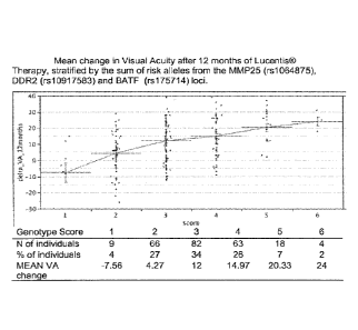
BRIEF DESCRIPTION OF THE DRAWINGS
FIGURE 1 shows the mean change in Visual Acuity after 12 months of Lucentis
therapy stratified by the sum of risk alleles from the MMP25, DDR2, and BATF
genes.
DETAILED DESCRIPTION OF THE PREFERRED EMBODIMENTS
The practice of the present invention will employ, unless otherwise indicated,
conventional techniques of molecular biology (including recombinant
techniques),
microbiology, cell biology, biochemistry, and immunology, which are within the
skill of the
art. Such techniques are explained fully in the literature, such as,
"Molecular Cloning: A
Laboratory Manual", second edition (Sambrook et al., 1989); "Oligonucleotide
Synthesis" (M.
J. Gait, ed., 1984); "Animal Cell Culture" (R. I. Freshney, ed., 1987);
"Methods in
Enzymology" (Academic Press, Inc.); "Current Protocols in Molecular Biology"
(F. M.
Ausubel et al., eds., 1987, and periodic updates); "PCR: The Polymerase Chain
Reaction",
(Mullis et al., eds., 1994).
Unless defined otherwise, technical and scientific terms used herein have the
same
meaning as commonly understood by one of ordinary skill in the art to which
this invention
belongs. Singleton et al., Dictionary of Microbiology and Molecular Biology
2nd ed., J. Wiley
& Sons (New York, N.Y. 1994), and March, Advanced Organic Chemistry Reactions,
Mechanisms and Structure 4th ed., John Wiley & Sons (New York, N.Y. 1992),
provide one
skilled in the art with a general guide to many of the terms used in the
present application.
DEFINITIONS
As used herein, the singular forms "a", "an" and "the" include the plural
unless the
context clearly dictates otherwise. For example, "a" cell will also include
"cells".
The term "comprising" is intended to mean that the compositions and methods
include
the recited elements, but do not exclude others.
The terms "VEGF" and "VEGF-A" are used interchangeably to refer to the 165-
amino
acid vascular endothelial cell growth factor and/or related 121-, 189-, and
206- amino acid
vascular endothelial cell growth factors, as described by Leung etal. Science,
246:1306
4
CA 02859193 2014-08-12
WO 2011/050034 PCT/US2010/053334
(1989), and Houck et al. Mot. Endocrin., 5:1806 (1991), together with the
naturally occurring
allelic and processed forms thereof.
An "anti-VEGF antibody" is an antibody that binds to VEGF with sufficient
affinity and specificity. Preferably, the anti-VEGF antibody of the invention
can be
used as a therapeutic agent in targeting and interfering with diseases or
conditions
wherein the VEGF activity is involved. An anti-VEGF antibody will usually not
bind
to other VEGF homologues such as VEGF-B or VEGF-C, or other growth factors
such
as P1GF, PDGF or bFGF. A preferred anti-VEGF antibody is a monoclonal antibody
that binds to the same epitope as the monoclonal anti-VEGF antibody A4.6.1
produced
by hybridoma ATCCO HB 10709 and is a high-affinity anti-VEGF antibody. A "high-
affinity anti-VEGF antibody" has at least 10-fold better affinity for VEGF
than the
monoclonal anti-VEGF antibody A4.6.1. Preferably the anti-VEGF antibody is a
recombinant humanized anti-VEGF monoclonal antibody fragment generated
according to WO 98/45331, including an antibody comprising the CDRs and/or the
variable regions of Y0317. More preferably, the anti-VEGF antibody is the
antibody
fragment known as ranibizumab (Lucentist)
The term "antibody" is used in the broadest sense and includes monoclonal
antibodies (including full length or intact monoclonal antibodies), polyclonal
antibodies, multivalent antibodies, multispecific antibodies (e.g., bispecific
antibodies), and antibody fragments so long as they exhibit the desired
biological
activity.
"Treatment" refers to both therapeutic treatment and prophylactic or
preventative
measures. Those in need of treatment include those already with the disorder
as well as
those in which the disorder is to be prevented or delayed.
The term "polymorphism" refers to a location in the sequence of a gene which
varies
within a population. A polymorphism is comprised of different "alleles". The
location of
such a polymorphism may be identified by its position in the gene and the
different amino
acids or bases that are found there. For example, Y402H CFH indicates that
there is variation
between tyrosine (Y) and histidine (H) at amino acid position 402 in the CFH
gene. This
amino acid change is the result of two possible variant bases, C and T, which
are two different
alleles. Because the genotype is comprised of two separate alleles, any of
several possible
variants may be observed in any one individual (e.g. for this example, CC, CT,
or TT).
5
CA 02859193 2014-08-12
WO 2011/050034 PCT/US2010/053334
Individual polymorphisms are also assigned unique identifiers ("Reference
SNP", "refSNP" or
"rs#") known to one of skill in the art and used, e.g., in the Single
Nucleotide Polymorphism
Database (dbSNP) of Nucleotide Sequence Variation available on the NCBI
website.
The term "genotype" refers to the specific alleles of a certain gene in a cell
or tissue
sample. In the example above, CC, CT, or 1-1 are possible genotypes at the
Y402H CFH
polymorphism.
The term "sample" includes a cell or tissue sample taken from a patient. For
example,
a sample may include a skin sample, a cheek cell sample, or blood cells.
Identification of the particular genotype in a sample may be performed by any
of a
number of methods well known to one of skill in the art. For example,
identification of the
polymorphism can be accomplished by cloning of the allele and sequencing it
using
techniques well known in the art. Alternatively, the gene sequences can be
amplified from
genomic DNA, e.g. using PCR, and the product sequenced. Several non-limiting
methods for
analyzing a patient's DNA for mutations at a given genetic locus are described
below.
DNA microarray technology, e.g., DNA chip devices and high-density microarrays
for
high-throughput screening applications and lower-density microarrays, may be
used. Methods
for microarray fabrication are known in the art and include various inkjet and
microjet
deposition or spotting technologies and processes, in situ or on-chip
photolithographic
oligonucleotide synthesis processes, and electronic DNA probe addressing
processes. The
DNA microarray hybridization applications has been successfully applied in the
areas of gene
expression analysis and genotyping for point mutations, single nucleotide
polymorphisms
(SNPs), and short tandem repeats (STRs). Additional methods include
interference RNA
microarrays and combinations of microarrays and other methods such as laser
capture
microdissection (LCM), comparative genomic hybridization (CGH) and chromatin
immunoprecipitation (ChiP). See, e.g., He et al. (2007) Adv. Exp. Med. Biol.
593:117-133 and
Heller (2002) Annu. Rev. Bionzed. Eng. 4:129-153. Other methods include PCR,
xIVIAP,
invader assay, mass spectrometry, and pyrosequencing (Wang et al. (2007)
Microarray
Technology and Cancer Gene Profiling Vol 593 of book series Advances in
Experimental
Medicine and Biology, pub. Springer New York).
Another detection method is allele specific hybridization using probes
overlapping the
polymorphic site and having about 5, or alternatively 10, or alternatively 20,
or alternatively
25, or alternatively 30 nucleotides around the polymorphic region. For
example, several
6
CA 02859193 2014-08-12
WO 2011/050034 PCT/1JS2010/053334
probes capable of hybridizing specifically to the allelic variant are attached
to a solid phase
support, e.g., a "chip". Oligonucleotides can be bound to a solid support by a
variety of
processes, including lithography. Mutation detection analysis using these
chips comprising
oligonucleotides, also termed "DNA probe arrays" is described e.g., in Cronin
et al. (1996)
Human Mutation 7:244.
In other detection methods, it is necessary to first amplify at least a
portion of the gene
prior to identifying the allelic variant. Amplification can be performed,
e.g., by PCR and/or
LCR or other methods well known in the art.
In some cases, the presence of the specific allele in DNA from a subject can
be shown
by restriction enzyme analysis. For example, the specific nucleotide
polymorphism can result
in a nucleotide sequence comprising a restriction site which is absent from
the nucleotide
sequence of another allelic variant.
In a further embodiment, protection from cleavage agents (such as a nuclease,
hydroxylamine or osmium tetroxide and with piperidine) can be used to detect
mismatched
.. bases in RNAIRNA DNA/DNA, or RNAJDNA heteroduplexes (see, e.g., Myers et
al. (1985)
Science 230:1242). In general, the technique of "mismatch cleavage" starts by
providing
heteroduplexes formed by hybridizing a control nucleic acid, which is
optionally labeled, e.g.,
RNA or DNA, comprising a nucleotide sequence of the allelic variant of the
gene with a
sample nucleic acid, e.g., RNA or DNA, obtained from a tissue sample. The
double-stranded
duplexes are treated with an agent which cleaves single-stranded regions of
the duplex such as
duplexes formed based on basepair mismatches between the control and sample
strands. For
instance, RNAIDNA duplexes can be treated with RNase and DNA/DNA hybrids
treated with
Si nuclease to enzymatically digest the mismatched regions. Alternatively,
either DNAJDNA
or RNA/DNA duplexes can be treated with hydroxylamine or osmium tetroxide and
with
piperidine in order to digest mismatched regions. After digestion of the
mismatched regions,
the resulting material is then separated by size on denaturing polyacrylamide
gels to determine
whether the control and sample nucleic acids have an identical nucleotide
sequence or in
which nucleotides they are different. See, for example, U.S. Pat. No.
6,455,249; Cotton et al.
(1988) Proc. Natl. Acad. Sci. USA 85:4397; Saleeba et al. (1992) Meth.
Enzpnol. 217:286-
295.
Alterations in electrophoretic mobility may also be used to identify the
particular
allelic variant. For example, single strand conformation polymorphism (SSCP)
may be used to
7
CA 02859193 2014-08-12
WO 2011/050034 PCT/US2010/053334
detect differences in electrophoretic mobility between mutant and wild type
nucleic acids
(Orita et al. (1989) Proc Natl. Acad. Sci USA 86:2766; Cotton (1993) Mutat.
Res. 285:125-144
and Hayashi (1992) Genet. Anal. Tech. App!. 9:73-79). Single-stranded DNA
fragments of
sample and control nucleic acids are denatured and allowed to renaturc. The
secondary
structure of single-stranded nucleic acids varies according to sequence, the
resulting alteration
in electrophoretic mobility enables the detection of even a single base
change. The DNA
fragments may be labeled or detected with labeled probes. The sensitivity of
the assay may be
enhanced by using RNA (rather than DNA), in which the secondary structure is
more sensitive
to a change in sequence. In another preferred embodiment, the subject method
utilizes
heteroduplex analysis to separate double stranded heteroduplex molecules on
the basis of
changes in electrophoretic mobility (Keen et al. (1991) Trends Genet. 7:5).
The identity of the allelic variant may also be obtained by analyzing the
movement of a
nucleic acid comprising the polymorphic region in polyacrylamide gels
containing a gradient
of denaturant, which is assayed using denaturing gradient gel electrophoresis
(DOGE) (Myers
et al. (1985) Nature 313:495). When DGGE is used as the method of analysis,
DNA will be
modified to ensure that it does not completely denature, for example by adding
a GC clamp of
approximately 40 bp of high-melting GC-rich DNA by PCR. In a further
embodiment, a
temperature gradient is used in place of a denaturing agent gradient to
identify differences in
the mobility of control and sample DNA (Rosenbaum and Reissner (1987) Biophys.
Chem.
265:1275).
Examples of techniques for detecting differences of at least one nucleotide
between 2
nucleic acids include, but are not limited to, selective oligonucleotide
hybridization, selective
amplification, or selective primer extension. For example, oligonucleotide
probes may be
prepared in which the known polymorphic nucleotide is placed centrally (allele-
specific
____________________________________________ probes) and then hybridized to
target DNA under conditions which pet mit hybridization only
if a perfect match is found (Saiki et al. (1986)Nature 324:163); Saiki et at.
(1989) Proc. Natl.
Acad. Sci. USA 86:6230). Such allele specific oligonucleotide hybridization
techniques may be
used for the detection of the nucleotide changes in the polymorphic region of
the gene. For
example, oligonucleotides having the nucleotide sequence of the specific
allelic variant are
attached to a hybridizing membrane and this membrane is then hybridized with
labeled sample
nucleic acid. Analysis of the hybridization signal will then reveal the
identity of the
nucleotides of the sample nucleic acid.
8
CA 02859193 2014-08-12
WO 2011/050034 PCMJS2010/053334
Alternatively, allele specific amplification technology which depends on
selective PCR
amplification may be used in conjunction with the instant invention.
Oligonucleotides used as
primers for specific amplification may carry the allelic variant of interest
in the center of the
molecule (so that amplification depends on differential hybridization) (Gibbs
et al. (1989)
Nucl. Acids Res. 17:2437-2448) or at the extreme 3' end of one primer where,
under
appropriate conditions, mismatch can prevent, or reduce polymerase extension
(Prossner
(1993) Tibtech 11:238 and Newton et al. (1989) Nucl. Acids Res. 17:2503). This
technique is
also termed "PROBE" for PRobe Oligo Base Extension. In addition it may be
desirable to
introduce a novel restriction site in the region of the mutation to create
cleavage-based
detection (Gasparini et al. (1992) Ala Cell. Probes 6:1).
In another embodiment, identification of the allelic variant is carried out
using an
oligonucleotide ligation assay (OLA), as described, e.g., in U.S. Pat, No.
4,998,617 and in
Laridegren, U. et al. Science 241:1077-1080 (1988). The OLA protocol uses two
oligonucleotides which are designed to be capable of hybridizing to abutting
sequences of a
single strand of a target. One of the oligonucleotides is linked to a
separation marker, e.g.,
biotinylated, and the other is detectably labeled, If the precise
complementary sequence is
found in a target molecule, the oligonucleotides will hybridize such that
their termini abut, and
create a ligation substrate. Ligation then permits the labeled oligonucleotide
to be recovered
using avidin, or another biotin ligand. Nickerson, D. A. et al. have described
a nucleic acid
detection assay that combines attributes of PCR and OLA (Nickerson, D. A. et
al. (1990)
Proc. Natl. Acad. Sci. USA 87:8923-8927). In this method, PCR is used to
achieve the
exponential amplification of target DNA, which is then detected using OLA.
The invention provides methods for detecting a single nucleotide polymorphism
(SNP)
in MMP25, DDR2 and BATF. Because single nucleotide polymorphisms are flanked
by
regions of invariant sequence, their analysis requires no more than the
determination of the
identity of the single variant nucleotide and it is unnecessary to determine a
complete gene
sequence for each patient. Several methods have been developed to facilitate
the analysis of
SNPs.
The single base polymorphism can be detected by using a specialized
exonuclease-
resistant nucleotide, as disclosed, e.g., in U.S. Pat. No. 4,656,127.
According to the method, a
primer complementary to the allelic sequence immediately 3' to the polymorphic
site is
permitted to hybridize to a target molecule obtained from a particular animal
or human. If the
9
CA 02859193 2014-08-12
W02011/050034 PCT/US2010/053334
polymorphic site on the target molecule contains a nucleotide that is
complementary to the
particular exonuclease-resistant nucleotide derivative present, then that
derivative will be
incorporated onto the end of the hybridized primer. Such incorporation renders
the primer
resistant to exonuclease, and thereby permits its detection. Since the
identity of the
.. exonuclease-resistant derivative of the sample is known, a finding that the
primer has become
resistant to exonueleases reveals that the nucleotide present in the
polymorphic site of the
target molecule was complementary to that of the nucleotide derivative used in
the reaction.
This method has the advantage that it does not require the determination of
large amounts of
extraneous sequence data.
A solution-based method may also be used for determining the identity of the
nucleotide of the polymorphic site (WO 91/02087). As above, a primer is
employed that is
complementary to allelic sequences immediately 3' to a polymorphic site. The
method
determines the identity of the nucleotide of that site using labeled
dideoxynucleotide
derivatives, which, if complementary to the nucleotide of the polymorphic site
will become
incorporated onto the terminus of the primer.
An alternative method is described in WO 92/15712. This method uses mixtures
of
labeled terminators and a primer that is complementary to the sequence 3' to a
polymorphic
site. The labeled terminator that is incorporated is thus determined by, and
complementary to,
the nucleotide present in the polymorphic site of the target molecule being
evaluated. The
method is usually a heterogeneous phase assay, in which the primer or the
target molecule is
immobilized to a solid phase.
Many other primer-guided nucleotide incorporation procedures for assaying
polymorphic sites in DNA have been described (Komher, J. S. et al. (1989)
Nucl. Acids. Res.
17:7779-7784; Sokolov, B. P. (1990) Nod. Acids Res. 18:3671; Syvanen, A.-C.,
et al. (1990)
Genomics 8:684-692; Kuppuswamy, M. N. et al. (1991) Proc. Natl. Acad. Sci. USA
88:1143-
1147; Prezant, T. R. et at. (1992) Hum. Mutat. 1: 159-164; Ugozzoli, L. at al.
(1992) GA TA
9:107-112; Nyren, P. et al. (1993) Anal. Biochem. 208:171-175). These methods
all rely on the
incorporation of labeled deoxynucleotides to discriminate between bases at a
polymorphic site.
Moreover, it will be understood that any of the above methods for detecting
alterations
in a gene or gene product or polymorphic variants can be used to monitor the
course of
treatment or therapy.
The methods described herein may be perfomied, for example, by utilizing pre-
CA 02 85 9193 2014-08-12
WO 2011/050034 PCT/US2010/053334
packaged diagnostic kits, such as those described below, comprising at least
one probe or
primer nucleic acid, which may be conveniently used, e.g., to determine
whether an individual
has an increased likelihood of developing AMD or whether a wet AMD patient has
an
increased likelihood of benefiting from treatment with an anti-VEGF antibody.
Sample nucleic acid for use in the above-described diagnostic and prognostic
methods
can be obtained from any cell type or tissue of a subject. For example, a
subject's bodily fluid
(e.g. blood) can be obtained by known techniques. Alternatively, nucleic acid
tests can be
performed on dry samples (e.g., hair or skin).
The invention described herein relates to methods and compositions for
determining
and identifying the allele present at several alleles, including the MMP25,
DDR2 and BATF
alleles at rs1064875, rs10917583 and rs175714, respectively. Probes can be
used to directly
determine the genotype of the sample or can be used simultaneously with or
subsequent to
amplification. The term "probes" includes naturally occurring or recombinant
single- or
double-stranded nucleic acids or chemically synthesized nucleic acids. They
may be labeled by
nick translation, Klenow fill-in reaction, PCR or other methods known in the
art. Probes of the
present invention, their preparation and/or labeling are described in Sambrook
et al. (1989)
supra. A probe can be a polynucleotide of any length suitable for selective
hybridization to a
nucleic acid containing a polymorphic region of the invention. Length of the
probe used will
depend, in part, on the nature of the assay used and the hybridization
conditions employed.
Labeled probes also can be used in conjunction with amplification of a
polymorphism.
(Holland et al. (1991) Proc. Natl. Acad. Sci. USA 88:7276-7280). U.S. Pat. No.
5,210,015
describes fluorescence-based approaches to provide real time measurements of
amplification
products during PCR. Such approaches have either employed intercalating dyes
(such as
ethidium bromide) to indicate the amount of double-stranded DNA present, or
they have
employed probes containing fluorescence-quencher pairs (also referred to as
the "TaqManCi"
approach) where the probe is cleaved during amplification to release a
fluorescent molecule
whose concentration is proportional to the amount of double-stranded DNA
present. During
amplification, the probe is digested by the nuclease activity of a polymerase
when hybridized
to the target sequence to cause the fluorescent molecule to be separated from
the quencher
molecule, thereby causing fluorescence from the reporter molecule to appear.
The TaqMan
approach uses a probe containing a reporter molecule--quencher molecule pair
that specifically
anneals to a region of a target polynucicotide containing thc polymorphism.
11
CA 02859193 2014-08-12
WO 2011/05003-1 PCT/US2010/053334
Probes can be affixed to surfaces for use as "gene chips." Such gene chips can
be used
to detect genetic variations by a number of techniques known to one of skill
in the art. In one
technique, oligonucleotides are arrayed on a gene chip for determining the DNA
sequence of a
by the sequencing by hybridization approach, such as that outlined in U.S.
Pat. Nos. 6,025,136
and 6,018,041. The probes of the invention also can be used for fluorescent
detection of a
genetic sequence. Such techniques have been described, for example, in U.S.
Pat. Nos.
5,968,740 and 5,858,659. A probe also can be affixed to an electrode surface
for the
electrochemical detection of nucleic acid sequences such as described in U.S.
Pat. No.
5,952,172 and by Kelley, S. 0. et al. (1999) Nucl. Acids Res. 27:4830-4837.
Additionally, the isolated nucleic acids used as probes or primers may be
modified to
become more stable. Exemplary nucleic acid molecules which are modified
include
phosphoramidate, phosphothioate and methylphosphonate analogs of DNA (see also
U.S. Pat.
Nos. 5,176,996; 5,264,564 and 5,256,775).
As set forth herein, the invention also provides diagnostic methods for
determining the
type of allelic variants of polymorphic regions present in MMP25, DDR2 or
BATF. In some
embodiments, the methods use probes or primers comprising nucleotide sequences
which are
complementary to a polymorphic region of MMP25, DDR2 or BATF. Accordingly, the
invention provides kits for performing these methods.
In some embodiments, the invention provides a kit for determining whether a
wet
AMD patient has an increased likelihood of benefiting from treatment with an
anti-VEGF
antibody, including a high-affinity anti-VEGF antibody. Such kits contain one
of more of the
compositions described herein and instructions for use. As an example only,
the invention also
provides kits for determining whether a wet AMD patient has an increased
likelihood of
benefiting from treatment with ranibizumab comprising a first oligonucleotide
and a second
oligonucleotides specific for a AG polymorphism in the MMP25 rs1064875 SNP.
Oligonucleotides "specific for" a genetic locus bind either to the polymorphic
region of the
locus or bind adjacent to the polymorphic region of the locus. For
oligonucleotides that are to
be used as primers for amplification, primers are adjacent if they are
sufficiently close to be
used to produce a polynucleotide comprising the polymorphic region. In one
embodiment,
.. oligonucleotides are adjacent if they bind within about 1-2 kb, e.g. less
than 1 kb from the
polymorphism. Specific oligonucleotides are capable of hybridizing to a
sequence, and under
suitable conditions will not bind to a sequence differing by a single
nucleotide.
12
CA 02859193 2014-08-12
WO 2011/050034 PCT/US2010/053334
The kit can comprise at least one probe or primer which is capable of
specifically
hybridizing to the polymorphic region of MMP25, DDR2 or BATF and instructions
for use.
The kits usually comprise at least one of the above described nucleic acids.
Kits for amplifying
at least a portion of MMP25, DDR2 or BATF generally comprise two primers, at
least one of
which is capable of hybridizing to the allelic variant sequence. Such kits are
suitable for
detection of genotype by, for example, fluorescence detection, by
electrochemical detection, or
by other detection.
Oligonucleotides, whether used as probes or primers, contained in a kit can be
detectably labeled. Labels can be detected either directly, for example for
fluorescent labels, or
indirectly. Indirect detection can include any detection method known to one
of skill in the art,
including biotin-avidin interactions, antibody binding and the like.
Fluorescently labeled
oligonucleotides also can contain a quenching molecule. Oligonucleotides can
be bound to a
surface. In some embodiments, the surface is silica or glass. In some
embodiments, the surface
is a metal electrode.
Yet other kits of the invention comprise at least one reagent necessary to
perform the
assay. For example, the kit can comprise an enzyme. Alternatively the kit can
comprise a
buffer or any other necessary reagent.
The kits can include all or some of the positive controls, negative controls,
reagents,
primers, sequencing markers, probes and antibodies described herein for
determining the
subject's genotype in the polymorphic region of MMP25, DDR2 or BATF.
The following example is intended merely to illustrate the practice of the
present
invention and is not provided by way of limitation.
EXAMPLE
Example 1. Genetic polymorphisms and their association with AMD occurrence
and treatment outcomes
Samples and genotyping
Peripheral blood samples from 250 de-identified subjects from Lucentis0
pivotal trials
(MARINA, ANCHOR, and FOCUS) who participated in the DAWN genetic substudy of
the
HORIZON extension trial were collected and genomic DNA was isolated. All
samples used
in the analysis had a self-identified race listed as "White" and had a
confirmed diagnosis of
neo-vascular AMD. The samples consisted of 104 males and 146 females, and the
average
13
CA 02859193 2014-08-12
WO 2011/050034 PCT/US2010/053334
age at baseline was 75.7 years of age. Written informed consent was obtained
from all
individuals in the study and the study protocols were approved by
institutional review boards.
In addition to the samples described above, 102 samples were collected as part
of the
DAWN study, resulting in a total of 352 total samples. The 352 samples were
genotyped using
the Illumina 550K Human HapMap Bead Array. We used stringent quality control
(QC)
criteria to ensure that high quality data was included in the final analysis.
Specifically, we a)
excluded individuals who had > 5% missing data and b) excluded individuals
based on cryptic
relatedness and duplicate samples based on IBS status (PI Hat >0.15, none
detected in this
data set). We included only SNPs with a) < 5 % missing data, b) HWE p-value >
1x10⁻⁶, and
c) MAF > 0.01 %. All QC tests were performed using PLINK (Purcell et al.
(2007)Am. J.
Hum. Genet. 81, 559-75).
Genome-wide association scan for response to Lucentis therapy
The DAWN samples were separated into 2 groups based on the treatment status
during
the MARINA, ANCIIOR and FOCUS trials. The Lucentis treated group included
individuals who received doses of 0.3mg, 0.5 mg or 0.5mg+PDT (N=242). The
SHAM/PDT
group consisted of individuals who received a mock injection (SHAM) or only
photodynamic
therapy (PDT) (N=110). We performed a genome-wide association scan to identify
genetic
variants significantly associated with change in visual acuity (VA, measured
in letters) after 12
months of Lucentis treatment (Table 1). From the list of top loci associated
with mean
change in VA, we identified 3 loci that contain genes that are likely to play
a role in wound
repair (Table 2) and created a "gene score" by summing the number of risk
alleles carried by
each individual from the MMP25 (rs1064875), DDR2 (rs10917583) and BATF
(rs175714)
loci (Figure 1).
14
CA 02859193 2014-08-12
WO 2011/050034 PCT/US2010/053334
Table 1: Rank ordered list of variants most associated with change in Visual
Acuity after
12 months of LucentisC therapy (P<2 x10⁻⁵).
Chromo- SNP Chromosomal Position (BP, Allele P value
Gene symbol
some based on NCBI build 36)
1 rs10917583 160897518 G 5.81E-06 DDR2
22 rs956548 25867502 A 7.10E-06 MIAT
1 rs10494373 160885986 C 7.98E-06 DDR2
19 rs7260544 51313304 C 8.24E-06 IGEL3
22 rs9608580 25862474 G 9.70E-06 MIAT
rs7913098 32014665 A 1.22E-05 L00646034
1 rs16843630 160936667 G 1.27E-05 DDR2
14 rs175714 75051609 A 1.63E-05 BAIT
16 rs1064875 3042210 A 1.68E-05 MMP25
1 rs1910339 174640705 A 1.69E-05 PAPPA2
CA 02859193 2014-08-12
WO 2011/050034 PCT/US2010/053334
Table 2: Mean change in Visual Acuity at 12 months of Lucentist treatment
stratified by
genotype at MMP25, DDR2 and BATF loci,
MMP25 (rs1064875) N % of population Mean change in VA
at 12 months
GIG 153 63% 8.4
A/G 72 30% 13.1
A/A 16 7% 22A
DDR2 (rs10917583) N % of population Mean change in VA
at 12 months
C/C 2 1% -22.0
C/A 35 15% 4.2
A/A 204 85% 12.3
BATF (rs175714) N % of population Mean change in VA
at 12 months
GIG 81 34% 5.4
A/G 118 49% 13.0
A/A 42 17% 15.2
Genome-wide association scan for Baseline clinical phenotypes
In all 352 DAWN samples we performed a genome-wide test for association to
several
baseline phenotypes, including: visual acuity at baseline (Table 3), Presence
of CNV in the
untreated Fellow eye (Table 4), and CNV classification (Predominantly classic
vs minimally
classic and occult classification, Table 5).
16
CA 02859193 2014-08-12
WO 2011/050034 PCT/US2010/05333-1
Table 3: Rank ordered list of variants associated with change in Visual Acuity
at baseline
in 352 individuals (P<2 x10⁻⁵).
Chromo- SNP Chromosomal Position (BP, Allele P value Permuted
some based on NCBT build 36) P value
2 rs707097 155027061 G .. 3.65E-07 0.00999
6 rs9295181 162401450 G 1.82E-06 0.05295
9 rs16927388 126159263 A 2.32E-06 0.05295
15 rs4316697 47517217 A 9.50E-06 0.1239
rs913035 29858610 C 1.04E-05 0.2218
17
CA 02859193 2014-08-12
WO 2011/050034 PCT/US2010/053334
Table 4: Rank ordered list of variants associated with presence of CNV in the
fellow eye
at baseline in 352 individuals (P<2 x10⁻⁵).
Chromo- SNP Chromosomal Allele P value Permuted Gene
some Position (BP, based P value symbol
on NCBI build 36)
8 rs2022976 107470989 A 1.20E-06 0.02498 OXR1
9 rs10813420 31013908 A 1.23E-06 0.02597 L00646753
17 rs7210510 24495724 A 2.27E-06 0.02098 MY018A
8 rs7828669 124007968 A 6.22E-06 0.1159 ZHX2
7 rs11764261 180194 A 8.15E-06 0.1568 L00645561
1 rs12741645 44335934 A 1.00E-05 0.2318 L00644743
7 rs2082744 99319352 G 1.03E-05 0.2008 TRIM4
9 rs10970046 30993452 G 1.08E-05 0.1828 L00646753
8 rs6991239 124006174 G 1.20E-05 0.2108 ZHX2
8 rs7819862 123993632 G 1.21E-05 0.2108 ZHX2
14 rs2144064 101188372 G 1.52E-05 0.1718 C14orf72
7 rs2571997 99352353 A 1.52E-05 0.2617 TR1M4
7 rs2572009 99326941 A 1.52E-05 0.2617 TRIM4
12 rs7297415 111145487 A 1.63E-05 0.2408 NULL
8 rs1368137 130401289 C 1.63E-05 0.2577 CCDC26
17 rs4986765 57118247 A 1.65E-05 0.1528 BRIP1
2 rs1344759 122755725 A 1.77E-05 0.3986 L00728241
18
CA 02859193 2014-08-12
WO 2011/050034 PCT/US2010/053334
Table 5: Rank ordered list of variants associated with CNV classification at
baseline in
352 individuals (P<2 x10⁻⁵).
Chromo- SNP Chromosomal Allele P value Permuted Gene
some Position (BP, based P value symbol
on NCRI build 36)
rs617738 84312795 A 3.53E-07 0.01199 NRG3
6 rs2076169 35496457 G 2.79E-06 0,06194 PPARD
10 rs6480533 73022225 G 3.73E-06 0.08192 CDH23
10 rs594612 84257806 G 4.77E-06 0.1009
NRG3
2 rs13029532 191584146 C 1.12E-05 0.2428
STAT1
10 rs11819553 73096471 G 1.48E-05 0,2567
CD1123
rs1291117 34935654 G 1.62E-05 0.1508
C20orf118
6 rs726281 152344271 G 1.87E-05 0.3357
ESR1
19