Note: Descriptions are shown in the official language in which they were submitted.
WO 2012/112755 PCT/US2012/025404
Optical Coupler For An Endoscope
[0001]
STATEMENT REGARDING FEDERALLY SPONSORED RESEARCH
[0002] Not Applicable.
BACKGROUND OF THE INVENTION
1. Field of the Invention
[0003] 'This invention relates to an optical coupler for improved optical
imaging of surfaces
covered with opaque fluids, semisolid materials or particulate matter.
2. Description of the Related Art
[0004] The demand for minimally invasive surgery continues to grow. The
ability to convert
open surgeries to minimally invasive procedures has been made possible with
video endoscopy,
but is limited when blood or other fluids are in the field of view. Other
technologies
(fluoroscopy, 3-D echo, MRI, etc.) currently are used to overcome the
challenge of performing
surgery in intravascular spaces, but each technology presents limitations.
[0005] Fluoroscopy has a two dimensional view and is used for diagnostic
procedures or
placement, and/or deployment of medical devices. Procedures are lengthy
creating increased
exposure to radiation for both patients and clinicians, increased expense, and
also may increase
morbidity due to extended anesthesia duration. Most importantly, images are
inferior to direct
vision, the gold standard in surgery.
[0006] Ultrasound three dimensional imaging systems have known problems as
well. Images
are created by transforming ultrasound waves to images. Images often contain
shadowing or
ghosting when instruments or devices are placed within the viewing field. MRI
surgical
procedures are very limited, costly and complex. Simple procedures take hours.
[0007] What is needed therefore is a device that allows diagnostic and
surgical procedures to
be performed in areas of the body where visibility is normally or has been
obstructed by blood,
stomach content, bowel content, or other opaque fluids and/or solid
particulate matter.
- 1 -
CA 2863587 2018-05-16
CA 02863587 2014-07-31
WO 2012/112755 PCT/US2012/025404
SUMMARY OF THE INVENTION
[0008] The foregoing needs are met by an optical coupler comprising a
clear gel in a semi-
solid state that attaches to the distal end (objective lens) of a conventional
or modified video
endoscope for performing diagnostic procedures and/or minimally invasive
surgical operations.
The optical coupler is biocompatible and for single use. It can be attached to
rigid or flexible
endoscopes (for example, gastroscopes thorascopes, laparoscopes, colonoscopes,
Natural Orifice
Transluminal Endoscopic Surgery (NOTES) endoscopes, esophagoscopes,
nasolarynoscopes,
arthroscopie scopes and ophthalmoscopes used in laparoscopic,
gastrointestinal, thorascopic,
colonoscopy and other endoscopic procedures). The optical coupler is an
important
advancement in minimally invasive surgeries and as a tool utilized in
diagnosis and treatment of
disease.
[0009] The optical coupler of the invention is used for visualization in
opaque fluids or
semisolids, and comprises a clear, soft, flexible gel coupled to the outer
distal portion of any
optical imaging device, such as an endoscope or a camera lens. When pressed in
contact with
the surface of an area to be viewed, the coupler creates an offset that allows
clear visualization
by mechanically displacing the opaque liquid, semisolids, or particulate
matter. This
displacement allows the optically clear coupler to come into contact with a
surface of interest,
thus producing an unobstructed view to the observer.
[0010] In a non-limiting medical embodiment, the coupler solves a long-
standing medical
challenge: keeping the tissue undergoing diagnostic or surgical repair free of
blood, bile, and/or
other opaque fluids that would obstruct the clinician's view. Because the
coupler comprises a
clear soft elastic gel, standard medical instruments can be maneuvered within
the area of the
offset, giving the clinician seamless access and a clear view of tissue in
situ. The coupler
provides for reduced surgical procedure time resulting in less invasive
effects and quicker patient
recovery, and potentially higher volume of scheduled procedures. The coupler
is intuitive to use
and would not require any learning curve for the clinician.
[0011] In a non-limiting industrial embodiment, the coupler can be
attached to a borescope,
pipe inspection or other imaging equipment to evaluate or repair surfaces
obstructed by opaque
fluids or solutions, semisolids, or particulate matter, such as oil, sewage or
silt.
- 2 -
CA 02863587 2014-07-31
WO 2012/112755 PCT/US2012/025404
[0012] In one aspect, the invention provides an optical coupler for
mounting at a distal end of
an optical imaging device for visualizing a surface area covered with an
opaque fluid and/or
particulate matter. The coupler includes a visualization section at one end of
the coupler and an
attachment section connected to and extending away from the visualization
section. The
attachment section is dimensioned to be mounted at the distal end of the
optical imaging device.
The visualization section includes a proximal surface for engaging the distal
end of the optical
imaging device. The visualization section includes an outer surface spaced
apart from the
proximal surface. The outer surface extends continuously from a first outer
side boundary across
to a second opposite outer side boundary of the visualization section. The
visualization section
may include a hollow instrument channel extending from the proximal surface
toward or through
the outer surface. The visualization section can be formed from an elastic
material capable of
transmitting an optical image of the surface area. In one form, the material
comprises a silicone
gel or a silicone elastomer.
[0013] In another aspect, the invention provides a device for visualizing
a surface area
covered with an opaque fluid and/or particulate matter. The device includes a
sheath having a
first lumen and a second lumen, a light guide positioned in the first lumen
for transmitting light
toward the surface area, an image carrying fiber positioned in the second
lumen, an object lens
positioned at a distal end of the image carrying fiber and optically connected
to the image
carrying fiber wherein the lens receives light that has been reflected from
the surface area, and an
optical coupler mounted at a distal end of the sheath. The coupler includes a
visualization
section at one end of the coupler. The visualization section includes a
proximal surface for
engaging the distal end of the optical imaging device, and the visualization
section includes an
outer surface spaced apart from the proximal surface wherein the outer surface
extends
continuously from a first outer side boundary across to a second opposite
outer side boundary of
the visualization section. The visualization section includes a hollow
instrument channel
extending from the proximal surface toward the outer surface. The
visualization section can
comprise an elastic material capable of transmitting an optical image of the
surface area. The
coupler includes an attachment section connected to and extending away from
the visualization
section wherein the attachment section is dimensioned to be mounted at the
distal end of the
optical imaging device.
- 3 -
CA 02863587 2014-07-31
WO 2012/112755 PCT/US2012/025.104
[0014] In yet another aspect, the invention provides a method for
visualizing a wall defining
a body cavity with an endoscope wherein the wall is covered or obstructed with
an opaque fluid
and/or particulate matter. The method uses an endoscope comprising a sheath
having a first
lumen and a second lumen, a light guide positioned in the first lumen, an
image carrying fiber
positioned in the second lumen, and an object lens positioned at a distal end
of the image
carrying fiber wherein the lens is optically connected to the image carrying
fiber. An optical
coupler is mounted on a distal end of the sheath. The coupler includes a
visualization section at
one end of the coupler. The visualization section includes a proximal
transverse surface
engaging the distal end of the sheath. The visualization section includes an
outer surface spaced
apart from the proximal surface wherein the outer surface extends continuously
from a first outer
side boundary across to a second opposite outer side boundary of the
visualization section, and
the visualization section comprises an elastic material capable of
transmitting an image of the
surface area. The endoscope is inserted into the body cavity, and the optical
coupler is
positioned in contact with a region of the wall of the body cavity thereby
displacing opaque fluid
and/or particulate matter adjacent the region. Light is transmitted through
the light guide and
optical coupler onto the region, and light that has been reflected from the
region is received at the
lens and an optical image is transmitted from the lens to the image carrying
fiber.
In still another aspect, the invention provides a method for visualizing a
wall defming a body
cavity with an endoscope where the wall is covered with an opaque fluid and/or
particulate
matter. The method includes the steps of: (a) providing an endoscope
comprising (i) a sheath
having a first lumen, a second lumen, a third lumen and a fourth lumen, (ii) a
light guide
positioned in the first lumen, (iii) an image carrying fiber positioned in the
second lumen, and
(iv) an object lens positioned at a distal end of the image carrying fiber
wherein the lens is
optically connected to the image carrying fiber; (b) inserting the endoscope
into the body cavity;
(c) feeding a first precursor through the third lumen and feeding a second
precursor through the
fourth lumen such that the first precursor and the second precursor react to
form an optical
coupler on a distal end of the sheath. The first precursor can be an optical
fluid and the second
precursor can be a cross linking agent.
[0015] The coupler includes a visualization section at one end of the
coupler. The
visualization section includes a proximal transverse surface engaging the
distal end of the sheath,
and the visualization section includes an outer surface spaced apart from the
proximal surface.
- 4 -
CA 02863587 2014-07-31
WO 2012/112755
PCT/US2012/025404
The outer surface extends continuously from a first outer side boundary across
to a second
opposite outer side boundary of the visualization section, and the
visualization section comprises
an elastic material capable of transmitting an image of the surface area. The
optical coupler is
positioned in contact with a region of the wall of the body cavity thereby
displacing opaque fluid
and/or particulate matter adjacent the region. Light is transmitted through
the light guide and
optical coupler onto the region, and light that has been reflected from the
region is received at the
lens and an optical image is transmitted from the lens to the image carrying
fiber.
[0016] In still another aspect, the invention provides for a method for
visualizing a surface of
a structure with a camera, the surface being covered with an opaque fluid
and/or particulate
matter. The method includes (a) providing a camera having a lens and a source
of light and (b)
mounting an optical coupler on the camera by engaging the optical coupler on
an outer surface of
the camera, the coupler including a visualization section at one end of the
coupler, the outer
surface extending continuously from a first outer side boundary across to a
second opposite outer
side boundary of the visualization section, and the visualization section
comprising an elastic
material capable of transmitting an image of the surface area. The method also
includes (c)
placing the camera and the optical coupler near the surface of the structure,
(d) positioning the
optical coupler in contact with a region of the surface of the structure
thereby displacing opaque
fluid and/or particulate matter adjacent the region, (e) transmitting light
from the light source
through the optical coupler onto the region, and (f) receiving at the lens,
light that has been
reflected from the region and capturing an optical image on the camera.
[0017] In yet another aspect, the invention provides for a method for
visualizing a surface of
a structure with a borescope, the surface being covered with an opaque fluid
and/or particulate
matter. The method includes (a) providing a borescope comprising (i) a sheath
having a first
lumen, a second lumen a third lumen and a fourth lumen, (ii) a light guide
positioned in the first
lumen, (iii) an image carrying fiber positioned in the second lumen, and (iv)
an object lens
positioned at a distal end of the image carrying fiber, the lens being
optically connected to the
image carrying fiber; (b)placing the borescope near the surface; and (c)
feeding a first precursor
through the third lumen and feeding a second precursor through the fourth
lumen such that the
first precursor and the second precursor react to form an optical coupler on a
distal end of the
sheath, the coupler including a visualization section at one end of the
coupler, the visualization
section including a proximal transverse surface engaging the distal end of the
sheath, the
- 5 -
CA 02863587 2014-07-31
WO 2012/112755 PCT/US2012/025404
visualization section including an outer surface spaced apart from the
proximal surface, the outer
surface extending continuously from a first outer side boundary across to a
second opposite outer
side boundary of the visualization section, and the visualization section
comprising an elastic
material capable of transmitting an image of the surface area. The method also
includes (d)
positioning the optical coupler in contact with a region of the surface of the
structure thereby
displacing opaque fluid and/or particulate matter adjacent the region; (e)
transmitting light
through the light guide and optical coupler onto the region; and (f) receiving
at the lens, light that
has been reflected from the region and transmitting an optical image from the
lens to the image
carrying fiber.
[0018] In yet another aspect, the invention provides for a hand-held device
that includes a
handle providing a user a portion to grip and a frame connected to the handle.
The frame has a
cavity. A transparent section is held within the cavity, the transparent
section can be punctured.
[0019] These and other features, aspects, and advantages of the present
invention will
become better understood upon consideration of the following detailed
description, drawings and
appended claims.
BRIEF DESCRIPTION OF THE DRAWINGS
[0020] Figure 1 is a side view of a first embodiment of an optical
coupler according to the
invention.
[0021] Figure 2 is a cross-sectional view of the optical coupler of
Figure 1 taken along line
2-2 of Figure 1.
[0022] Figure 3 is a cross-sectional view of the optical coupler of
Figures 1 and 2 taken
along line 3-3 of Figure 2, the optical coupler being attached to an
endoscope.
[0023] Figure 4 is a cross-sectional view similar to Figure 3 of a second
embodiment of an
optical coupler according to the invention, the optical coupler being attached
to an endoscope.
[0024] Figure 5 is a cross-sectional view similar to Figure 4 of the second
embodiment of the
optical coupler according to the invention engaging an inner wall of a body
cavity.
[0025] Figure 6 is a cross-sectional view similar to Figure 4 of the
second embodiment of the
optical coupler according to the invention engaging an inner wall of a body
cavity wherein a
medical instrument has been advanced through an instrument lumen of the
endoscope, an
- 6 -
CA 02863587 2014-07-31
WO 2012/112755 PCT/US2012/025404
instrument channel of the optical coupler, a solid body of the optical
coupler, and against the
inner wall of the body cavity.
[0026] Figure 7 is a cross-sectional view similar to Figure 3 of a third
embodiment of an
optical coupler according to the invention, the optical coupler being attached
to an endoscope.
[0027] Figure 8 is a side view of another embodiment of an optical coupler
according to the
invention similar to the coupler in Figures 1 and 2, but with the instrument
channel extended
through the outer surface.
[0028] Figure 9 is a cross-sectional view of the optical coupler of
Figure 8 taken along line
9-9 of Figure 8.
[0029] Figure 10 is a side view of another embodiment of an optical coupler
according to the
invention where there is not an instrument channel in the coupler, and an
electrode and wire are
molded into the coupler.
[0030] Figure 11 is a cross-sectional view of the optical coupler of
Figure 10 taken along line
11-11 of Figure 10.
[0031] Figure 12a is a cross-sectional view of another embodiment of an
optical coupler
attached to an endoscope with a biopsy forceps placed through the endoscope
and into the optical
coupler, the jaws of the biopsy forceps being opened.
[0032] Figure 12b is a cross-sectional view of the embodiment in Figure
12a, with the jaws
of the biopsy forceps closed to take a biopsy sample.
[0033] Figure 12c is a cross-sectional view of the embodiment in Figures
12a and 12b, with
the biopsy forceps being withdrawn after having taken the biopsy sample.
[0034] Figure 12d is a detailed view of the embodiment shown in Figure
12a.
[0035] Figure 13a is a side view of another embodiment of an optical
coupler where the
outer surface of the optical coupler is angled.
[0036] Figure 13b is a side view of the embodiment in Figure 13a, where the
optical coupler
is inspecting a weld.
[0037] Figure 14a is a perspective view of an optical coupler similar to
the optical coupler
shown in Figure 12a - Figure 12c, but the optical coupler is attached to a
camera.
[0038] Figure 14b is a front view of the optical coupler and camera in
Figure 14a, with the
.. optical coupler and camera placed in a pipe filled with a liquid.
- 7 -
CA 02863587 2014-07-31
W02012/112755 PCT/US2012/025404
[0039] Figure 15a is perspective view of an optical coupler and camera
similar to the
embodiment shown in Figures 14a and 14b, with a semi-rigid tube placed
parallel to the camera
and through the optical coupler.
[0040] Figure 15b is a front plan view of the optical coupler and camera
in Figure 15a, with
the optical coupler and camera examining a defect in a pipe filled with
liquid.
[0041] Figure 16 is a cross-sectional view of another embodiment of an
optical coupler
attached to an endoscope that has an auxiliary fluid channel.
[0042] Figure 17a is a cross-sectional view of a coupler having a concave
outer surface that
is attached to an endoscope approaching tissue covered in blood.
[0043] Figure 17b is a cross-sectional view of the coupler and endoscope
from Figure 17a,
with the coupler pressed against a wall.
[0044] Figure 17c is the cross-sectional view of the coupler and
endoscope from Figure 17b,
with fluid from the instrument channel flushing the trapped opaque liquid
between the outer
surface of the coupler and the wall.
[0045] Figure 18a is a side plan view of a coupler attached to an endoscope
with the use of a
cap.
[0046] Figure 18b is an exploded view of the coupler of Figure 18a.
[0047] Figure 19 is a coupler attached to a rigid endoscope having a 00
end surface, with the
coupler having an angled instrument channel.
[0048] Figure 20 is a coupler attached to a rigid endoscope having a 300
end surface, with
the coupler having a straight instrument channel.
[0049] Figure 21 is another embodiment of a coupler attached to an
endoscope, the coupler
being made of multiple materials.
[0050] Figure 22a is a perspective view of a coupler used in a handheld
device.
[0051] Figure 22b is a cross-sectional view of the coupler of Figure 22a,
with a stapler
passing through the coupler to treat a laceration.
[0052] Figure 23a is a front plan view of a mold that can be used to make
a coupler.
[0053] Figure 23b is an exploded view of the mold of Figure 23a.
[0054] Like reference numerals will be used to refer to like parts from
Figure to Figure in the
following description of the drawings.
- 8 -
CA 02863587 2014-07-31
WO 2012/112755 PCT/US2012/025404
DETAILED DESCRIPTION OF THE INVENTION
[0055] The invention provides an optical coupler for improved optical
imaging of surfaces
covered with opaque fluids, semisolid materials or particulate matter. In one
form, the optical
coupler is a clear gel attached to the outer distal portion of any optical
imaging or image
capturing device, such as an endoscope or camera lens. When pressed in contact
with the surface
of an area to be viewed, the gel creates an offset that allows clear
visualization by mechanically
displacing the opaque liquid or soft semisolids.
[0056] An attachment section of the optical coupler can be mounted on the
distal portion of
the insertion part of the endoscope. A visualization section of the optical
coupler comprises a
soft, elastic, flexible, optically clear gel, and covers the distal end of the
endoscope. The
visualization section of the optical coupler may be thicker if a wider field
of view is needed, or
thinner if a closer working distance is needed. The attachment section of the
optical coupler can
be a sleeve continuous to the visualization section of the optical coupler.
This sleeve is slipped
over the distal portion of the endoscope until the inner surface of the
attachment section of the
optical coupler makes contact with a lens of the endoscope. The elastic
properties of the sleeve-
like attachment section of the optical coupler, and its smaller internal
diameter provide a secure
hold on the endoscope.
[0057] The optical coupler may have a hollow instrument channel or
channels that extend
through the visualization section of the optical coupler. The channel(s) can
be the same diameter
and align with the working instrument lumen(s) or channels(s) of the
endoscope. This allows
probes or instruments to be passed from the endoscope lumens or channels
through the
visualization section of the optical coupler.
[0058] Often during endoscopic examinations and procedures, the tissue or
an object being
viewed is or can be obstructed by blood or other opaque bodily fluids. With
the optical coupler
attached to the endoscope, the optical coupler pushes through the opaque
fluid, displacing it.
Since the coupler is soft, contact can be safely made with the tissue or
object needed to be
viewed. The optical coupler, with slight compression, will keep in contact
with the tissue and
because the visualization section of the optical coupler is clear, visibility
is not impeded thereby
providing a clear view to image with the endoscope.
- 9 -
CA 02863587 2014-07-31
WO 2012/112755 PCT/US2012/025404
[0059] The object being viewed may have steep undulations. In one embodiment
of the
coupler, a very low durometer (soft) surface of the coupler conforms to the
shape of the object.
The coupler has a very elastic nature so vibrations and movements of the
endoscope are
dampened, also improving the ability to visualize. The optical coupler's soft,
flexible, elastic
properties will cause minimal deformation or damage to soft tissue. Additional
force beyond
what is needed to displace the opaque fluid can be applied to the endoscope to
flatten or unfold
tissue that may be in a contracted state. This would reveal areas of the
tissue that would not be
seen without the coupler. In another embodiment, the coupler can be comprised
of a high
durometer (stiff) material to allow the tissue to conform to the shape of the
coupler. Because the
tissue conforms to the shape of the coupler, fluids are displaced and allow
clear visualization. In
both embodiments, medical instruments can be passed through the aligned
working instrument
lumen(s) of the endoscope and instrument channel(s) of the coupler, making
surgical repairs,
taking biopsies, etc., possible with endoscopic instruments and methods.
[0060] Depending on the endoscopic procedure, the optical coupler
properties may vary. For
example, a high tensile strength and high tear resistance for the coupler
material may be suitable.
In certain applications, a totally elastic material that provides an
elastomeric coupler may be
beneficial.
[0061] Couplers composed of hydrophobic materials may be the best choice
for use in a
water or blood environment. For example, a coupler composed of silicone
material repels blood,
staying clear for extended periods of use, additionally the silicone repels
lipids. The coupler, or a
portion of the coupler, being composed of a hydrophobic substance will not
swell from the
uptake of water or fluids in its working environment.
[0062] In an oil, grease, and water environment, a coupler coated with a
super hydrophilic
would be advantageous. After the coupler has an initial exposure to water,
water molecules push
away other molecules, gaining access to the surface of the coupler forming
stable hydrogen
bonds that are reluctant to break. This keeps contaminants away from the
coupler so it remains
clear longer.
[0063] Couplers composed of hydrophilic materials may be advantageous in
the coupler. If
the coupler or portions of the coupler are composed of a hydrophilic
substance, it will swell in its
working environment increasing the area of displacement or swell to a
predetermined shape.
- 10
CA 02863587 2014-07-31
WO 2012/112755 PCT/US2012/025404
[0064] High thermal resistance can be beneficial in the material of the
coupler. For example,
the material will not melt with heat (e.g., 500 F-1200 F) from electrocautery
or radio ablation
procedures. Electrical isolative, high corona resistance materials are also
beneficial when the
coupler is used in electro, radio frequency, cautery, or harmonic scalpel
procedures.
[0065] The coupler also provides for various safety improvements in
endoscopic procedures.
In both laparoscopic and general surgery, smoke emitted from the burning
tissue while using
electro cauterization instruments often cause poor or zero visibility in the
surgical field. The
procedures must be stopped until the smoke is dissipated. When the electro
cauterization is used
in conjunction with the coupler constant visualization of the tissue remains,
the coupler displaces
the smoke. The coupler can be dimensioned so that it is soft with no sharp
edges to cause
dissections. The coupler can be dimensioned with a larger surface area to
dissipate force when
an endoscope is pushed forward in a body lumen. The coupler is dimensioned
with a large outer
surface area as compared to the objective lens of the endoscope. This is
advantageous as one
small drop of blood can totally obscure vision from an endoscope objective
lens. Small drops of
blood on the outer surface of coupler will only partially obscure visibility.
The coupler can be
dimensioned with a domed shape, smooth slippery outer surface that will allow
better
maneuvering in tube-like structures such as the esophagus, colon, veins, and
arteries. The
coupler also corrects wide-angle curvature created by the common lens used on
videoscopes.
[0066] The coupler gel can be composed with a variety of materials
including
polydimethylsiloxane, hydrogels, polyurethanes, albumin based gels, mineral
oil based gels,
polyisoprene, polybutadiene, or other clear composite. One preferred material
is
polydimethylsiloxane because of its biocompatibility in medical applications,
low price, and it is
easy to mold and cure. Clear, flexible hydrogels that have extreme resistance
to tearing are
another preferred material.
[0067] The material used to form the optical coupler can be comprised of
two or more
compounds, for example an opaque compound attaches and holds two visualization
portions of a
coupler in position, the first visualization portion is an inner clear semi
rigid compound shaped to
match the field of view and minimum depth field of the imaging system, and the
second portion
is attached to the outer boundary of the first visualization portion and is
composed of very soft
gel providing additional area of fluid displacement for maneuvering and
positioning instruments
-11-
CA 02863587 2014-07-31
WO 2012/112755
PCT/US2012/025404
under direct vision. Methods described in U.S. Patent Nos. 7,235,592 and
7,205,339 can be
utilized to produce a coupler with portions or areas of the gel with different
physical properties.
[0068] The invention can be used in various applications. With Natural
Orifice
Translumenal Endoscopic Surgery (NOTES), the coupler enables procedures to
continue when
unexpected bleeding or other fluids such as bile or stomach contents obstruct
the view. Also, the
coupler can create or increase working space by pushing organs out of the
field of view. With a
laryngoscope in trauma and emergency situations, the coupler would push blood,
foreign objects,
or food away to increase visibility to allow visualizing of the trachea. When
taking biopsies is
required, the coupler isolates the intended biopsy target, the tumor or area
to be biopsied from
surrounding tissue. Close focusing and contact with the tissue with the aid of
the coupler can
improve reliability by allow multiple biopsies to taken in exact locations
defining borders of the
tumor, and minimize tumor cells from entering the blood stream or lymph
channels. A cautery
probe or electrode can be used simultaneously or in conjunction with the
biopsy forceps,
minimizing bleeding and length of procedure.
[0069] The coupler can be used in various endoscopic intra-cardiac
procedures such as: (1)
myocardial biopsy (for transplant monitoring or tumor sampling); (2) valve
repair or
reconstruction; (3) patent foramen ovale (PFO) closure; (4) ventricle septal
defect (VSD)
closure; (5) pacing wire placement or removal; (6) stem cell injection; (7)
coronary sinus
cannulations (8) and maze procedure. In cryoablation, a specialized composite
coupler could be
made that has warming channels to warm the external surface of the coupler to
protect
surrounding tissue from freezing. In radiofrequency ablation, insulating and
isolating properties
of the coupler would concentrate power, protecting surrounding tissue.
[0070] The coupler can be used in various vascular procedures. The
coupler can be used to
guide proper placement of covered stents in dissected aortas, or visualize an
infra-vascular laser.
The coupler could be used to inspect the suture line of a large or small
vessel anastomosis to
evaluate the quality of the suturing and or determine the location of any
bleeding.
[0071] In certain surgical or trauma situations there is severe arterial
bleeding from a wound
or vessel. Often the' first action taken is to compress a finger or sponge on
the area of bleeding.
After time passes the finger or sponge is removed. If the bleeding continues
either more
compression or other actions are taken such as blind clamping, suctioning the
blood away and
then clamping and suturing, or homeostatic materials are applied. Blood loss
can be substantial.
- 1 2 -
CA 02863587 2014-07-31
WO 2012/112755 PCT/US2012/025404
An embodiment of the invention mounted at the end of a finger shaped wand can
be compressed
over a bleeding site, both clearing the field of blood and creating a view to
locate the point of
bleeding. Since the coupler is clear, soft and biocompatible, a suture or
staple can be passed
though the coupler to repair the bleeding site.
[0072] The coupler is also beneficial in non-medical applications.
Embodiments of the
coupler can be attached to the distal end (objective lens) of a borescope or
attached to micro or
conventional video cameras, inspection scopes, or still cameras. This allows
viewing and/or
making repairs inside pipes, holding tanks, containers, etc when the fluid is
opaque, such as
petroleum products, sewerage, food products, paint, etc, eliminating the need
to empty the pipes
or containers (e.g., oil tanks). The size of the coupler or the amount of
flexibility can be scaled
for specific applications, for example, displacing large volumes of fluid when
examining large
areas. The shape of the coupler can be generally flat, convex (with varying
levels of curvature),
or shaped for specific tasks. For example, the coupler may be shaped as a
square, or as an
angular shape to displace opaque fluids in the corners of a tank to inspect
the seams.
Examination of joints, welds, seams for corrosion or cracks could be performed
in pipes that
contain moving fluid. A coupler could be used in conjunction with a video
camera and a robotic
vehicle to view remote locations. Large couplers with large working channels
will allow devices
to be passed though a coupler to make repairs using screws, adhesive patches,
etc. The coupler
can be formed from materials that resist acid, alkalinity, high heat, or
viscosity of the fluid being
displaced by the coupler. As opposed to medical usage (disposable, single
use), the coupler
preferred embodiment with industrial applications would be reusable.
[0073] The working channels within the coupler or parallel to the coupler
allow surgical
instruments, probes, biopsy needles, needles, sutures etc. to be passed to the
area being viewed.
Since the coupler is flexible, the channels can move within or around the
coupler without
compromising its function. One enabling property of the coupler is its soft
flexible shape that
conforms to the tissue or object being viewed. This characteristic reduces
damage to delicate
tissues or structures.
[0074] Another advantage of the coupler is that only the specific area
being viewed through
the coupler attached to the endoscope requires illumination and therefore, the
targeted view
requires less light to be supplied by the endoscope lighting system. Because
the number of light
fibers required for illumination is less, endoscopes can be smaller or less
expensive to
- 13-
CA 02863587 2014-07-31
WO 2012/112755 PCT/US2012/025404
manufacture. Also, since it is only necessary to illuminate the area of the
coupler at its outer
boundary, endoscopes of smaller diameter would be required to view a targeted
area.
[0076] An external light source for the endoscope can increase the
functionality of the
coupler. When the coupler does not contact the surface of tissue in a large
chamber, such as the
stomach inflated with air, light emitting from the distal end of the endoscope
can be reflected
from the outer surface of the coupler back to the camera lens, degrading the
endoscope view.
Using an external light source and turning off the endoscope light source
reduces the reflection.
Alternatively, a light fiber placed through the instrument channel which stops
at the outer
boundary will provide lighting while viewing objects in the inflated stomach.
After the coupler
contacts the tissue covered by opaque fluid or blood, the external light is
shut off or the light
fiber is withdrawn from the instrument channel.
[0076] The coupler can be a semi-solid gel, which is transparent and
flexible, that attaches to
a wide variety of endoscopes. For minimally invasive procedures, the smallest
possible scope is
used. The optimal shape and size of the coupler can be determined by the field
of view of the
endoscope, or conversely an endoscope can be chosen that will match the size
and shape of the
coupler. The shape of the coupler can be manufactured with a preformed shape
matched to the
contour of the object that will be examined, for example an endoscope coupler
could be made in
the shape of the blood pool at the apex of the heart. This coupler can be used
in conjunction with
a 2mrn angioscope maneuvered into the apex of the heart and displace the blood
to visualize the
inside wall of the ventricle of the beating heart.
[0077] The coupler can be attached to the endoscope with a clear adhesive
material. The
coupler can be attached as a screw on auxiliary lens or filter allowing
different couplers with
different purposes or functions to be utilized with the same scope. The
coupler can be attached
and held in place with suction. The coupler can be attached by sewing on with
sutures. The
coupler can be attached with wire, nylon or other braid material. The coupler
can be attached to
endoscopes with mesh or pliable membranes. When using a mesh net to attach the
coupler to the
endoscope, gel strength and viscosity must be high enough to prohibit gel flow
through holes in
the outer layer of mesh.
[0078] A coupler can be compressed in a tube fixed to the end of the
scope. A coupler
attached to the endoscope can be compressed in a retractable sheath.
- 14 -
CA 02863587 2014-07-31
WO 2012/112755
PCT/US2012/025404
[0079] The coupler can be made in situ by injecting an optical fluid
(e.g., a siloxane
polymer) and a cross linking agent (e.g., a multifunctional slime) into two
separate lumens of the
endoscope. The liquid components combine and crosslink to form a cured
viscoelastic solid
(e.g., a silicone gel or silicone elastomer) inside a pliable membrane
attached to the distal end of
the scope. The solid body of the coupler can be reinforced with micro thin
strands of
biocompatible fibers, carbon fiber, Nitinol, suture materials, and/or light
fibers. In situ formation
of the coupler allows a larger coupler to be formed inside the body,
increasing the area of
visibility. The coupler can be chemically or mechanically dissolved for
removal after use.
[0080] If the coupler is confined inside a balloon, membrane, mesh or a
tube-like structure
with higher wall tension than the systolic blood pressure, tunnels formed by
moving the
instruments, probes, needles or other devices within the coupler will be
refilled with gel keeping
the gel transparent. To keep the gel contained within the coupler would
require a gel strength
high enough to prohibit flow through holes made in the outer balloon, membrane
or mesh by the
needles or devices.
[0081] Embodiments of the coupler can have one, two or more working
channels that align
with the endoscope's working lumens. Other versions of the coupler allow for
additional internal
channels or along the edges of the device for use in more complex procedures,
such as suturing.
[0082] The coupler can be used in any minimally invasive procedure.
Biopsies in the body,
for example, could be taken under direct view, reducing the need for CO2
inflation. The coupler
allows exact placement of needles and medical devices in situations where
active bleeding or
other bodily fluids impede visibility. The coupler can be held with pressure
over an active
bleeding site to stop bleeding until the suturing process, stapling, clamping
or medical device
placement is complete.
[0083] Other instruments or devices can be pushed through the coupler,
not compromising
its form or transparency. Channels are created by piercing the coupler with
needles, probes or
instruments and the channel will reseal as the medical instruments are
withdrawn.
[0084]
Attaching the coupler to endoscopes that contain working lumens, the coupler
and
endoscope work in unison. Transparent or semi- transparent soft flexible tubes
are passed
though these channels penetrating the coupler, creating continuous channels
that allow probes to
be passed to the targeted area. These probes may include sensors, hypodermic
needles,
- 15-
CA 02863587 2014-07-31
WO 2012/112755 PCT/US2012/025404
instruments, light fibers or medical devices that can be passed in and out of
the coupler to exact
repeatable positions.
[0085] Fixed channels external to the endoscope can be added to steer or
direct probes
around the coupler to be seen and maneuvered within the viewing area. The
device could be
fixed to a 45 degree scope or mirror set 45 degrees to a lens to permit
viewing from the side of a
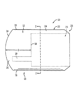
scope. This allows viewing of the side of vessels or tube as the scope is
pushed forward. When
used in conjugation with wide angle optics, the coupler yields a
circumferential view in a pipe or
vessel.
[0086] Turning now to Figures 1-3, there is shown a first example
embodiment of an optical
coupler 10 according to the invention. The optical coupler 10 includes a
visualization section 12
at a distal end 13 of the optical coupler 10. The visualization section 12 has
a generally slightly
curved, convex outer surface 14 that extends from a first outer side boundary
15 to a second
opposite outer side boundary 16 of the optical coupler 10. The outer surface
14 may be
constructed to be generally flat, but a curved outer surface 14 is preferable
because the curvature
helps to clear the field of view by pushing any fluid or matter from the
center of the outer surface
14 to the outer boundaries 15, 16. A flat outer surface 14 may be more
difficult to clear since the
pressure is equal across the entire area of contact and fluid can become
trapped between the lens
and a surface in which it is desired to view or perform work. A curved outer
surface 14 is also
preferable to correct any curvature distortion created by an objective lens 40
that may be used in
conjunction with the coupler 10. The optical coupler 10 has a proximal surface
18, and a hollow
instrument channel 19 extends from the proximal surface 18 toward the outer
surface 14.
[0087] The hollow instrument channel 19 may be constructed such that the
channel 19 does
not extend all the way through the visualization section 12 to the outer
surface 14. In such a
case, a barrier section 20 of material is provided between a distal end 21 of
the hollow instrument
channel 19 and the outer surface 14 of the optical coupler 10. Alternatively,
the instrument
channel 19 may extend the full length of the visualization section 12,
extending through the
optical coupler 10, as shown in Figures 8 and 9. Such a configuration may
allow for the free and
unencumbered exchange of instruments. A water tight seal or valve 29, such as
a Tuohy-Borst
type valve, may be employed on the proximal end 17 of the endoscope instrument
channel 19 to
prevent or minimize air, fluid, and/or foreign matter from flowing through the
instrument
channel 19.
- 16-
CA 02863587 2014-07-31
WO 2012/112755 PCT/US2012/025404
[0088] While an instrument channel 19 is shown in the optical coupler 10
of Figures 1-3, the
visualization section 12 may be constructed without an instrument channel 19.
In such a case,
instruments may be passed directly through the visualization section 12 as the
visualization
section 12 may be constructed of a material that is self-sealing and elastic
enough to permit
instruments to be passed through the entire length of the visualization
section 12 of the optical
coupler 10. An example of an optical coupler 10 without an instrument channel
19 is shown in
Figures 10 and 11, and is described in more detail below.
[0089] The optical coupler 10 also includes an attachment section 22
connected to and
extending away from the visualization section 12. The attachment section 22 is
at the proximal
end 23 of the optical coupler 10. The proximal end 23 of the optical coupler
may be angled to
lessen the chance that the optical coupler 10 may catch on any surfaces when
the optical coupler
10 is being removed from its environment of use. In the embodiment shown, the
attachment
section 22 is in the form of a cylindrical wall 24. The proximal surface 18
and the cylindrical
wall 24 of the optical coupler 10 define a hollow cylindrical opening 25 of
the optical coupler 10
within the sleeve-like cylindrical wall 24.
[0090] Referring to Figure 3, the optical coupler 10 can be mounted on an
endoscope 30.
The endoscope 30 has a distal end 31 that is inserted in the hollow
cylindrical opening 25 of the
optical coupler 10. In one form, the cylindrical wall 24 of the coupler 10 has
a diameter one to
three millimeters larger than the endoscope 30. The endoscope 30 has a sheath
32 with an outer
surface 33 that snugly engages the cylindrical wall 24 of the optical coupler
10. In a non-
limiting example, the sheath 32 has an outside diameter of 7-15 millimeters.
An end surface 34
of the endoscope 30 sealingly engages the proximal surface 18 of the optical
coupler 10. The
endoscope 30 includes a first lumen 35 and a second lumen 36 and a third lumen
37 that extend
from the end surface 34 of the endoscope 30 to a proximal end (not shown) of
the endoscope.
Lumen internal diameters of 2-4 millimeters are typical. A light guide 39 is
positioned in the
first lumen 35 for transmitting light toward a surface area at or beyond the
outer surface 14 of the
optical coupler 10. An object lens 40 is positioned at a distal end of an
image carrying fiber 42,
and the lens 40 is optically connected to the image carrying fiber 42 for
receiving light that has
been reflected from the surface area being viewed. The object lens 40 and the
image carrying
fiber 42 are located in the second lumen 36. The third lumen 37 aligns with
the hollow
instrument channel 19 of the optical coupler 10 when the optical coupler 10 is
mounted on the
- 17-
CA 02863587 2014-07-31
WO 2012/112755 PCT/US2012/025404
endoscope 30. In the embodiment shown, the instrument channel 19 and the third
lumen 37 have
the same size inner diameter within a tolerance of 5%. The optical coupler
10 can also include
a Light Emitting Diode (LED) 11 near the outer surface 14 of the coupler to
provide illumination
prior to the coupler contacting any fluids, tissue, or structure. The LED 11
may be provided
power via a wire (not shown) in the endoscope 30 or from an external source.
[0091] In one example configuration, the endoscope 30 may be a fixed-
focus endoscope
having a specific depth of field. The outer surface 14 may be spaced apart
from the proximal
surface 18 of the optical coupler 10 by a length D (see Figure 1) equal to a
reference distance
selected from values in the depth of field distance range of the endoscope 30.
In one example
configuration, the endoscope 30 may have a depth of field in the range of 2 to
100 millimeters.
In this case, the outer surface 14 is spaced apart from the proximal surface
18 of the optical
coupler 10 by a length in the range 2 to 100 millimeters. Preferably, the
length D equals a
reference distance that is in the lower 25% of values in the depth of field
distance range of the
endoscope 30. In one example configuration, the endoscope 30 may have a depth
of field in the
range of 2 to 100 millimeters. In this case, the length D equals a value of 2-
26 millimeters.
More preferably, the length D equals a reference distance that is in the lower
10% of values in
the depth of field distance range of the endoscope 30. In one example
configuration, the
endoscope 30 may have a depth of field in the range of 2 to 100 millimeters.
In this case, the
length D equals a value of 2-13 millimeters. Most preferably, the length D
equals a reference
distance that is greater than or equal to the lowest value (e.g., 2
millimeters) in the depth of field
distance range of the endoscope 30. In one version of the coupler 10, the
length D is 7-10
millimeters, or a typical distance that the endoscope 30 is held from tissue
that would be
receiving an endoscopic treatment or therapy.
[0092] The design of the length D for the optical coupler 10 should also
take into
consideration the characteristics of the materials that compose the coupler
10, such as any
possible compression of the coupler 10 when it is held against a surface. For
example, if the
coupler 10 may be compressed 1 millimeter when held against a surface and the
lowest value in
the depth of field distance range of the endoscope 30 is 2 millimeters, then
the length D should
be greater than or equal to 3 millimeters to compensate for this possible
compression.
[0093] The optical coupler 10 can be formed from a variety of materials. In
one version of
the optical coupler 10, the optical coupler 10 is molded from a material
selected from silicone
-18-
CA 02863587 2014-07-31
WO 2012/112755 PCT/US2012/025404
gels, silicone elastomers, epoxies, polyurethanes, and mixtures thereof. The
silicone gels can be
lightly cross-linked polysiloxane (e.g., polydimethylsiloxane) fluids, where
the cross-link is
introduced through a multifunctional silane. The silicone elastomers can be
cross-linked fluids
whose three-dimensional structure is much more intricate than a gel as there
is very little free
fluid in the matrix. In another version of the optical coupler 10, the
material is selected from
hydrogels such as polyvinyl alcohol, poly(hydroxyethyl methacrylate),
polyethylene glycol,
poly(methacrylic acid) , and mixtures thereof. The material for the optical
coupler 10 may also
be selected from albumin based gels, mineral oil based gels, polyisoprene, or
polybutadiene.
Preferably, the material is viscoelastic.
[0094] Turning now to Figures 23a and 23b, fully functional couplers 10 can
be made by
combining an uncured silicone material with an additive/ heat curing agent.
Various silicone
material and additives can be used to produce couplers of differing degrees of
softness. The
material can be premixed in a 20 cc vial and placed in a vacuum chamber to
remove air entrained
in the silicone during the mixing process. Next, the silicone is poured into a
chamber 1101 of
Part A of a four piece mold 1100 and placed in the vacuum chamber if any
bubbles were visible.
After the silicone material in Part A was clear, Part B of the mold was
screwed to Part A via
threading 1102, 1103 on Parts A and B, respectively. The chamber 1104 in Part
B is then filled
and de-bubbled as described for part A. Mold parts C and D are pre assembled
using a set screw
1105 to ensure the resulting lens have the proper shape. The leading portion
1106 of assembled
Part C/D is dipped in the silicone material, then centered over the silicone
in Part A/B with the
aid of the alignment pins 1107 and dropped and or pushed downward in
respective holes 1108
until full seated against Part B. The leading portion 1106 includes an
instrument channel pin
1109 to form an instrument channel in the coupler-. The assembly is cured in
an oven at 90 C
for at least one hour. After curing, the mold 1100 is disassembled by
unscrewing Part A from
Part B, pulling Part C/D from Part B. A thick waled polyvinyl tube (not shown)
can be placed
over the outer surface of the coupler, after applying a vacuum to the tubing
the coupler is pulled
out of Part B.
[0095] Referring back to Figures 1-3, in the optical coupler 10, the
material is optically clear
such that the light guide 39 can transmit light through the optical coupler 10
toward a surface
area at or beyond the outer surface 14 of the optical coupler 10 and such that
the optical coupler
10 is capable of transmitting an optical image of the surface area being
viewed back to the lens
- 19 -
CA 02863587 2014-07-31
WO 2012/112755 PCT/US2012/025404
40. In one version of the optical coupler 10, the material has a degree of
light transmittance
greater than 80% based on test standard ASTM D-1003 (Standard Test Method for
Haze and
Luminous Transmittance of Transparent Plastics). In another version of the
optical coupler 10,
the material has a degree of light transmittance greater than 90% based on
test standard ASTM
D-1003. In another version of the optical coupler 10, the material has a
degree of light
transmittance greater than 95% based on test standard ASTM D-1003. In another
version of the
optical coupler 10, the material has a degree of light transmittance grater
than 98% based on test
standard ASTM D-1003. Preferably, the material has an optical absorption of
less than 0.1% in
the visible light range, and more preferably the material has an optical
absorption of less than
0.01% in the visible light range. The material has an index of refraction of
about 1.3 to about
1.7, and preferably, the index of refraction of the material matches the index
of refraction of the
light guide 39, or is as low as possible.
[0096] The optical coupler 10 may also be coated with different materials
to reduce the
amount of adherence properties. Additionally, some coatings of the optical
coupler 10 improve
with light reflections. Sample coatings that may be used on the optical
coupler include
thermoplastic film polymer based on p-xylylene such as Parylene C, which is an
optically clear
biocompatible polymer having abrasion resistant and hydrophobic properties.
[0097] The hardness of the material of the optical coupler 10 can be
varied depending on the
application. If the surface being viewed has steep undulations, a very low
durometer (soft)
surface of the coupler will form to the shape of the object. Alternatively,
the coupler could
comprise a high durometer (stiff) material to allow the tissue to conform to
the shape of the
coupler. In one form, the material has a durometer ranging from 2-95 on the
Shore 00 scale. In
another form, the material has a durometer ranging from 2-20 on the Shore 00
scale. In another
form, the material has a durometer ranging from 40-80 on the Shore 00 scale.
In another form,
the material has a durometer ranging from 60-80 on the Shore 00 scale. As
alluded to above,
the material in some applications may preferably have a durometer outside of
the ranges of the
Shore 00 scale just discussed. Although materials having a hardness of 80 or
more on the Shore
00 scale may not technically be considered a "gel", this specification
generally refers to the
materials that can compose the coupler 10 by using the term "gel." The use of
the term "gel" is
not meant to limit the invention to specific materials or specific ranges of
hardness on the Shore
00 scale.
- 20 -
CA 02863587 2014-07-31
WO 2012/112755
PCT/US2012/025404
[0098] Turning now to Figures 4-6, there is shown a second example embodiment
of an
optical coupler 210 according to the invention. The optical coupler 210 can be
formed from any
of the same materials as the optical coupler 10. The optical coupler 210
includes a visualization
section 212 at a distal end 213 of the optical coupler 210. The visualization
section 212 has an
outer surface 214 with a greater degree of curvature than the embodiment shown
in Figures 1-3.
The convex, generally dome shaped outer surface 214 extends from a first outer
side boundary
215 to a second opposite outer side boundary 216 of the optical coupler 210.
The optical coupler
210 has a proximal surface 218, and a hollow instrument channel 219 extends
from the proximal
surface 218 toward the outer surface 214. A barrier section 220 of material is
provided between
a distal end 221 of the hollow instrument channel 219 and the outer surface
214 of the optical
coupler 210. Preferably, all of the visualization section 212 (other than the
hollow instrument
channel 219) is a non-porous solid viscoelastic material.
[0099] The
optical coupler 210 also includes an attachment section 222 connected to and
extending away from the visualization section 212. The attachment section 222
is at the
proximal end 223 of the optical coupler 210. In the embodiment shown, the
attachment section
222 is in the form of a cylindrical wall 224. The proximal surface 218 and the
cylindrical wall
224 of the optical coupler 210 define a hollow cylindrical opening 225 of the
optical coupler
210.
[00100] The optical coupler 210 can be mounted on an endoscope 30. The
endoscope 30
has a distal end 31 that is inserted in the hollow cylindrical opening 225 of
the optical coupler
210. The endoscope 30 has a sheath 32 with an outer surface 33 that snugly
engages the
cylindrical wall 224 of the optical coupler 210. An end surface 34 of the
endoscope 30 sealingly
engages the proximal surface 218 of the optical coupler 210. The endoscope 30
includes a first
lumen 35 and a second lumen 36 and a third lumen 37 that extend from the end
surface 34 of the
endoscope 30 to a proximal end (not shown) of the endoscope. A light guide 39
is positioned in
the first lumen 35 for transmitting light toward a surface area at or beyond
the outer surface 214
of the optical coupler 210. An object lens 40 is positioned at a distal end of
an image carrying
fiber 42, and the lens 40 is optically connected to the image carrying fiber
42 for receiving light
that has been reflected from the surface area. The object lens 40 and the
image carrying fiber 42
are located in the second lumen 36. The third lumen 37 aligns with the hollow
instrument
channel 219 of the optical coup1er2 10 when the optical coupler 210 is mounted
on the
- 21 -
CA 02863587 2014-07-31
WO 2012/112755 PCT/US2012/025404
endoscope 30. In the embodiment shown, the instrument channel 219 and the
third lumen 37
have the same size inner diameter within a tolerance of 5%.
[00101] The endoscope 30 can have a field of view of A degrees (e.g.,
90-170 ) as shown
in Figure 4. In Figure 4, a portion of the outer surface 214 of the
visualization section 212 is
dome-shaped, and the portion of the outer surface 214 of the visualization
section 212 that is
dome-shaped is within the field of view of the endoscope 30. This provides for
improved
imaging with an increased working space as organs can be pushed out of the
field of view.
[00102] Still referring to Figures 5 and 6, after the physician mounts
the optical coupler
210 on the endoscope 30, the endoscope is inserted into a body cavity 51. The
optical coupler
210 is placed in contact with a region 52 of the wall 54 of the body cavity 51
thereby displacing
opaque fluid and/or particulate matter in contact with or adjacent the region.
Light is transmitted
from a light source through the light guide 39 in a conventional manner. The
light then passes
through the optical coupler 210 and onto the region 52. Reflected light then
passes back through
the optical coupler 210 and the lens 40 receives the reflected light from the
region 52. The lens
40 transmits an optical image to the image carrying fiber 42 which transmits
the optical image to
an eyepiece or video display in a conventional manner.
[00103] The physician then inserts a medical instrument 60 in direction
B (see Fig. 5) in
the third lumen 37 of the sheath 32 of the endoscope 30. The medical
instrument 60 is passed
through the instrument channel 219 in the coupler 210 and then the medical
instrument 60 is
pierced through the barrier section 220 and the outer surface 214 of the
coupler 210. A medical
procedure can then be performed using the medical instrument 60 on the region
52 of the wall 54
of the body cavity 51. Non-limiting examples of the medical instrument 60
include a biopsy
forceps, an electrocauterization device, an ablation device, and a suturing or
stapling device.
Optionally, viewing optics can be pierced through the barrier section 220 and
the outer surface
214 of the coupler 210.
[00104] Turning now to Figure 7, there is shown a third example
embodiment of an
optical coupler 310 according to the invention. The optical coupler 310 can be
formed from any
of the same materials as the optical coupler 10. The optical coupler 310 can
be mounted on an
endoscope 30. The optical coupler 310 includes a visualization section 312 at
a distal end 313 of
the optical coupler 310. The visualization section 312 has a generally dome
shaped outer surface
314 that extends from a first outer side boundary 315 to a second opposite
outer side boundary
- 22 -
CA 02863587 2014-07-31
WO 2012/112755
PCT/US2012/025404
316 of the optical coupler 310. The optical coupler 310 has a proximal surface
318, and a hollow
instrument channel 319 extends from the proximal surface 318 toward the outer
surface 314.
The optical coupler 310 also includes an attachment section 322 connected to
and extending
away from the visualization section 312. The attachment section 322 is at the
proximal end 323
of the optical coupler 310. In the embodiment shown, the attachment section
322 is in the form
of a cylindrical wall 324. The proximal surface 318 and the cylindrical wall
324 of the optical
coupler 310 define a hollow cylindrical opening 325 of the optical coupler
310.
[00106] In the
optical coupler 310, a narrowed passage 373 is provided at the distal end
321 of the hollow instrument channel 319. A self-sealing membrane 371 seals
the narrowed
passage 373 of the hollow instrument channel 319. The membrane 371 can be
pierced by the
medical instrument 60 and the membrane 371 reseals after withdrawal of the
instrument 60 from
the membrane 371.
[00106] Turning
now to Figures 10 and 11, an optical coupler 10 similar to the coupler
displayed in Figures 1-3 is shown, however, the coupler 10 does not have an
instrument channel
19 and has an electrocauterization device 75. The electrocauterization device
75 in the optical
coupler 10 includes a wire 27 extending through the visualization section 12
which connects with
an electrode 26 on the outer surface 14. The wire 27 and electrode 26 may be
molded into the
materials forming the optical coupler 10 during the manufacturing process of
the coupler 10.
Other instruments may also be molded into the optical coupler 10 in this
fashion as well. Doing
so would provide an optical coupler 10 that is simple and inexpensive to
manufacture, as well as
a coupler 10 with a lesser chance that air, fluid, and/or foreign matter from
the surrounding
environment will enter the coupler 10 when it is attached to an endoscope,
camera, or other
device. Instead of molding the wire 27 and electrode 26 into the coupler 10,
the wire 27 and
electrode 26 may be delivered through the visualization section 12 of the
coupler 10 after the
coupler 10 is formed, due to the properties and characteristics of the coupler
10. Of course,
instruments other than or in addition to the electrocauterization device 75
may be delivered
through the coupler 10 when the coupler 10 does not have an instrument channel
19.
[00107] However,
the wire 27 attached to the electrode 26 may also be configured in an
optical coupler 10 that also includes one or more instrument channels 19. The
wire 27 may be
embedded in the visualization section 12 and run parallel and close to the
hollow instrument
- 23 -
CA 02863587 2014-07-31
WO 2012/112755 PCT/US2012/025404
channel 19. Alternatively, the wire 27 may pass through the visualization
section 12 in an
instrument channel 19.
[00108] In one non-limiting example coupler for use in endoscopic
gastrointestinal
procedures, the durometer of the coupler is about 15 on a Shore 00 scale, if
the tissue is
delicate. Necrotic friable tissue requires a softer durometer and therefore, a
durometer less than
6 may be desired. The coupler requires enough compression and flexural
strength to displace
fluids. If examining a stomach with multi folds, a durometer of 50 on a Shore
00 scale may be
desirable. The coupler should have optical clarity in the visible light range
400-750 nanometers.
For Photodynamic Therapy, IR or florescence studies different ranges of light
transmission,
absorption or refraction may be beneficial.
[00109] Other non-limiting example specifications for a coupler used as
an adjunct for
gastrointestinal procedures are as follows: Biocompatible, single use; or made
for multiple uses.
Flexibility: durometer range from 2-80, Shore 00 scale; Minimal optical
absorption (<0.1%);
Index of refraction: approximately 1.40 -1.50, but may be matched to the
endoscope light
transmission, water, air, or whatever Index of refraction bests reduces lens
surface reflections;
Tensile strength: minimum, strong enough to displace fluid and tough enough to
resist tearing;
Elastic and self-sealing ; Hydrophobic: surface repellant; Hydrophilic: within
the matrix
structure; High thermal resistance: will not melt with heat (500 F-1200 F)
from electrocautery or
radio ablation; and Autoclavable: at 250 F-273 F.
[00110] Turning now to Figures 12a-12c, another embodiment of an optical
coupler 410 is
shown mounted on an endoscope 30. The optical coupler 410 can be formed from
any of the
same materials as the optical couplers previously described 10, 210, 310. The
optical coupler
410 includes a visualization section 412 with a first outer boundary 415 and a
second outer
boundary 416 and a hollow instrument channel 419 that extends through the
visualization section
412 to an outer surface 414. As shown in Figures 12a-12c, the first and second
outer boundaries
415, 416 extend at an angle a from the outer surface of the endoscope 30.
[00111] A biopsy forceps 60 is inserted into a first lumen 35 in the
endoscope 30 and is
passed through the instrument channel 419 in the optical coupler 410. The
endoscope 30 may be
configured to have other lumens as described in previous embodiments. In
Figure 12a, the jaws
61 of the biopsy forceps 60 are opened near the outer surface 414 of the
visualization section
412. Because the visualization section 412 is composed of elastic materials,
the visualization
- 24-
CA 02863587 2014-07-31
WO 2012/112755 PCT/US2012/025404
section 412 may expand when the jaws 61 are opened to take a biopsy sample of
tissue from a
wall 54 of the body cavity 51, as illustrated in Figure 12a. The forceps 60
cannot be opened in
the fixed diameter of the lumen 35 of the endoscope 30. When the jaws 61 of
the forceps 60 are
opened, the hinged jaws 61 can trap material comprising the coupler 410,
possibly hindering
functionality of the forceps 60. To alleviate this hindrance, the instrument
channel 419 may be
lined with a clear, flexible tube 419a and/or the jaws 61 of the forceps 60
may be covered with a
soft, flexible sleeve 61a, as illustrated in Figure 12d.
[00112] The biopsy sample is captured and removed from the wall 54 of
the body cavity
51 as shown in Figures 12b and 12c. Figure 12b shows the jaws 61 of the biopsy
forceps 60
closing and taking a biopsy sample of tissue from a wall 54 of the body cavity
51. Then, as
shown in Figure 12c, the biopsy forceps 60 with the biopsy sample may be
removed from the
endoscope 30 by passing through the instrument channel 419 and the lumen 35.
After the biopsy
sample is withdrawn and inspected, the instrument channel 419 can be used to
place a
coagulation device at the exact biopsy site on the wall 54 or to reinsert the
biopsy forceps 60 to
obtain an additional biopsy sample.
[00113] Other types of biopsy forceps and graspers that can be used
with the couplers
described herein include, but are not limited to: oval cups, long oval cups,
long oval cups with
spike, serrated cups, serrated cups with spike, alligator graspers, elongated
rat tooth "stent
remove", rat tooth graspers, three nail graspers, tripod graspers, fork 1x2
graspers. Of course,
other medical tools 60 other than biopsy forceps and graspers can be used with
the couplers
described herein.
[00114] As previously mentioned, the optical coupler could be used in
non-medical
applications. Figures 13-15 show two such examples of environments and
applications of where
the optical coupler may be employed.
[00115] Turning first to Figures 13a and 13b, an optical coupler 510 is
shown mounted to
a borescope 77. The optical coupler 510 may be used for inspecting surfaces or
objects covered
by opaque liquids or particulate materials. The optical coupler 510 can be
formed from any of
the same materials as referenced for the optical couplers previously described
10, 210, 310, 410.
The optical coupler 510 has a visualization section 512 that has a first outer
boundary 515, a
second outer boundary 516, and an outer surface 514 that extends continuously
from the first
outer boundary 515 to the second outer boundary 516. The first and second
outer boundaries
- 25 -
CA 02863587 2014-07-31
WO 2012/112755 PCT/US2012/02540.1
515, 516 extend outwards from the borescope 77 at an angle, similar to the
boundaries 415, 416
shown in Figures 12a-12c above. The outer surface 514 is angled, such that it
is composed of a
first segment 514a and a second segment 514b. If desired, the optical coupler
510 may be
configured with other features as previously described, such as an instrument
channel.
[00116] As shown in Figure 13b, the optical coupler 510 is designed such
that the first and
second segments 514a, 514b of the outer surface 514 will displace opaque
liquid or particulate
materials in a corner of two plates 78, 80 that may render viewing the plates
78, 80 difficult.
This coupler 510 design may be beneficial for viewing a weld 82 between plates
78, 80, or for
viewing the surfaces of the plates 78, 80 for defects. Of course, the optical
coupler 510 may be
configured with other features, as previously described.
[00117] Figures 14a, 14b, 15a, and 15b depict an optical coupler 610
mounted on a
camera 84. As shown in Figure 14a, the camera 84 has a lens 85 and may take
still images,
videos, or both. The optical coupler 610 can be formed from any of the same
materials as
referenced for the optical couplers previously described 10, 210, 310, 410,
510. The optical
coupler 610 has a visualization section 612, a first outer boundary 615, a
second outer boundary
616, arid an outer surface 614 that extends continuously from the first outer
boundary 615 to the
second outer boundary 616. A light ring 686 is attached to an outer surface 87
of the lens 85 of
the camera 84, near the proximal end 623 of the coupler 610. As shown in
Figure 15a, the
optical coupler 610 may also include an instrument channel 619 such that a
semi rigid tube 88
may be placed parallel to or through the camera 84 and through the instrument
channel 619.
Alternatively, the coupler 610 may not have an instrument channel 619 and the
tube 88 may be
pierced through the visualization section 612. The tube 88 may extend to the
outer surface 614
of the optical coupler 610.
[00118] As shown in Figures 14b and 15b, the camera 84 and optical
coupler 610 may be
placed in a pipe 90 that is filled with an opaque liquid 91, such as oil. The
camera 84 may be
moved through the use of motorized platform 92 to view the internal surface 93
of the pipe 90 to
search for defects 94. As shown in Figure 15b, the semi rigid tube 88, fixed
parallel to the
camera 84 and placed through the coupler 610, may be used to deliver
adhesives, cements, or the
like to the defect area 94 to repair the defect.
[00119] Another optical coupler 710 is shown in Figure 16. The optical
coupler 710 is
mounted on an endoscope 30. A first lumen 35 of the endoscope 30 aligns with
an instrument
- 26 -
CA 02863587 2014-07-31
WO 2012/112755
PCT/US2012/025404
channel 719 in the coupler 710. A second lumen 36 provides access for an image
carrying fiber
42 connected to a lens 40, which contacts the coupler 710. A third lumen 37
provides an
auxiliary fluid channel 41in the endoscope 30. A nozzle 43 is provided at the
distal end of the
auxiliary fluid channel 41. The optical coupler 710 includes an annular
chamber 45 that can
receive fluid 47 from the auxiliary fluid channel 41 and nozzle 43, and allow
the fluid to pass
through the instrument channel 719 in the coupler 710. Fluid 47 can be a clear
fluid, such as a
water or saline to rinse away debris in the field of view or to clean the
outer surface 714 of the
coupler 730.
[00120] Figures 17a-17c illustrate a coupler 810 having a concave outer
surface 814 and a
first lumen 835 that is mounted to an endoscope 30. As shown in Figures 17a-
17c, as the coupler
30 is moved toward the wall 54 of a body cavity 51, an opaque liquid 91 (such
as trapped blood)
can become trapped between the outer surface 814 and the wall 54 and restrict
the field of view
of the endoscope 30. The coupler 810 includes an instrument channel 819 that
is aligned with
the first lumen 835. Thus, fluid 47 can be flushed through the endoscope 30
via the first lumen
835 and through the instrument channel 819 in coupler 810. Although not shown,
fluid 47 can
alternatively and/or additionally be flushed through the endoscope 30 via an
auxiliary fluid
channel in the endoscope 30, similar to that as described above with respect
to Figure 16. When
the pressure of the introduced fluid 47 exceeds the pressure exerted by the
coupler 810 against
the wall 54 of the body cavity 51, the fluid 47 will flush the trapped opaque
liquid 91 from the
area between the outer surface 814 and the wall 54 to allow for unrestricted
view of the wall 54.
Additionally, because the coupler 810 can be comprised of soft materials, the
coupler 810 can
allow the endoscope 30 to view and perform activities on the wall 54 of the
body cavity 51 with
an unrestricted field of view with the application of less force being applied
to the wall 54. With
a gel lens attached to the endoscope, injection of a therapeutic can be
accomplished after tissue
contact. After the lesion has been cut free by an endoscopic tool such as an
electro cautery knife
or wire snare, the detached lesion can be removed by applying suction through
the instrument
channel.
[00121] To facilitate attachment to an endoscope 30, the coupler 10 can
be packaged with
a cap 70 as illustrated in Figures 18a and 18b. The cap 70 can be secured over
the coupler 10 as
illustrated in Figure 18a. The cap 70 protects the coupler 10 from dust and
finger prints when
handling and the cap preferably has a clear top 76. The cap 70 also includes a
rod 74 that
- 27 -
CA 02863587 2014-07-31
WO 2012/112755 PCT/US2012/025404
extends away from the cap 70 in a direction opposite the cover 76. The rod 74
is used to align
the coupler to the endoscope 30 and can enter the instrument channel 19 of the
coupler 10. Once
the cap 70 is placed on the coupler 10, the o-ring 72 that resides in a
circumferential groove 71 in
the cap 70A can be slid down to the visualization section 22 of the coupler 10
to assist in
retaining the coupler 10 on the endoscope 30. An alternative cap (not shown)
for the coupler 10
could be to shrink wrap clear plastic wrap over the coupler 10 after the rod
74 has been placed
through the instrument channel 19 of the coupler 10.
[00122] Coupler according to the present invention can also be used
with rigid endoscopes
30. As illustrated in Figure 19, a coupler 10 is attached to a rigid endoscope
having a 00 end
surface 34 and a field of view A. Due to the optical structure of most rigid
endoscopes, internal
lumens for instruments or fluid channels are not possible. A channel 49 that
runs parallel to the
outer surface of the endoscope 30 can be attached to the endoscope, directed
though the sides of
the coupler 10, and exit at an angle though the outer surface 14 of the
coupler 10. Due to the
angled nature of the channel 49, instruments passed through the channel 49 may
remain visible
by the operator and closer to the center of the filed of view A of the
endoscope 30. If the
endoscope 30 has an angled end surface 34, as illustrated in Figure 20, the
channel 49 may be
straight in order to remain closer to the center of the field of view B of the
endoscope 30. A
small lens imbedded in the surface of the gel lens that contacts the endoscope
fibers could re-aim
the light or diffuse the light to reduce surface reflections on gel lens.
[00123] Turning now to Figure 21, a coupler 910 can be comprised of more
than one
material. The coupler 30 can include a first lumen 35, a second lumen 36 with
an image carrying
fiber 42 and a lens 40, and a third lumen 37. The coupler 910 can be include a
clear plastic or
glass lens 40 within the field of view portion 981 of the coupler 910. The
field of view portion
981 of the coupler 910 is preferably 30-40 Shore on the 00 Scale and can be
used to reduce light
loss, magnify, decrease magnification, redirect the image, or change the focal
length of the
endoscope 30. Small lens or mirrors (not shown) placed in the coupler 30 near
the endoscope
light lens 40 will re-aim the light output, reducing reflections in the field
of view A or
concentrate light within the field of view A. The coupler 910 also includes an
instrument
channel portion 983, which is preferably 6-15 Shore on the 00 Scale, and a
structure portion
985, which is preferably 80 or more Shore on the 00 Scale.
Examples
- 28 -
CA 02863587 2014-07-31
WO 2012/112755
PCT/US2012/025404
[00124] The following Examples have been presented in order to further
illustrate the
invention and are not intended to limit the invention in any way.
Example 1
[00125] A coupler in a shape similar to that of Figure 4 was formed
from Sylgard 184
silicone elastomer available from Dow Coming Midland, Michigan USA. This
silicone has an
index of refraction of 1.43, and a durometer of about 80 on the Shore 00
scale. A monopolar
electro cauterization wire was pre molded into the coupler and wire pulled
through the
endoscope working channel of a Pentax EG3430 11.4min gastroscope. The wire was
connected
to a Bovie electro cauterization unit. The coupler was slipped over the distal
end of the
gastroscope. In the open chest of a sheep, the colono scope was advanced in
blood approaching
an area to be electro coagulated and video images showed a yellow flame/spark
from
electrocautery with no smoke visible.
Example 2
[00126] A coupler in a shape similar to that of Figure 4 was formed
from Sylgard 184
silicone elastomer. The coupler was attached to the end of a Pentax EG3430
11.4 mm
gastroscope. Suitable video images were obtained in an electrocauterization
procedure on a
sheep esophagus wall.
Example 3
[00127] A coupler in a shape similar to that of Figure 4 was formed from
Sylgard 184
silicone elastomer. The coupler was attached to the end of a Pentax EG3430
11.4 mm
gastroscope. Suitable video images were obtained in a sheep stomach.
Example 4
[00128] A coupler in a shape similar to that of Figure 4 was formed
from Sylgard 184
silicone elastomer. The coupler was attached to the end of a Pentax 2931 9.8
mm gastroscope.
Suitable video images were obtained inside a sheep inferior vena cava. The
portal vein entrance
into the vena cava was indentified. A small thrombus was also identified.
Example 5
[00129] A coupler in a shape similar to that of Figure 1 was formed
from Curing Gel OC-
451A-LPH 15, a silicone-based optical curing gel available from Nye
Lubricants, Inc.,
Fairhaven, Massachusetts, USA. The silicone gel has an index of refraction of
1.51, and a
- 29 -
CA 02863587 2014-07-31
WO 2012/112755 PCT/US2012/025404
durometer of 15 on the Shore 00 scale. The coupler was attached to the end of
a Pentax 9.8 mm
gastroscope. Suitable video images were obtained for a swine stomach wall.
Example 6
[00130] A coupler in the shape similar to Figure 1 was formed from a
polyvinyl alcohol
(PVA) solution in a mixed solvent consisting of water and dimethyl sulfoxide
(DMSO). Suitable
images were obtained inside the swine stomach.
[00131] Other working prototypes of couplers were made with: OCK- 451 -
80, OCK-451-
LPH, and OCK-451-LPH-15 silicone-based optical curing gels available from Nye
Lubricants,
Inc.; curable dimethylvinyl-terminated dimethyl siloxane available as Dow
Coming CY 52-276;
hydro gel lens; transparent poly (vinyl alcohol) hydrogel; a two component,
low viscosity
silicone compound available as Master Sil 151MED; and mineral oil and a
powdered plastic.
[00132] Handheld Device
[00133] Turning now to Figures 22a and 22b, a handheld device 1010 is
shown. The
handheld device 1010 includes a handle 1012, a frame 1014, and a cavity 1016
within the frame
1014. Within the cavity 1016 is a transparent section 1018 that is
puncturable, or easily pierced,
with surgical instruments. The transparent section 1018 can be comprised of
similar materials as
described above with respect to the optical couplers, and provides many of the
same benefits and
advantages as described above.
[00134] The handheld device 1010 may be used in a variety of
circumstances. For
example, the handheld device 1010 may be used to push aside an opaque liquid
91, such as
blood, from the wall 54 of a body cavity 51, such as near a laceration in the
skin of a patient. In
doing so, the transparent section 1018 allows the physician to view the wall
54 of the body cavity
51. The pressure of the transparent section 1018 may also help coagulation.
The upper and
lower surfaces 1020, 1022 of the transparent section 1018 may be slightly
convex as shown, flat,
or concave as described above with respect to the couplers, or provided in any
other desired
shape. Because the transparent section 1018 can be pierced, a medical tool 60
(such as a stapler)
may pass through the transparent section 1018, allowing the physician to treat
the wall 54 of the
body cavity 51 while the opaque liquid 91 is removed from the area of
treatment. As illustrated
in Figure 22b, the handle 1012 may have an angle with respect to the frame
1014 to provide
more room for a physician's hand when the handheld device is held near an area
of treatment.
-30-
CA 02863587 2014-07-31
WO 2012/112755 PCT/US2012/025404
[00135] The transparent section can also be self-sealing such that the
medical tool 60 can
be removed without opaque liquids 91 filling the punctured section of the
transparent section
1018 previously occupied by the tool 60. Additionally, the transparent section
1018 can be
detachable from the handheld device 1010 such that after one use a new
transparent section 1018
can be installed after sanitizing the handheld device 1010.
[00136] The transparent section can comprise a material selected from
the group
consisting of silicone elastomers, silicone gels, albumin based gels, mineral
oil based gels,
epoxies, polyurethanes, polyisoprene, polybutadiene, and mixtures thereof. The
material can be
a crosslinked polysiloxane. The material can be a hydrogel selected from the
group consisting of
.. polyvinyl alcohol, poly(hydroxyethyl methacrylate), polyethylene glycol,
and poly(methacrylic
acid).
[00137] Of course, the handheld device 1010 is not restricted to
medical applications and
may be used for other purposes, such as industrial applications discussed
above with respect to
the optical couplers.
[00138] Thus, the invention provides an optical coupler for mounting on an
endoscope,
borescope, camera, or the like. The coupler provides improved optical imaging
of surfaces
covered with opaque fluids, semisolid materials or particulate matter.
[00139] Although the invention has been described in considerable
detail with reference to
certain embodiments, one skilled in the art will appreciate that the present
invention can be
practiced by other than the described embodiments, which have been presented
for purposes of
illustration and not of limitation. Therefore, the scope of the appended
claims should not be
limited to the description of the embodiments contained herein.
- 31 -