Note : Les descriptions sont présentées dans la langue officielle dans laquelle elles ont été soumises.
1
APPARATUS AND METHOD FOR IMAGING FEET
RELATED CASES
BACKGROUND
a. Field of the Invention
The present invention relates generally to apparatus and methods for obtaining
measurements of human feet, and, more particularly, to an apparatus and method
for
obtaining measurements of the contours of human feet with the feet held in a
preferred
physical configuration, for use in the manufacture of orthotic devices or for
other purposes.
b. Related Art
Obtaining accurate measurements of the human foot, and more particularly an
accurate determination of its shape and contours, is desirable for many
purposes. Perhaps
the most basic reason is for the sizing and fitting of shoes, but beyond this
are more
particular purposes such as for constructing specialized shoe inserts and
other orthotic
devices. In general terms, the purpose of=such orthotic devices is to optimize
functions of
the foot and/or to correct functional problems that result from deficiencies
in the bone
structure and/or associated soft tissues of the foot.
Although in many cases substantial benefits can be achieved using inserts and
other
orthotic devices constructed on the basis of one or more standardized or
idealized models of
feet, the characteristics of feet naturally vary from person to person, so
that in general the
maximum benefits can only be provided by a custom-fitted device. This is
particularly true
in the case of individual feet that differ significantly from the "norm" in
terms of shape,
CA 2809826 2017-09-25
CA 02809826 2013-02-27
WO 2012/030373 - 2 -
PCT/US2011/001337
structure and/or functional abnormalies. The construction of custom orthotics
and similar
devices in turn depends on obtaining an accurate representation of the
person's foot and of
the plantar (lower) surface of the foot in particular.
One traditional technique for obtaining a representation of a patient's foot
has been
to obtain a direct mold of the foot. For example, the foot may be placed in or
covered with a
material (e.g., plaster- or resin-laden cloth) that hardens to maintain its
shape, in order to
obtain a negative mold of the foot. The mold is subsequently filled with
plaster or other
hardenable material to form a positive representation of the foot, over which
the orthotic
device is then molded, with corrections being made to the shape of the cast as
appropriate.
Although the traditional cast-molding system described in the preceding
paragraph
can yield excellent results, it is by nature highly labor intensive and time-
consuming in
practice; furthermore, the process of applying the material to the patient's
foot and allowing
it to take a set while holding the foot in position requires a minimum of
several minutes to
complete, during which the foot must be kept essentially immobile, causing
inconvenience
and potential discomfort to the patient as well as being fatiguing for the
clinician.
Moreover, common practice is for the molds of the patient's feet to be
obtained by
podiatrists and other practitioners in various locales and then sent to a
specialist laboratory
for actual manufacture of the orthotic devices, resulting in significant
delays as well as
shipping costs.
An alternative to forming a mold directly from the foot is to reduce the
shape/contour of the foot to some form of data that can be transmitted to the
laboratory for
construction of the orthotic device. In some instances, this has been done by
using one or
more probes or other members that physically contact the foot at a series of
locations to
determine its contours; for example, certain devices have utilized an array of
pin-like probes
that are displaced when pressed against the plantar surface of the foot (or
vice versa), with
various distances by which individual pins/members are displaced representing
the contours
of the foot.
Other approaches have utilized optics in one manner or another; for example,
some
systems employ laser scanning mechanisms, with the location of points along
the plantar
surface of the foot being calculated from an angular relationship between the
laser and or
other sensor, while other systems project a pattern of lines or other
geometric images onto
CA 02809826 2013-02-27
3
WO 2012/030373 - - PCT/US2011/001337
the plantar surface from which the contours can be calculated; with currently
available
technology, a complete laser scan of the plantar surface of the foot requires
only about
fifteen seconds to complete, while digital imaging of the foot using projected
lines requires a
mere fraction of a second. The resulting data, typically digital, can then be
conveniently
transmitted to the laboratory for manufacture of orthotic devices, for example
using a
computer-controlled milling machine to form positive casts for molding of the
orthotics, or
even to form the orthotics themselves.
Systems that are able to produce digitized data accurately representing the
contours
of the foot, such as those noted above, offer significant advantages in terms
of speed,
efficiency, economy and patient comfort. However, despite these advantages
such systems
have on whole failed to provide entirely satisfactory results in terms of the
end product,
especially by comparison to the traditional molding process. One of the
principal reasons,
the inventor has found, is that in general such systems have necessarily
imparted a degree of
distortion to the foot during operation: For example, many prior optical
scanners and
imagers involve the patients standing on or otherwise placing their feet
against a panel of
glass or other transparent material, via which the plantar surfaces are
exposed to the light
source/sensor; pressing the foot against the panel causes the soft tissues of
the foot to flatten
and spread out in the areas of contact, so that when imaged the surface may be
in a
configuration that is far from optimal in terms of the function and comfort of
the foot.
In addition to distortion of the soft tissues, a serious but somewhat more
subtle
problem relates to positioning of the bone structure of the foot. As is known
to those skilled
in the relevant art, the bone structure of the human foot transitions through
a series of phases
between heel strike and toe off, over what is referred to as the "gait cycle."
In particular the
foot transitions from an adaptive phase at heel strike, in which the bone
structure is
comparatively yielding and is able to collapse somewhat to absorb impact and
conform to
the underlying surface, to a "rigid lever" phase, as weight begins to be
transferred onto the
forefoot, in which the bone structure becomes more-or-less locked so that the
foot can
provide stability and effective propulsion at toe off. The correct "locking"
of the bone
structure, and more particularly of the midtarsal joint, is critical for the
foot to function
properly, and is therefore a central goal of functional orthotic devices.
Accurately
configuring an orthotic device to meet this goal, however, requires being able
to ascertain
CA 02809826 2013-02-27
4
WO 2012/030373 - - PCT/US2011/001337
the contours of the foot with the bone structure in the correct end-point
condition,
specifically with the subtalar joint of the foot in what is referred to as the
"neutral position"
and with the midtarsal joint locked, which is generally difficult or even
impossible to
accomplish using prior systems such as those noted above. The matter is
greatly
complicated by the fact that individual feet vary greatly in terms of overall
orientation (e.g.,
in the amount of pronation) when the joints of the foot are in the correct
condition.
Accordingly, there exists a need for an apparatus and method for obtaining
data
representing contours of a foot, accurately and without distortion of the soft
tissues or bone
structure of the foot. Moreover, there exists a need for such an apparatus and
method that is
able to obtain the data representing the contours of the foot with the
structure of the foot
being held in the predetermined correct condition. Still further, there exists
a need for such
an apparatus and method that can be employed simply, efficiently and
effectively in a
clinical environment, and that in use is also convenient and comfortable for
the patient.
CA 02809826 2013-02-27
WO 2012/030373 - 5 - PCT/US2011/001337
SUMMARY OF THE INVENTION
The present invention addresses the problems cited above, and provides an
apparatus
for determining contours of the plantar surfaces of a patient's foot, with the
foot optimally
positioned and configured and without distortion of the soft tissues or bone
structure of the
foot.
In a broad sense, the apparatus comprises (a) an imaging section that
optically
measures the contours of the plantar surface of the foot; (b) an alignment
section that
orientates the foot relative to the imaging section, the alignment section
comprising at least
one support member that engages the plantar surface of the foot substantially
only beneath a
lateral forefoot area of the foot, with the plantar surface of the foot
directed towards the
imaging section; and (c) means for moving the foot relative to the alignment
section so that
the lateral metatarsal head area of the foot is reactively loaded in a dorsal
direction by the at
least one adjustable support member so as to lock the midtarsal joint.
The at least one support member may comprise at least one support member for
engaging the plantar surface of the foot substantially only beneath an area of
the fourth and
fifth metatarsal heads of the foot, and preferably may comprise a support
member for
engaging the plantar surface of the foot substantially only beneath the fifth
metatarsal head
of the foot.
The at least one support member may further comprise a substantially
transparent
pad portion that engages the plantar surface of the foot, so that the engaged
area of the foot
is exposed through the transparent pad to an optical sensor of the imaging
section. The at
least one support member may be linearly adjustable to accommodate feet having
different
lengths, and laterally adjustable to accommodate feet having different widths.
The at least one support member may comprise first and second support members
mounted on right and left sides of an imaging area of the imaging section of
the apparatus.
The imaging area may be located proximate a predetermined focal length of the
imaging
section, so that when a foot is supported on one of the first or second
support members the
plantar surface of the foot will be located proximate the focal length of the
imaging section.
The alignment section of the apparatus may further comprise a heel rest for
centering
the rearfoot and also the distal aspect of the leg relative to the imaging
section of the
CA 02809826 2013-02-27
WO 2012/030373 - 6 - PCT/US2011/001337
apparatus. The heel rest may comprise a generally V-shaped heel stirrup. The
heel stirrup
may be adjustably mounted so as to accommodate feet and legs of different
lengths.
The alignment section of the apparatus may further comprise a laser pointer
located
at the distal aspect of the patient's foot that generates a reference beam for
alignment of the
patient's foot in the alignment section. The reference beam may be aligned
from the laser
pointer to a center of the heel rest of the apparatus. The beam may be
centered over a
viewing area for the imaging section of the apparatus.
The apparatus may further comprise a wheeled carriage for rolling away from
the
patient in response to pressure exerted in a distal direction by a foot
resting in the apparatus.
The wheeled carriage may comprise means for allowing the carriage to be freely
moveable
over a floor in the transverse plane. The means for allowing the carriage to
be moveable in
the transverse plane may comprise one or more casters mounted on the carriage,
or one or
more ball transfer units.
The invention also provides a method for determining the contours of the
plantar
surface of a patient's foot. In a broad aspect, the method comprises the steps
of: (a)
providing a support proximate an imaging device for determining contours of
the plantar
surface of the foot; (b) moving the support relative to the foot so as to
apply a dorsally-
directed reactive force substantially only to a lateral forefoot area of the
foot so as to lock a
midtarsal joint of the foot; and (c) aligning the foot so that a subtalar
joint of the foot is
substantially in its neutral condition.
The step of moving the support relative to the foot may comprise moving the
support
relative to the foot so as to apply a dorsally-directed reactive force to
substantially only an
area of a fourth and fifth metatarsal head of the foot, preferably to
substantially only an area
of a fifth metatarsal head of the foot.
The step of applying the reactive force in the dorsal direction may comprise
placing
the foot into the heel rest with the forefoot dorsiflexed, and then
plantarflexing the forefoot
onto the support member so that the support member engages the lateral
forefoot area so as
to generate the dorsally-directed reactive force. The step of plantarflexing
the foot may
comprise lowering the associated knee into extension and allowing the ankle to
plantarflex
the foot, preferably to a position in which the foot extends at an angle of
about 90 to the
ankle.
CA 02809826 2013-02-27
WO 2012/030373 - 7 - PCT/US2011/001337
The step of aligning the foot may further comprise the step of positioning the
foot
substantially in alignment with a central plane of a viewing area of the
imaging section of
the apparatus. The step of positioning the foot substantially in alignment
with the central
plane of the viewing area of the imaging section may comprise the steps of
providing a
visual reference line that is substantially in alignment with the central
plane of the viewing
area, and aligning the second metatarsal head area of the foot and the distal
one-third of the
lower leg with the visual reference line. The visual reference line may be
aligned
substantially with a center of the heel stirrup of the alignment section. The
step of providing
a visual reference line may comprise providing a visible beam from a laser
pointer device.
The step of aligning the foot so that the subtalar joint is substantially in a
neutral
condition may comprise aligning the second metatarsal head of the foot with
the distal one-
third of the lower leg so as to place the subtalar joint of the foot in the
neutral position. The
step of aligning the second metatarsal head with the distal one-third of the
lower leg may
comprise placing a rearfoot portion of the foot in engagement with the imaging
device, and
adjusting a forefoot portion of the foot relative to the rearfoot portion so
as to bring the
second metatarsal head into alignment with the distal one-third of the lower
leg. The step
of adjusting the forefoot portion of the foot relative to the rearfoot portion
so as to bring the
second metatarsal head into alignment with the distal one-third of the lower
leg may
comprise extending the patient's leg from a bent configuration in which a knee
thereof is
raised to a straightened configuration in which the knee is lowered, so as to
move the
imaging device distally and medially relative to the patient to a position in
which the device
is in coalignment with the second metatarsal head and the distal one-third of
the lower leg.
These and other features and advantages of the present invention will be more
fully
understood and appreciated from a reading of the following detailed
description with
reference to the accompanying drawings.
CA 02809826 2013-02-27
WO 2012/030373 - 8 - PCT/US2011/001337
BRIEF DESCRIPTION OF THE DRAWINGS
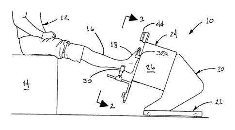
FIG. 1 is a side elevational, environmental view of a foot imaging apparatus
in
accordance with a preferred embodiment of the present invention, showing the
apparatus
with the right foot of a patient in position for imaging and measurement of
the contours of
the plantar surface;
FIG. 2 is a front elevational, environmental view of the foot imaging
apparatus of
FIG. 1, showing the manner in which the foot is aligned with a laser pointer
and also
reactively loaded by a support under the fifth metatarsal head so that the
foot is held in the
correct condition and orientation for imaging;
FIG. 3 is a front elevational view of the apparatus of FIGS. 1-2, with the
patient's
foot removed, showing the supporting structure and also the face of the
optical imaging
section of the apparatus;
FIG. 4 is a partial cross-sectional view of the foot imaging apparatus of
FIGS. 1-3,
taken along line 4-4 in FIG. 3, showing the structure of the adjustable
support in greater
detail;
FIG. 5 is an enlarged side elevational view of the imaging apparatus of FIG. 1-
3,
partially in cutaway, showing the manner in which the optical imaging section
of the
apparatus projects a pattern of lines onto the plantar surface of the foot,
that are viewed at an
angle by a camera to determine the contours of the plantar surface;
FIGS. 6-8 are sequential side elevational, environmental views of the foot
imaging
apparatus of FIGS. 1-3, showing the manner in which a patient's foot is placed
in the heel
support portion of the apparatus with the knee first raised and the ankle
dorsiflexed, and the
leg then straightened and the ankle plantarflexed;
FIG. 9 is a perspective view of a foot imaging apparatus in accordance with
another
preferred embodiment of the present invention;
FIG. 10 is a side elevational view of the foot imaging apparatus of FIG. 9,
showing
the configuration and locations of the components thereof in greater detail;
FIG. 11 is a bottom plan view of the apparatus of FIGS. 9-10, taken along line
11-11
in FIG. 10, showing the configuration of the wheeled chassis of the apparatus
in greater
detail;
CA 02809826 2013-02-27
WO 2012/030373 - 9 - PCT/US2011/001337
FIG. 12 is a rear elevational view of the apparatus of FIGS. 9-10, taken along
line
12-12 in FIG. 10, showing the configuration of the alignment components of the
apparatus
in greater detail;
FIGS. 13-15 are sequential elevational, environmental views of the imaging
apparatus of FIGS. 9-10 with a patient's foot placed therein, showing the
manner in which
the foot is set in the alignment section of the apparatus and the leg then
extended and the
ankle joint plantarflexed, similar to FIGS. 6-8;
FIG. 16 is a first top plan, environmental view of the apparatus of FIGS. 9-10
with
the patient's foot placed therein, in the position shown in FIG. 14 with the
leg bent and the
knee raised and with the ankle joint dorsiflexed;
FIG 17 is a second top plan, environment view of the apparatus of FIGS. 9-10
with
the patient's foot placed therein, in the position shown in FIG. 16 with the
ankle
plantarflexed and the knee lowered and the leg extended so as to push the
apparatus away
from the patient;
FIG. 18 is a side elevational, environmental view of a foot imaging apparatus
in
accordance with another embodiment of the present invention, in which the foot
is moved
vertically relative to the alignment section of the apparatus to reactively
load the area of the
fifth metatarsal head to lock the midtarsal joint, and the chassis of the
apparatus is moved
medially/laterally and/or proximally/distally as needed to align the foot and
place the
subtalar joint in a neutral condtion;
FIG. 19 is a second side elevational, environmental view of the foot imaging
apparatus of FIG. 9, showing the position of the foot when it has been lowered
onto the
alignment section of the apparatus;
FIG. 20 is a plan view of the foot imaging apparatus of FIGS. 18-19, taken
along line
20-20 in FIG. 18, showing the relationship of the foot to the alignment
section and also to
the aperture for the optical imaging section of the apparatus;
FIG. 21 is a cross-sectional view of the alignment section of the foot imaging
apparatus of FIGS. 18-19, taken along line 21-21 in FIG. 20, showing the
structure of the
supports for reactively loading the fifth metatarsal head of the foot in
greater detail; and
FIG. 22 is a cross-sectional view, similar to FIG. 21, of the alignment
section of a
foot imaging apparatus in accordance with another embodiment of the invention,
in which
CA 02809826 2013-02-27
WO 2012/030373 - 10 - PCT/US2011/001337
the support members include adjustable height pads for engaging the fifth
metatarsal head
areas of the feet.
CA 02809826 2013-02-27
WO 2012/030373 - 11 - PCT/U S2011/001337
DETAILED DESCRIPTION
FIG. 1 shows a foot imaging apparatus 10 in accordance with a first preferred
embodiment of the present invention. The apparatus is shown in use in
conjunction with a
patient 12 in a seated position on a chair 14 or other support, with a leg 16
and foot 18
outstretched.
As can be seen with further reference to FIG. 1, the imaging apparatus 10
includes an
optical imaging section 20 mounted on a rolling chassis section 22, and an
alignment section
24 that is spaced from the imaging section 20 towards the patient's foot 18.
The alignment
section 24 in the illustrated embodiment includes a spacer frame 26, which in
the
embodiment illustrated in FIG. 6-7 is formed by a somewhat box-shaped
structure, with a
through passage and open ends that define an aperture 28 via which the plantar
surface of
the foot is exposed to the optics of the imaging section 20. A principal
function of the
spacer frame is to support the alignment components, as described below, such
that the
plantar surface of the patient's foot will be held proximate a predetermined
focal length of
the camera in the imaging section 20; it will therefore be understood that the
shape and
construction of the spacer frame are somewhat arbitrary and may vary
significantly
depending on design factors.
As can be seen referring again to FIG. 1 and also FIG. 2, the alignment
section 24
includes a set of cooperating foot alignment components that are mounted to
the outer end of
the spacer frame (i.e., the end facing towards the patient), namely, a heel
stirrup 30, right
and left adjustable supports 32a, 32b for engaging the plantar surface of the
foot, and a laser
pointer 44 for projecting a visual reference line onto the foot. As will be
described in
greater detail below, the alignment components serve to position and load the
foot such that
the bone structure is held steady with the subtalar joint in the "neutral"
position and with the
midtarsal joint locked, which as explained above is critical for properly
determining
contours of the foot needed to construct an effective orthotic device. As
noted above, the
bone structure of a functional human foot transitions through a series of
phases beginning
with heel strike (when the heel makes initial contact with the ground or other
surface), with
the bone structure initially being somewhat loose and free to collapse and
spread to a degree
in order to absorb shock and conform to the underlying surface. Then as weight
moves
, a,
- 12 -
forwardly on the foot, with forward motion of the body, the bone structure
transitions to a
comparatively rigid configuration: In particular the center of weight, as
borne by the plantar
surface of the foot, initially follows a somewhat forward and lateral path, as
the rearfoot
simultaneously undergoes eversion, with the midtarsal joint becoming "locked"
as the center
of weight transfers onto the area of the fifth metatarsal head (generally in
the area beneath
the base of the small toe). The midtarsal joint remains locked for the
remainder of the gait
cycle, so that the foot forms a substantially rigid "lever" for efficiently
transmitting force to
the ground during toe-off. A more complete explanation of the gait cycle and
the locking
and unlocking of the metatarsal joint is found in U.S. Patent No. 5,960,566.
The alignment components of the present invention exploit the characteristics
of the
foot as a rigid lever, as described in the preceding paragraph, to locate the
foot in position
for imaging of its plantar surface; moreover, in the present invention this is
accomplished
without distorting the soft tissue or bone structure of the foot.
As can be seen in FIGS. 1-2, the heel rest or "stirrup" 30 is preferably
somewhat V-
shaped so as to have a centering effect on the rearfoot, and therefore also
the distal portion
of the leg, and is spaced somewhat away from the general plane of the plantar
surface, the
latter being located proximate aperture 28, so as to retainingly engage the
foot in the area
located near the top of the heel area/ bottom of the distal one-third of the
lower leg, with the
size and angle of the V-shaped area being configured to hold this area of the
leg firmly but
without discomfort to the patient. The V-shaped stirrup 30 is generally
located along the
centerline of the aperture 28 and therefore along the axis of the imaging
section, thus
allowing it to be used with either right or left feet.
As can be seen with further reference to FIG. 2 and also FIGS. 3-4, the right
and left
adjustable support members 32a, 32b project inwardly towards the centerline of
the aperture
28 from respective sidewalls 34a, 34b of the spacer frame 24. Pad members 36a,
36h are
mounted on the inboard ends of the adjustable arms 32a, 32b, and are
preferably formed of a
rigid yet transparent material that is capable of applying a dorsally-directed
force to the
plantar surface of the foot without obscuring it from view by the imaging
section, such as
plexiglass or LexanTM for example. The pad members 36a, 36b are preferably
sized to
CA 2809826 2018-02-28
CA 02809826 2013-02-27
WO 2012/030373 - 13 - PCT/US2011/001337
engage only the area of the foot immediately beneath the fifth metatarsal
head, with
dimensions of about 1" by 0.5" being generally suitable.
The arm members 32a, 32b are adjustable to accommodate different lengths and
widths of feet; in the embodiment that is illustrated in FIGS. 1-4, the arm
members 32a, 32b
are held in sliding engagement against the end surfaces of the sidewalls 34a,
34b of the
spacer frame 24, by guide strips 38a, 38b that are secured to the end surfaces
so as to define
slots sized to form a friction-fit but slidable engagement with the respective
arm members.
The arm members can therefore be selectively slid both longitudinally and
laterally (in and
out) with respect to the centerline of the aperture 28, so as to position the
pads 36a, 36b
beneath the fifth metatarsal head area of feet having different sizes, the
right side arm and
pad being used for right feet and the left side arm and pad being used for
left feet. As can be
best seen in FIG. 4, the arm members are preferably provided with upturned tab
portions 40
on their outer ends that facilitate manual adjustment of the arm members, as
well as
upturned end plates 42 located at the junctions where the transparent end pads
are mounted
to the arm members, the latter serving to engage the sides of the foot lateral
of the
transparent pads so that only the transparent material extends below the
plantar surface of
the feet.
The position of the heel stirrup 30 is also adjustable to accommodate feet and
legs of
different sizes. First, as can be seen in FIG. 3, the stirrup is supported on
the upper end of a
generally vertical arm 44 that is joined to a second, generally horizontal bar
by a bracket 48
that is in frictional engagement with the latter. The heel stirrup can
therefore be selectively
slid towards and away from the support arms 32a, 32b at aperture 28, as
indicated by arrow
49 in FIG. 5, to accommodate feet having smaller/shorter or bigger/taller
rearfoot areas
and/or difference in the size of the distal one-third of the lower leg.. The
end of horizontal
bar 46 is mounted to a second, generally vertical bar 50, that passes through
a friction-fit
sleeve 52 in sliding engagement therewith, the sleeve being fixedly mounted to
the spacer
frame 24 by a bracket 56; friction through the sleeve 52 is controlled by a
knob 54, so that
the position of the stirrup is adjustable in a generally vertical direction as
indicated by arrow
58 in FIG. 5.
Also mounted at the end of the spacer frame 24 proximate aperture 28 is the
laser
pointer 60, held in place by a support bracket 62, that projects a visible
beam 64 generally
CA 02809826 2013-02-27
WO 2012/030373 - 14 - PCT/US2011/001337
along the centerline of the aperture 28 and also in alignment with the center
of the V-shaped
heel stirrup 30 as well as the central plane of the camera 98, as indicated by
the dotted-line
image in FIG. 3. The laser beam thus provides a visual reference line for the
center plane of
the aperture 28 and the imaging area as a whole.
As was noted above, the components of the alignment section serve to orientate
the
bone structure of the foot with the midtarsal joint in the locked position,
employing
alignment of the bone structure in conjunction with a dorsally-directed
(upward) loading of
the fifth metatarsal head, essentially mimicking the reactive force of gravity
experienced by
the fifth metatarsal head at the corresponding point in the gait cycle.
The steps in accomplishing the positioning and locking of the foot are best
seen in
FIGS. 2 and 6-8. As an initial step, the imaging apparatus 10 is brought into
proximity with
the seated patient, so that the centerline that is established by the laser
pointer and V-shaped
heel stirrup is in general alignment with and towards the user's hip on the
side of the foot
that is to be imaged (e.g., in general alignment with the right portion of the
hip if the right
foot is to be imaged). The patient's foot is then placed in the stirrup 30 as
shown in FIG. 6,
with the knee slightly bent (raised), and with the ankle dorsiflexed and the
heel thrust
forward as indicated by arrow 70 in FIG. 6, so that the plantar surface of the
heel is located
closely proximate the plane that is defined by the adjustable pads 36a, 36b at
aperture 28. In
so doing, the stirrup takes the majority of the weight off of the extremity,
which
simultaneously centralizing the rearfoot and distal one-third of the lower leg
relative to the
aperture and imaging section. The clinician (operator) adjusts the respective
arm member
32a, 32b so that the associated pad 36a, 36b is positioned beneath the lateral
forefoot, and in
particular the fifth metatarsal head of the bone structure as shown in FIG. 2,
and the patient
then plantarflexes the ankle joint so as to lower the forefoot as indicated by
arrow 72 in
FIG. 7. In so doing, the plantar surface of the forefoot in the area beneath
the fifth metatarsal
head conies into contact with the pad 36a/36b on the support arm, so that the
fifth metatarsal
head is held against further movement in the plantar direction; plantarflexing
the forefoot
merely requires the patient to relax the ankle from holding the foot from the
"heel forward"
condition in which the foot is initially set in the stirrup, so that when the
forefoot is fully
relaxed and lowered, the fifth metatarsal head is subject to an upward
(dorsally-directed)
force mimicking the loading of the fifth metatarsal head created by the force
of gravity
CA 02809826 2013-02-27
WO 2012/030373 - 15 - PCT/US2011/001337
during the corresponding phase of the natural gait cycle. A dorsally-directed
force sufficient
to load the fifth metatarsal head to resistance is created merely by the
tension exerted by the
muscles of the lower leg when in a relaxed condition, acting through the
Achilles tendon and
with the ankle joint serving as the fulcrum, so that the midtarsal joint
assumes the locked
configuration without the patient having to purposely press down on the
forefoot using the
muscles and ligaments in a manner that might cause distortion of the foot or
deviation from
the correct shape, and without the area of the fifth metatarsal head having to
bear excessive
weight that might also cause distortion of the tissues and/or patient
discomfort.
To centralize the foot relative to the central axis of the viewing area and
place the
subtalar joint in a neutral condition, while keeping the midtarsal joint
locked, the leg is next
adjusted to position the second metatarsal head (in the area proximate the
base of the second
toe) with the beam 64 that is projected by the laser pointer 44, the beam
being aligned with
the center of the heel stirrup as noted above; in the embodiment of FIGS. 1-8,
centralization
of the foot is achieved by sliding the adjustable arm members 32a, 32b in or
out as
necessary. The patient's knee is then lowered and the ankle joint concurrently
plantarflexed
to about 90 , as indicated by arrow 74 in FIG. 8, so as to push the apparatus
10 away from
the chair or other support on which the patient is seated. In response, the
apparatus rolls
away from the patient over the floor on its wheeled chassis, as indicated by
arrow 76 in
FIG. 8; wheeled chassis 22 is supported by a pair of casters 80 at its
trailing end (toward the
patient) and a single caster 82 at its leading end (away from the patient), so
as to permit the
chassis to turn inwardly in an arc towards a patient's centerline as the
apparatus moves away
from the patient, thus accommodating the natural inward deviation (angle
towards the
midline of the body) that is present in most lower legs. The effect of the
combined distal
and medial motion of the apparatus is to bring the second metatarsal head of
the foot into
general alignment with the distal one-third of the lower leg so as to place
the subtalar joint in
the neutral condition, with the alignment being verified visually by the line
of the laser beam
pausing over the top of the second metatarsal head and up the distal portion
of the lower leg.
In practice, it has been found that with casters and a floor surface selected
for minimal
rolling resistance, the inward turning action of the cart as it rolls away
from the patient very
effectively obtains the correct alignment of the foot to the leg (as shown in
FIG. 2) with little
or no intervention or subsequent adjustment being required by the clinician;
to the extent
CA 02809826 2013-02-27
W020121030373 -16- PCT/US2011/001337
that minor corrections or "fine tuning" of the alignment is needed, this is
easily performed
by simply sliding the support arms in or out in the manner described above, to
bring the
second metatarsal head and lower third of the lower leg back into alignment
with the beam
of the laser.
It will be understood that other arrangements of casters or wheels may be used
on the
cart to allow the rolling and turning action, in addition to the "tricycle"
caster arrangement
described, and furthermore that in some instances the patient may be seated on
a chair or
other support that rolls away from and/or turns relative to the imaging
apparatus rather than
vice versa.
Positioned and locked in the manner described, the pad 36a/36b on which the
fifth
metatarsal head rests effectively establishes the transverse plane of the
foot, at a position
proximate the focal length of the camera of the imaging section of the
apparatus. Since, in
the illustrated embodiment, the V-shaped heel stirrup holds the rearfoot and
distal one-third
of the lower leg essentially perpendicular to the plane of the metatarsal
support pads 36a,
36b, the two pads effectively establish a transverse plane of the foot at
essentially 00
eversion/inversion relative to the frontal plane. However, as noted above,
individual feet
vary greatly, and depending on the degree of eversion exhibited by the foot
(e.g., 6 everted,
8 everted, and so on), the medial aspect of the forefoot may in some
instances be positioned
above the 00 transverse plane or below the 00 transverse plane when the
midtarsal joint is
locked and the subtalar joint is in the neutral position. Therefore, another
significant
advantage of the present invention, in which a support exists only under the
lateral forefoot
and preferably only under the fifth metatarsal head rather than all the way
across the foot, is
that the medial aspect of the foot is free to elevate above or depress beyond
the 00 transverse
plane as the nature of the particular foot dictates, which is not possible in
the case of devices
in which the entire width of the foot is pressed against a plate of glass or
other continuous
support or surface.
With the foot aligned and held in the manner described, the entire plantar
surface of
the foot is exposed to the optical system of the imaging section of the
apparatus, the area
under the fifth metatarsal head being "visible" to the optics by virtue of the
transparent
material of which the support pads are formed. Furthermore, since the foot is
centered on
the central plane of the camera (at aperture 28), the camera is able to
capture the image a
CA 02809826 2013-02-27
WO 2012/030373 - 17 - PCT/US2011/001337
sufficient distance up both sides (medial and lateral) of the foot, so that
adequate contour
data can be obtained without need for views at multiple angles or using
multiple cameras. In
the preferred embodiment that is illustrated in FIGS. 1-5, the imaging section
utilizes a
three-dimensional measurement instrument that operates on the basis of
projecting a pattern
of parallel lines onto the plantar surface of the foot and then capturing the
resulting image
using a camera set at a predetermined angle to the axis of projection, the
image then being
analyzed to determine the contours of the plantar surface. Such three-
dimensional digitizers
are available, for example, from Virtual 3-D Technologies Corp., Cutchogue,
New York,
, USA, systems of this general type sometimes being referred to as "white
light" digitizers.
As compared with systems based on scanning lasers, "white light" digitizers
offer significant
advantages, including almost instantaneous operation and therefore the ability
to effectively
"freeze" the image of the foot and eliminate the effects of movement, greatly
simplifying
operation in a clinical environment. It will be understood, however, that in
some
embodiments other forms of three-dimensional imaging systems may be utilized
in the
imaging section of the apparatus, including but not limited to scanning laser
systems, for
example.
Inasmuch as the "white light" three-dimensional digitizer alone is a more-or-
less
"off the shelf- component, its operation will be described herein only
briefly. As is shown
in FIG. 3, the digitizer employed in the illustrated embodiment includes a
transparent or
semi-transparent face plate 90 on which a series of opaque, parallel,
transverse orientation
lines 92 are formed. As can be seen in FIG. 5, a bulb 94 or other light source
is positioned
in the housing of the digitizer behind the faceplate 90, generally along an
axis substantially
perpendicular to the plantar surface of the foot 18. Operation of the light
source 94
illuminates the plantar surface of the foot, with the images of the opaque
lines 92 being
projected against the plantar surface, as indicated by dotted lines 96, to
create a
corresponding pattern of lines 78 on the surface of the foot. The resulting
image is captured
by a camera 98 set to view the surface along an axis 99 a predetermined acute
angle to the
axis at which the pattern is projected onto the foot. Thus, although the lines
97 appear
generally parallel as viewed along the projection axis from plate 90, the
contours of the
plantar surface of the foot cause the lines to deviate from parallel in the
image that is
captured by camera 98. The deviation from parallel, combined with the known
angle
CA 02809826 2013-02-27
WO 2012/030373 - 18 - PCT/US2011/001337
between the axis of projection and the axis of the camera, and other factors,
permits the
contours of the foot to be accurately calculated by associated software, with
the data being
outputted in suitable digital form.
The data representing the contours of the patient's foot can therefore be
obtained
quickly and conveniently in a clinical environment using the apparatus of the
present
invention. The patient may be seated in a suitable chair and place his or her
foot into the
alignment section of the apparatus in the manner described and then push away,
with the
attendant clinician making minor adjustments as necessary and simply
activating the switch
to digitize the contours of the foot. Not only are clinical efficiency and
patient comfort
greatly enhanced, but the opportunities for error are greatly reduced as
compared with prior
techniques.
FIGS. 9-18 illustrate a second preferred embodiment of the present invention,
which
is generally similar to the embodiment described above in terms of overall
operation and
layout, but which differs somewhat in its carriage and alignment sections.
As can be seen in FIG. 9, the apparatus 100 includes an imaging section 102
that is
substantially the same as described above and includes a face plate 104 for
projecting a
pattern of parallel lines onto the plantar surface of the foot. Rather than
being supported on
a box-like spacer frame, however, the alignment section 106 of the apparatus
is supported on
a pair of rigid flange portions 108a, 108b that extend upwardly from the
sidewalls 110a,
110b of the wheeled chassis 112. Also, rather than the pivoting casters of the
embodiment
described above, the wheeled chassis includes a pair of horizontal axis wheels
114a, 114b on
the trailing end disposed towards the patient, and a single ball transfer unit
(ball roller) 116
on the opposite end that is free to roll in any direction; it will be
understood that in some
instances there may be multiple ball transfer units rather than the single
unit that is shown.
A particular advantage of the arrangement of wheels and ball transfer unit
employed in
chassis 112, as compared with conventional casters, is that this avoids the
initial pivoting
motion or "jog" that is created by the offset vertical axes of casters, which
allows the
apparatus to follow a comparatively smooth, unbroken arc as it moves away and
pivots
towards the centerline of the body as the patient's leg is extended, and which
also facilitates
free movement of the chassis in the transverse plane of the floor in order to
perform
adjustments as necessary.
CA 02809826 2013-02-27
WO 2012/030373 - 19 - PCT/US2011/001337
As can be seen with further reference to FIG. 10 and also FIG. 11, the chassis
112
includes an optional extensible (e.g., telescoping) handle 118, on the side
disposed away
from the patient, both to provide an aid to the clinician in adjusting the
position of the
chassis and also to facilitate transportation of the assembly between
locations, such as
between examination rooms.
Referring again to FIGS. 9-10, it can be seen that the alignment section 106
of the
embodiment illustrated therein includes a V-shaped heel stirrup 120, right and
left adjustable
plantar support members 122a, 122b, and a laser pointer 125, that perform
functions
similarly to the corresponding elements described above. Rather than sliding
bars, however,
the adjustable support members 122a, 122b are rotatable units slidingly
mounted in
generally vertical channels 124a, 124b. As can better be seen in FIG. 12, the
rotatable
supports include hand grip portions 126, and inwardly disposed somewhat
semicircular
projecting flange portions 128 formed of a transparent material, similar to
the transparent
support pads described above. Thus, as can be seen in FIG. 12, the transparent
support
flanges 128 are positioned beneath fifth metatarsal areas of the feet simply
by sliding the
respective (right or left) support member 122a, 122b through its associated
track to the
general location and then rotating the flange inwardly, by turning handgrip
126. As can be
seen the handgrips 126 preferably have an enlarged oval form, with the
distance between the
edge of the handle and the outer edge of the clear projecting flange 128 being
selected such
that the flange will be positioned beneath the fifth metatarsal head when the
handle portion
is moved to be adjacent or in contact with the side of the foot. The
semicircular shape of the
transport flanges 28 in combination with the slide channels, also facilitates
rapid and
convenient positioning of the supports beneath the heads, so that this can be
accomplished
without excessive manipulation or "fiddling."
The V-shaped heel stirrup 120, in turn, is supported on a platform 130 that
projects
towards the patient, in sliding engagement with a pair of tracks 132a, 132b
that permit the
stirrup to be moved towards or away from the aperture 134 in a manner similar
to that
described above, but with a simplified sliding motion. The sliding interfit
between the
tracks and the cooperating portions of the heel stirrup 120 preferably
includes a slight
frictional resistance, as do tracks 124a, 124b and the cooperating portions of
the adjustable
members 122a, 122b, so that the members can be conveniently slid to the
desired locations
CA 02809826 2013-02-27
WO 2012/030373 - 20 - PCT/US2011/001337
but will then remain in place without assistance once released. As with the
heel stirrup
described above, stirrup 120 is centered on the central plane of the imaging
section of the
apparatus, as can be seen from its relationship to beam 136 and camera 138 in
FIG. 12.
Use of the apparatus 100 and the manner in which it cooperates with a
patient's foot
and leg is generally similar to the embodiment described above, and is
illustrated in
FIGS. 13-18.
As can be seen in FIG. 13, the user's foot is first placed in the heel stirrup
with the
knee raised and the forefoot dorsiflexed and heel pressed forward, as
indicated by arrow
140. The respective lateral forefoot support member 122a, 122b is adjusted
into position
along its track, and then rotated inwardly to move the clear support flange
128 thereof into
the area beneath the fifth metatarsal head of the foot. The patient next
relaxes the foot and
allows the ankle to plantarflex the forefoot, as indicated by arrow 142, so
that the fifth
metatarsal head is subjected to a mild reactive force in the dorsal direction,
mimicking the
force of gravity so as to lock the midtarsal joint in the manner described
above. The support
member 122a/122b is then rotated inwardly/outwardly as needed in order to
align the second
metatarsal head of the forefoot with the centerline beam 134 projected by
laser 125.
The patient then lowers the knee and extends the leg, as indicated by arrow
144 in
FIG. 16, causing the apparatus to roll outwardly and turn inwardly (towards
the patient's
centerline), as can be seen by comparison of FIGS. 17-18. In particular, FIG.
17 shows the
orientation of the apparatus 100 relative to the patient when in the position
of FIG. 14, i.e.,
with the forefoot plantarflexed 90 to the lower leg but with the knee still
raised; as can be
seen therein, the apparatus, including its wheeled chassis at this point, lies
in substantially
coaxial alignment with the patient's lower leg and the associated side of the
hip. However,
as the patient lowers the knee and straightens the leg so as to push the
apparatus away, the
inward deviation of the lower leg causes the leading (distal) end of the
apparatus to turn
inwardly towards the centerline of the patient's body, as is indicated by the
shift between the
dotted and solid reference lines 146, 146' in FIGS. 17 and 18, allowing the
second
metatarsal head and distal one-third of the lower leg to come into alignment
with one
another and with the beam 150 projected by the laser 125. In short, as the
patient pushes the
apparatus away the wheeled chassis allows the apparatus, and in particular the
central plane
of the imaging section, to align itself with the distal one-third of the lower
leg such that the
CA 02809826 2013-02-27
W020121030373 -21- PCT/US2011/001337
subtalar joint is in the neutral position. Minor adjustments can then be made
by the clinician
if necessary, rotating the handgrip of the associated support and/or using
handle 118 and
also a crossbar 152 at the top of the alignment section. To move the foot into
alignment
with the center plane of the imaging section, as indicated by the beam 136 of
the laser
pointer passing over the second metatarsal head of the foot and onto the
distal one-third of
the lower leg; in so doing, the neutral position of the subtalar joint can
be
approximated/verified by the clinician manually rotating the lower leg
internally and
externally and observing the resultant movement of the laser beam 136, to one
side and the
other from alignment with the second metatarsal head and distal one-third of
the lower leg.
The image of the plantar surface of the foot is then captured for digitization
by simply
pressing one of the switches 154 that actuate the imaging section 102 of the
apparatus.
It will be understood that in some cases or embodiments the dorsally-directed
load
may be applied to the area of the fifth metatarsal head in a direct manner,
rather than by first
setting the foot into the stirrup or other support with the heel projected and
then
pantarflexing the forefoot onto the support as described. However, it has been
found that
such an approach generally leads to the ankle joint being in a plantarflexed
position and the
remainder of the foot in an inverted position relative to the transverse plane
at the viewing
area, and therefore less than optimal results when imaged. This problem is
avoided by
placing the foot/leg on the stirrup with the ankle dorsiflexed and then
plantarflexing the foot,
in the manner that has been described.
The embodiments described above employ wheeled chassis to achieve relative
movement between the patient and alignment section in order to position the
foot with the
midtarsal joint locked and the subtalar joint in the neutral position. FIGS.
18-22, in turn,
illustrate an embodiment in which the patient's foot is lowered onto the
support member of
the apparatus in order to establish the requisite reactive force acting
dorsally on the fifth
metatarsal head.
Accordingly, as can be seen in FIG. 18, the patient 12 is seated on a
vertically
movable platform 158, such as an examination table for example, with the foot
18
positioned more-or-less directly over the apparatus 160. The apparatus 160
includes an
imaging section 162 and spacer frame 164 similar to the corresponding
components of the
embodiment shown in FIGS. 1-2, and also an alignment section 166, all
supported on a
CA 02809826 2013-02-27
WO 2012/030373 - 22 - PCT/US2011/001337
wheeled chassis 167. However, rather than moving horizontally to load the
foot, the
assembly remains in position on the floor as the patient is lowered to bring
the fifth
metatarsal head area of the foot into contact with the adjustable support 168,
as indicated by
arrow 170 in FIG. 18.
Since relative movement is provided by the table 158 or other vertically
moveable
support, the patient need not dorsiflex the foot before placing it in the
apparatus; instead, the
heel is simply positioned in the heel stirrup 174 and reactive force is
generated as the fifth
metatarsal head area of the foot comes into contact with and is then
reactively lifted by the
transparent pad 172 at the end of the support member 168a/168b; in so doing,
the heel
stirrup 174 is allowed to move vertically with the foot by the sliding
engagement formed
with its upwardly projecting support 176, similar to the stirrup 120 and
support 130
described above. The position of the apparatus can then be adjusted in the
transverse plane
of the floor to place the subtalar joint in the neutral configuration and
bring the foot into
alignment, with the beam 178 of laser 180 aligned with the second metatarsal
head and
distal one-third of the lower leg, by moving the apparatus on the floor in the
necessary
direction or directions using wheeled chassis 167. It will be understood that
relative vertical
movement between the apparatus and the patient's foot may in some instances be
established by raising the apparatus, or an operative portion thereof,
relative to the patient's
foot, rather than lowering the patient's foot onto the apparatus as shown.
In the embodiment illustrated in FIGS. 18-21, the adjustable members are
mounted
for pivotable adjustment in brackets 182a, 182b at the sides of the aperture
184, as indicated
by dotted line images 168a' and 168b' in FIG. 20 (see also FIG. 21). FIG. 22,
in turn, shows
an arrangement in which the alignment section includes adjustable supports
190a, 190b
having head members 192 at their inboard ends that are vertically adjustable
by means of
shafts 194 that are in threaded engagement with cooperating nuts 196, to aid
in adjusting the
position of the plantar surface of the foot, and in particular with respect to
the focal length of
the camera of the imaging section. It will be understood that other forms of
adjustable
support members may occur to those skilled in the relevant art, and
furthermore that
although having the support members formed of a transparent material is
preferable in terms
of imaging accuracy, it is anticipated that in some instances opaque supports
may be used
instead and the obscured contours established by interpolation or other
suitable means.
CA 02809826 2013-02-27
wo 2012/030373 - 23 - PCT/US2011/001337
It is to be recognized that various alterations, modifications, and/or
additions may be
introduced into the constructions and arrangements of parts described above
without
departing from the spirit or ambit of the present invention as defined by the
appended
claims.