Note : Les descriptions sont présentées dans la langue officielle dans laquelle elles ont été soumises.
DEVICES AND METHODS FOR THE TREATMENT OF
VASCULAR DEFECTS
[00011
Background
[0002] The invention relates generally to medical devices and more
particularly to expandable
medical devices and methods for treating vascular defects. For example, the
invention can relate to
expandable medical devices and methods for treating an aneurysm. Aneurysms are
dilations in a
blood vessel caused from weakening of a blood vessel wall. The dilation is
produced by the pressure
exerted by normal blood flow, which can cause the weakened segment of the
blood vessel to swell. In
some cases, this swelling results in a sac, or balloon-like polyp protruding
from the main or parent
vessel. Continued growth and/or eventual rupture of the ballooned arterial
wall can have devastating
results for a patient. As such, unruptured aneurysms should be treated to
prevent hemorrhage.
Additionally, ruptured aneurysms can be treated to avert a subsequent rupture
and/or additional
damage.
100031 Some known medical devices and treatment methods used for treating
an aneurysm
include delivering a platinum coil to the sac of the aneurysm. The platinum
coil is electrolytically
separated from a delivery wire, thus inducing a charge in the coil which can
cause a thrombotic effect
in the aneurysm. In known procedures, about 30% of the volume of the aneurysm
is packed with
coils. Such known devices and methods, however, often have an about 30%
recanalization rate,
meaning blood flow returns to the aneurysm again and can cause the coil-packed
aneurysm to swell
further. Additionally, such known devices and methods require prolonged
procedure times for the
patient and correspondingly increased exposure to radiation for the patient.
Moreover, such devices
and methods do not treat the neck of the aneurysm, which is the area between
the parent blood vessel
and the sac of the aneurysm.
1
CA 2812012 2017-08-16
CA 02812012 2013-03-08
WO 2012/034135 PCT/US2011/051268
[0004] Another known treatment method includes the use of both a coil and a
stent. The
coil is delivered to the sac of the aneurysm as described above, and the stent
is positioned
within the parent blood vessel such that a portion of the stent is disposed
over the neck of the
aneurysm. Such procedures have several drawbacks. For one, delivery of two
separate types
of devices (i.e., coil(s) and a stent) is a more complex procedure, often
resulting in a longer
procedure time for the patient. The stent may lead to intra-stent stenosis of
the blood vessel.
Additionally, a patient would likely be required to take a blood thinner
indefinitely following
the procedure. Moreover, such devices and methods are not suitable for
treatment of
aneurysms positioned at a bifurcation of the blood vessel (i.e., between
adjacent branches of a
vessel).
[0005] Another known device and treatment method includes the use of a flow
diverter
delivered to the parent blood vessel adjacent the neck of the aneurysm.
Generally, the flow
diverter is positioned within the parent blood vessel over the neck of the
aneurysm to prevent
additional blood flow into the aneurysm from the vessel. In current
procedures, more than
one flow diverter is required per aneurysm to ensure blood flow is
appropriately diverted
from the aneurysm. Such a device and treatment method has similar drawbacks to
the use of
a stent, described above. Specifically, the flow diverter may lead to stenosis
of the blood
vessel and the patient would likely be required to take a blood thinner
indefinitely following
the procedure. Additionally, known flow diverters are not suitable for
treating an aneurysm
positioned at a bifurcation of the blood vessel. Moreover, long term follow-up
of patients
treated using a flow diverter is showing an increased rate of recanalization
to the aneurysm.
[0006] Thus, there is a need for improved systems, devices and methods for
treating
vascular defects, such as balloon-type aneurysms, as described herein.
Summary of the Invention
[0007] Devices and methods for treating vascular defects, such as, for
example, balloon-
type aneurysms, are described herein. In one embodiment, an apparatus includes
an insertion
portion and an expandable implant. The expandable implant is configured to be
deployed in
an aneurysm and is coupled to the insertion portion. The expandable implant
has a first
portion and a second portion coupled to the first portion. The expandable
implant is movable
between a first configuration in which the first portion and the second
portion are
2
CA 02812012 2013-03-08
WO 2012/034135 PCT/US2011/051268
substantially linearly aligned and a second configuration in which the second
portion at least
partially overlaps the first portion.
Brief Description of the Drawings
[0008] FIG. 1 is a schematic illustration of a medical device according to
an embodiment
in a first configuration.
[0009] FIG. 2 is a schematic illustration of a medical device according to
an embodiment
in a second configuration.
[0010] FIG. 3 is a side view of a medical device according to an embodiment
in a first
configuration.
[0011] FIG. 4 is a side view of a medical device according to an embodiment
in a second
configuration.
[0012] FIG. 5A is a view of the medical device of FIG. 3 in a first
configuration during
insertion into an aneurysm.
[0013] FIG. 5B is a view of the medical device of FIG. 3 in a second
configuration during
insertion into an aneurysm.
[0014] FIG. 5C is a view of the medical device of FIG. 3 in a third
configuration during
insertion into an aneurysm.
[0015] FIG. 6 is a view of a portion of a medical device in an expanded
configuration,
according to an embodiment.
[0016] FIGS. 7-13 are views of a medical device in an expanded
configuration, according
to embodiments.
[0017] FIG. 14 is a view of a medical device in a partially collapsed
configuration,
according to an embodiment.
[0018] FIG. 15 is a view of the medical device of FIG. 14 in an expanded
configuration,
according to an embodiment.
3
CA 02812012 2013-03-08
WO 2012/034135 PCT/US2011/051268
[0019] FIG. 16 is a view of a portion of a medical device in an expanded
configuration
according to an embodiment, with a first portion spaced apart from a second
portion.
[0020] FIG. 17A is a view of a portion of a medical device in a collapsed
configuration
according to an embodiment.
[0021] FIG. 17B is a view of a portion of a medical device in an expanded
configuration
according to an embodiment.
[0022] FIG. 18 is a flowchart of a method according to an embodiment.
Detailed Description
[0023] Medical devices and methods of treatment are described herein to
treat patients
experiencing a vascular defect, such as an aneurysm, in a circulatory blood
vessel and the
effects of that defect, including hemorrhagic stroke. For example, the devices
and methods
described herein can be useful for treating vascular defects present in
vasculature that is
tortuous, of small-diameter, and/or that is otherwise difficult to access.
More specifically, the
devices and methods described herein can be useful for treating saccular (also
referred to as
balloon-type or berry) aneurysms, bifurcate aneurysms, fistulas, and other
defects in
vasculature, including defects in neurovasculature. The medical devices and
methods of
treatment described herein can reduce hemorrhagic events while promoting
endothelialization
of an opening between an aneurysm and a parent blood vessel from which the
aneurysm
bulge formed (e.g., at a neck of the aneurysm).
[0024] Various embodiments of a medical device for occupying all or
substantially all of
the volume of an aneurysm and/or promoting endothelialization at or proximate
to the
aneurysm are described herein. In some embodiments, the medical device
includes an
expandable implant including an electropositive woven or braided material. The
filaments or
strands forming the braid or weave are configured to encourage recruitment
and/or retention
of endothelial cells to the device and therefore within the defect. The
expandable implant is
configured to assume a non-linear pre-determined three-dimensional shape
within a sac of the
aneurysm upon release from a tubular or other delivery constraint (e.g., a
catheter or
cannula). The electropositive woven or braided material has a particular
porosity and
includes multiple openings between the filaments or strands when the
expandable implant is
in the expanded configuration. Such openings are ideal in the blood
environment for
4
CA 02812012 2013-03-08
WO 2012/034135 PCT/US2011/051268
harboring endothelial cells recruited to the site. The electropositivity of
the material
encourages endothelialization in the presence of the electronegative charges
of the blood and
body tissues. Said another way, the electropositivity of the expandable
implant in relation to
a charge of blood and tissue (which is electronegative in comparison) provides
an
environment in the defect that promotes endothelialization. Endothelialization
within the
defect can ultimately result in the defect walling-off from the parent vessel.
For example, the
growth and development of an endothelial layer over a neck of an aneurysm can
wall off the
aneurysm from the parent vessel and allows flow dynamics to equilibrate at the
defect. As
such, the device can be configured to facilitate healing the defect and
preventing
recanalization because tissue is created from within the body that resists
aberrant blood flow
and redistributes the flow pressure that may have created the defect. Upon
healing with
endothelialization, the pressure is evenly distributed along the parent vessel
in a manner that
precludes recanalization at the defect post-treatment. Furthermore, blood from
within the
parent vessel no longer has access to the walled off defect once the
endothelialization process
is complete. Additionally, at least a portion of the expandable implant can be
positioned over
the neck of the aneurysm once the implant is deployed within the aneurysm such
that the
portion disrupts the flow of blood from the parent vessel into the aneurysm.
As such, the
expandable implant provides blood flow disruption in advance of and in
addition to growth
and development of the endothelial layer over the neck of the aneurysm.
[0025] A medical device described herein can include an insertion portion
(e.g., a guide
wire) and an expandable implant formed with, for example, woven or braided
filaments in a
mesh-like configuration. The terms mesh and braid can each refer herein to a
fabric or
material of woven or braided filaments or strands of wire or polymer. The
expandable
implant of the medical device can be configured to compress or collapse for
delivery into a
blood vessel. In some embodiments, the medical device can be inserted while in
a collapsed
or compressed configuration through a delivery device, such as, for example, a
microcatheter,
cannula, delivery tube or sheath. In some embodiments, the medical device can
be deployed
without the use of such a delivery device.
[0026] The expandable implant of the medical device can have a collapsed or
compressed
configuration such that the expandable implant has a diameter that can fit
within the narrow
constraints of the neurovasculature and/or within a lumen of a delivery
catheter. The
expandable implant of the medical device can be formed with, for example, an
arrangement
of strands (e.g., a mesh or braid arrangement of strands or filaments) that
can compress and expand.
Such materials include Nitinol, MP35N, stainless steel, cobalt chromium,
titanium, platinum,
tantalum, tungsten, or alloys thereof, or polyester, polyethylene (PET),
DacronTM, PEEK, vectronTM,
and suture materials, and are available from Fort Wayne Metals of Fort Wayne,
Indiana, California
Fine Wire Company of Grover Beach, California, other metal manufacturers,
Ethicon Inc. of
Somerville, New Jersey, Genzyme of Cambridge, Massachusetts, Poly-Med, Inc. of
Anderson, South
Carolina, and/or other medical grade suture and fiber manufacturers. The
expandable implant can be
compressed over and/or along the insertion portion of the medical device. The
insertion portion can
be, for example, a wire. In some embodiments, a medical device includes an
insertion portion
movably disposable within a lumen of a delivery device. A distal portion of
the insertion portion can
be coupled to the expandable implant. The expandable implant can be moved from
a collapsed
configuration to an expanded configuration while disposed within, or as it is
being inserted into, a
defect (e.g., an aneurysm).
[0027] In some embodiments, the expandable implant can be formed with
filaments of
superelastic or shape memory material (such as, e.g., nitinol) and the braid
or mesh can be set in a
predefined shape prior to attaching the expandable implant to the insertion
portion of the medical
device. In such an embodiment, when the expandable implant is deployed and
expands, it assumes a
biased predetermined shape. The predetermined shape can be a generic shape,
such as that of a
sphere, or can be a custom-made shape based on a shape of a target aneurysm
within a patient.
Suitable materials are described in more detail herein.
[0028] The medical devices described herein can include one or more
expandable implants
formed with a woven mesh or braid that has variably sized apertures (also
referred to herein as
"openings" or "pores"). Said another way, the devices are formed with a
material that has a particular
porosity or pore density. In some embodiments, an expandable implant can have
sections of mesh or
braid having variation in density of the filaments and may include portions or
bands of densely spaced
filaments (i.e., lower porosity) spaced by portions or bands that are less
dense (i.e., higher porosity).
The less dense braid portion can have larger openings in the braid, while the
more dense braid portion
can have smaller openings in the braid. Material (e.g., bodily tissue such as
endothelial cells) can be
encouraged to enter and/or attach to interstices of the mesh of the expandable
implant. For example,
the more dense braid portion can be used to encourage greater endothelial cell
6
CA 2812012 2017-08-16
CA 02812012 2013-03-08
WO 2012/034135 PCT/US2011/051268
attachment and the less dense braid portion can be used to reduce the overall
weight and or
material to be implanted in the patient. The less dense sections can also
direct the final shape
of the expandable implant. For example, sections of less dense (more open)
mesh or braid
can direct the effects of expansion of the implant.
[0029] In some embodiments, a medical device can be delivered to a desired
treatment
site within a vasculature by inserting the medical device through a lumen of a
delivery
catheter (e.g., a microcatheter). The expandable medical device can be
inserted through the
delivery catheter in a collapsed or compressed configuration. The expandable
implant of the
expandable medical device can be moved out through a distal end of the
delivery catheter at
the treatment site (e.g., into a sac of an aneurysm) and moved to an expanded
configuration.
In some embodiments, the delivery catheter is used to compress or collapse the
expandable
implant. For example, the expandable implant can be formed with a biased
expanded
configuration and when it is placed within a lumen of a catheter it is
compressed. When the
expandable implant is moved outside of the catheter, it can assume its biased
expanded
configuration. In the expanded configuration, a first portion of the
expandable implant
substantially overlaps a second portion of the expandable implant. The first
and second
portions of the expandable implant can be discrete structures or can be
portions of a unitary
or monolithically constructed device.
[0030] A medical device, such as an expandable implant, described herein
can include a
first porous member and a second porous member coupled to the first porous
member. Each
of the first and second porous members include a first end and a second end.
The first and
second porous members each have a collapsed configuration for insertion
through a blood
vessel and an expanded configuration for occupying at least a portion of the
volume defined
by the sac of an aneurysm. In some embodiments, the first porous member is
substantially
elongate and has a greater width in its expanded configuration than in its
collapsed
configuration. The second porous member is substantially elongate and has a
greater width
in its expanded configuration than in its collapsed configuration. In some
embodiments, the
width of the first porous member is greater than the width of the second
porous member, for
example, when each of the first and second porous members are in their
expanded
configurations.
[0031] In some embodiments, the first porous member is configured to occupy
a first
volume in its collapsed configuration and a second, greater, volume in its
expanded
7
CA 02812012 2013-03-08
WO 2012/034135 PCT/US2011/051268
configuration. For example, the first porous member can have a substantially
spherical,
oblong, or other suitable shape in its expanded configuration that occupies a
greater volume
than the substantially elongate shape of the first porous member in its
collapsed
configuration. The second porous member can be configured to move or curve
into a three
dimensional configuration in the expanded configuration such that a first
segment of the
second porous member overlaps with a second segment of the second porous
member. In its
expanded configuration, the second porous member can define an interior region
configured
to receive the first porous member in its expanded configuration. For example,
in some
embodiments, the second porous member has a substantially spherical shape with
an open
interior region configured to receive the first porous member.
[0032] In some embodiments, a medical device, such as an expandable
implant, described
herein can include a first porous member and a second porous member. Each of
the first and
second porous members include a first end and a second end. The first and
second porous
members each have a collapsed configuration for insertion through a blood
vessel and an
expanded configuration for occupying at least a portion of the volume defined
by a sac of an
aneurysm. The first and second porous members are each substantially elongate
in the
collapsed configuration. In its expanded configuration, the first porous
member has a three-
dimensional shape including a first segment configured to overlap with a
second segment and
defining an interior region. The second porous member is configured to be
disposed in the
interior region of the first porous member when each of the first and second
porous members
are in their respective expanded configurations. In some embodiments, the
second porous
member can be formed integrally or monolithically with the first porous
member. In some
embodiments, the second porous member can be woven or braided using the same
filaments
that form the first porous member.
[0033] In some embodiments, the expandable implant is in the form of a
braided tube that
includes fibers of a super elastic shape memory alloy, or polymeric fibers. In
some
embodiments, the expandable implant can effect a shape deformation inducing a
substantially
spherical contour. In some embodiments, the expandable implant can effect a
shape
deformation inducing a helical contour. In some embodiments, the shape
deformation can
include inducing radial expansion and/or axial shortening.
100341 The medical devices described herein can be used to occupy at least
a portion of
the volume defined by a sac of an aneurysm and/or to promote
endothelialization of the neck
8
CA 02812012 2013-03-08
WO 2012/034135 PCT/US2011/051268
of the aneurysm to inhibit or stop blood flow into the aneurysm, which can
lead to, for
example, hemorrhagic stroke. In some embodiments, wire or polymer filaments
can be used
to form a woven mesh or braided strands that can be expandable, and have
apertures sized to
promote endothelial cell attachment at the aneurysm.
[0035] It is noted that, as used in this written description and the
appended claims, the
singular forms "a," "an" and "the" include plural referents unless the context
clearly dictates
otherwise. Thus, for example, the term "a lumen" is intended to mean a single
lumen or a
combination of lumens. Furthermore, the words "proximal" and "distal" refer to
direction
closer to and away from, respectively, an operator (e.g., surgeon, physician,
nurse, technician,
etc.) who would insert the medical device into the patient, with the tip-end
(i.e., distal end) of
the device inserted first inside a patient's body. Thus, for example, the end
first inserted
inside a patient's body would be the distal end of the medical device, while
the end outside of
or inserted later into a patient's body would be the proximal end of the
medical device.
Additionally, the terms "first," "second," "third," and so on, used to
describe similarly
identified elements is for purposes of clarity only, and are not meant to
imply a priority or
that such numerical identifier must be associated with that particular element
in the claims.
[0036] FIGS. 1 and 2 are schematic illustrations of a vascular medical
device 100
according to an embodiment in a first configuration and a second
configuration, respectively.
The medical device is configured to promote healing of an aneurysm. More
specifically, at
least a portion of the medical device is configured to occupy at least a
portion of the volume
defined by a sac of the aneurysm and, in some embodiments, at least a portion
of the medical
device is configured to promote endothelial cell attachment over a neck of the
aneurysm.
Once endothelialization over the aneurysm neck is complete, blood flow into
the aneurysm
sac from a parent blood vessel (i.e., the vessel on which the aneurysm formed)
is prevented.
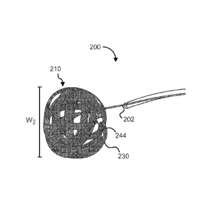
[0037] The medical device 100 can include an insertion portion 102 and an
expandable
implant 110. The insertion portion 102 is coupled to the expandable implant
110, such as, for
example, at a proximal portion 112 of the expandable implant 110. In some
embodiments,
the insertion portion 102 is removably coupled to the expandable implant 110.
In this
manner, the insertion portion 102 can be separated from the expandable implant
110
following delivery of the expandable implant to the aneurysm and removed from
a patient's
vasculature. The insertion portion 102 can be, for example, a guide wire or a
distal end
portion of a wire. The medical device 100 can be used with a cannula or
catheter 104 (shown
9
CA 02812012 2013-03-08
WO 2012/034135 PCT/US2011/051268
in dashed lines in FIGS. 1 and 2) to, for example, deliver the expandable
implant 110 to the
aneurysm.
[0038] The expandable implant 110 is configured to be deployed in the
aneurysm (e.g., in
a sac of an aneurysm). The expandable implant 110 has a first portion 120 and
a second
portion 130. As shown in FIG. 1, the expandable implant 110 has a first
configuration in
which the first portion 120 and the second portion 130 are substantially
linearly aligned. In
its first configuration, the expandable implant 110 is configured for
insertion through a blood
vessel. The expandable implant 110 is also configured for insertion through a
neck of the
aneurysm when in its first configuration.
[0039] The expandable implant 110 is movable between its first
configuration and a
second configuration in which the second portion 130 at least partially
overlaps the first
portion 120, as shown in FIG. 2. For example, the second portion 130 can be
configured to
bend, curve and/or twist in multiple turns such that multiple segments of the
first portion 120
and the second portion 130 are overlapped. Additionally, at least one of the
first portion 120
and the second portion 130 can be configured to bend or curve in multiple
turns such that the
respective first or second portion is overlapped with itself. In some
embodiments, the
expandable implant 110 can be understood to have multiple first portions and
multiple second
portions. In other words, the expandable implant can continually overlap
itself in its
deployed configuration to occupy all or substantially all of the volume of the
aneurysm.
[0040] In its second configuration, the expandable implant 110 is
configured to occupy at
least a portion of the volume defined by the sac of the aneurysm. In some
embodiments,
when the expandable implant 110 is in its second configuration, at least a
portion of the
expandable implant is configured to be positioned over the neck of the
aneurysm. For
example, the portion of the expandable implant 110 at which the second portion
130 overlaps
the first portion 120 can be configured to be positioned over the neck of the
aneurysm. As
such, the portion of the expandable implant 110 disposed over the aneurysm
neck has an
increased density (e.g., a dual density compared to the first portion 120 or
the second portion
130 individually), which helps to limit or prevent blood flow from entering
the sac of the
aneurysm. The portion of the expandable implant 110 positioned over the
aneurysm neck can
be a scaffold for endothelial cell attachment at the aneurysm neck. For
example, the portion
of the expandable implant 110 positionable over the aneurysm neck can be
porous, such as by
including a porous mesh, as described in more detail herein. In some
embodiments, the first
CA 02812012 2013-03-08
WO 2012/034135 PCT/US2011/051268
portion 120 and the second portion 130 of the expandable implant 110 arc
biased to the
second configuration.
[0041] As noted above, in some embodiments, at least a portion of the
expandable
implant 110 is porous. For example, in some embodiments, at least a portion of
the
expandable implant 110 can include and/or be constructed of a mesh (e.g.,
woven, braided, or
laser-cut) material such that a wall or layer of the expandable implant 110
defines multiple
openings or interstices 118. More specifically, in some embodiments, at least
one of or both
the first portion 120 and the second portion 130 of the expandable implant 110
can include
the porous mesh. The porous mesh can have a first porosity when the expandable
implant
110 is in its first configuration and a second porosity when the expandable
implant is in its
second configuration. More specifically, in some embodiments, the porous mesh
can have a
greater porosity when the expandable implant 110 is in its second
configuration than when
the expandable implant is in its first configuration. The porosity of the
porous mesh can be
increased, for example, because one or more individual pores or openings are
larger when in
the second configuration than in the first configuration. For example, the
porous mesh can be
expanded in the second configuration, thereby increasing the space between
filaments of the
mesh (and thus the size of one or more openings of the mesh). In other words,
an overall
volume of pore openings can be increased. In another example, the porosity of
the porous
mesh can be increased because one or more openings that were closed off when
the
expandable implant 110 was collapsed into its first configuration are reopened
when the
expandable implant is moved to its second configuration. In other words, a
number of open
pores can be increased.
[0042] In some embodiments, the first portion 120 and the second portion
130 can have
one of the same or different porosities. For example, the first portion 120
can have a porosity
greater than a porosity of the second portion 130. In another example, the
second portion 130
can have a porosity greater than the porosity of the first portion 120. In
still another example,
the first and second portions 120, 130 can have substantially equivalent
porosities in the
expanded configuration.
100431 In some embodiments, at least one of the first portion 120 and the
second portion
130 includes one, two, three, or more layers. For example, in some
embodiments, the first
portion 120 of the expandable implant 110 includes a first layer (not shown in
FIGS. 1 or 2)
of porous mesh and a second layer (not shown in FIGS. 1 or 2) of porous mesh.
The first
11
CA 02812012 2013-03-08
WO 2012/034135 PCT/US2011/051268
layer and the second layer can have the same or different porosities. In some
embodiments,
the first layer is offset from the second layer. As such, the porosity of the
first portion is
determined by the porosities of the first and second layers and the manner in
which the first
layer is offset from the second layer.
[0044] In some embodiments, at least a portion of the expandable implant
110, such as at
least one of the first portion 120 or the second portion 130 can include a
shape-memory
material, such as, for example, nitinol, and can be preformed to assume a
desired shape.
Thus, in such an embodiment, the portion of the expandable implant 110 (e.g.,
the first
portion 120 and/or the second portion 130) can be biased into an expanded
second
configuration and moved to a collapsed first configuration by restraining or
compressing the
portion of the expandable implant.
[0045] In some embodiments, at least a portion of the expandable implant
110, such as at
least one of the first portion 120 or the second portion 130 can include an
electropositive
material, described in more detail below.
100461 The expandable implant 110 when in the expanded configuration can
have a
variety of different shapes, sizes and configurations. For example, in some
embodiments,
when in the expanded configuration the expandable implant 110 can be
substantially
spherical. In some embodiments, the expandable implant 110 can be
substantially helical. In
some embodiments, the expandable implant 110 can be substantially circular,
disc-shaped, or
ring-shaped. In some embodiments, the expandable implant 110 can be a custom-
made shape
based on a shape of a target aneurysm within a patient; for example, a shape
modeled after
the shape of the target aneurysm as detected by an imaging device. For
example, an image of
the aneurysm shape can be acquired using an angiogram, and the expandable
implant 110 can
be modeled after the shape of the aneurysm shown in the angiogram. In some
embodiments,
the expandable implant 110 can include multiple portions having varying outer
perimeters or
outer diameters. For example, in some embodiments, when in the expanded
configuration the
expandable implant 110 can include a first portion having a first outer
perimeter, a second
portion having a second outer perimeter and a third portion having a third
outer perimeter. In
such an embodiment, the second outer perimeter can be smaller than each of the
first outer
perimeter and the third outer perimeter.
12
CA 02812012 2013-03-08
WO 2012/034135 PCT/US2011/051268
[0047] In one
example use of the medical device 100, a catheter 104 can be inserted into
a blood vessel and directed to a desired treatment site near a vascular
defect, such as the
aneurysm. The expandable implant 110 is inserted into an elongate lumen of the
catheter 104
for delivery to the treatment site. A distal portion of the catheter 104 is
positioned adjacent
the aneurysm within the blood vessel. The expandable implant 110 is moved from
a first
position inside the catheter to a second position outside the catheter. When
the expandable
implant 110 is in its first position, each of the first portion 120 and the
second portion 130 are
in a first configuration. For example, in the first configuration, each of the
first and second
portions 120, 130 can be compressed or collapsed within the lumen of the
catheter 104 and
are substantially linear in configuration.
[0048] The
expandable implant 110 can be oriented with respect to an opening in the
vessel wall in fluid communication with the aneurysm such that the expandable
implant can
enter a sac of the aneurysm when the expandable implant 110 is moved to its
second position.
The expandable implant 110 can be moved from its first position to its second
position with
the assistance of the insertion portion 102 such that the expandable implant
110 directed into
and positioned within a sac of the aneurysm. When the expandable implant 110
is in its
second position, the first and second portions each have a second
configuration. For
example, in the second configuration, each of the first and second portions
120, 130 can be
expanded into a three-dimensional shape. The three-dimensional shape of the
first portion
120 in the second configuration can be similar to or different from the three-
dimensional
shape of the second portion 130. In the second configuration, the first
portion 120 of the
expandable implant 110 substantially overlaps the second portion 130. In
some
embodiments, the second portion 130 is disposed in an interior region defined
by the first
portion when each of the first portion and the second portion are in their
respective second
configurations.
[0049] The
first and second portions 120, 130 can be moved to their respective second
configurations concurrently or sequentially. For example, in some embodiments,
the second
portion 130 is moved to its second configuration before the first portion 120
is moved to its
second configuration. The expandable implant 110 can assume a biased
expandable
configuration such that the walls of the expandable implant 110 contact at
least a portion of
the wall of the aneurysm and/or such that a portion of the expandable implant
is disposed
over the neck of the aneurysm. The presence of the expandable implant 110 over
the neck of
13
CA 02812012 2013-03-08
WO 2012/034135 PCT/US2011/051268
the aneurysm can substantially reduce and/or prevent further blood flow from
the parent
vessel into the aneurysm sac because the expandable implant can act as a
physical flow
disruptor for blood flowing from the parent vessel and as a scaffold for
endothelial cell
attachment at the aneurysm neck to promote endothelialization of the
neck/vessel wall. The
insertion portion 102 can then be disconnected from a proximal end of the
expandable
implant 110 and removed through the catheter 104.
[0050] FIGS. 3, 4, 5A, 5B and 5C illustrate a medical device according to
an
embodiment. The medical device 200 can include all or some of the same
features and
functions as described above for medical device 100. The medical device 200
includes an
insertion portion 202 and an expandable implant 210. The expandable implant
210 is
removably coupled at its proximal end to a distal end of the insertion portion
202.
[0051] The expandable implant 210 includes a first portion 220 and a second
portion 230.
As shown in FIGS. 3 and 5A, the expandable implant 210 has a first, or
collapsed,
configuration in which the first and second portions 220, 230 are
substantially linearly
aligned. In this manner, the expandable implant 210 can be disposed within a
lumen of a
catheter 204 for delivery through a blood vessel V to a treatment site, such
as to an aneurysm
A. In its first configuration, the expandable implant 210 has a first width
WI, as shown in
FIG. 2. As shown in FIGS. 4 and 5B-5C, the expandable implant 210 is moveable
to a
second, or expanded or deployed, configuration. The insertion portion 202 is
configured to
move the expandable implant 210 from the first configuration to the second
configuration.
The insertion portion 202 can be disconnected from the expandable implant 210
when the
expandable implant 210 is in its second configuration.
[0052] In its second configuration, the expandable implant 210 is
configured to occupy at
least a portion of the volume defined by a sac of the aneurysm A. As such, the
expandable
implant 210 has a second width W2 in the second, expanded, configuration
greater than its
first width W1. For example, the expandable implant 210 can be substantially
narrow and
elongate in its first configuration and can assume a three-dimensional shape
in its second
configuration. In the embodiments illustrated in FIGS. 3-5C, the expandable
implant 210 has
a substantially spherical shape in its second configuration. The expandable
implant 210 can
be compliant such that its three-dimensional shape can accommodate any
irregularities in the
shape of the aneurysm. In the second configuration, the second portion 230 of
the
expandable implant 210 at least partially overlaps the first portion 220. At
least a portion of
14
CA 02812012 2013-03-08
WO 2012/034135 PCT/US2011/051268
the expandable implant 210 is configured to be positioned over a neck N of the
aneurysm A
when the expandable implant is in its second configuration within the sac of
aneurysm A.
The expandable implant 210 is configured to facilitate endothelial cell
attachment at the neck
N of the aneurysm A, as described in more detail herein.
[0053] In the embodiment illustrated in FIG. 3, the first portion (or
member) 220 is a first
ribbon-like strand and the second portion (or member) 230 is a second ribbon-
like strand
discrete from the first portion. In other embodiments, an expandable implant
can include a
first portion and a second portion from a single ribbon-like strand (e.g.,
integrally or
monolithically constructed), instead of discrete portions. A first end 222 of
the first portion
220 is coupled to a first end 232 of the second portion 230. Any suitable
mechanism for
coupling the first end 222 of the first portion 220 to the first end 232 of
the second portion
230 can be used, such as an adhesive, a mechanical coupler, a weld, or the
like, or any
combination of the foregoing. For example, the first ends 222, 232 can be
coupled by a band
240. The band 240 can also be configured to help couple the insertion portion
202 to the
expandable implant 210. The band 240 can be or can include, for example, a
radiopaque
marker.
[0054] A second end 224 of the first portion 220 and a second end 234 of
the second
portion 230 each have a radiopaque marker 242, 244, respectively, coupled
thereto. The
radiopaque markers 242, 244 are configured to facilitate imaging of the
expandable implant
210 during delivery to the treatment site and/or subsequent to implantation.
The markers
242, 244 are configured to be wholly disposed within the sac of the aneurysm A
when the
expandable implant 210 is in its second configuration. As such, the markers
242, 244 will
not puncture the a wall of the aneurysm A or the vessel V, and the markers
242, 244 will not
interfere with endothelial cell attachment at the aneurysm neck. This is also
beneficial
because if the markers 242, 244 were positioned at or proximate to the neck of
the aneurysm,
blood from a parent blood vessel could have a tendency to clot around the
marker.
[0055] When the expandable member 210 is moved between its first
configuration and its
second configuration, at least one of the first portion 220 and the second
portion 230 is also
moveable between a first configuration and a second configuration. The first
portion or
member 220 has a first, collapsed, configuration in which the first portion
220 is substantially
elongate and has a first width. The first portion 220 has a second, expanded,
configuration, in
which the first portion 220 has a second width greater than the first width.
For example, the
CA 02812012 2013-03-08
WO 2012/034135 PCT/US2011/051268
first portion 220 can be moveable from a substantially linear, elongate
collapsed
configuration to a multi-dimensional (e.g., three-dimensional) shape in the
expanded or
deployed configuration. As shown in FIGS. 4 and 5C, the first portion 220 can
have a three-
dimensional shape in the expanded configuration that lends an overall
spherical shape to the
expandable implant 210. The first portion 220 can be biased to its second,
expanded,
configuration.
[0056] The first portion or member 220 is porous and, for example, can
include or be
constructed of a porous mesh. The porous mesh can be formed using filaments
that are
woven or braided together in a manner that openings or interstices are present
between
portions of the filaments at least when the expandable implant 210 is in its
second
configuration. For example, the porous mesh can include a plurality of braided
wires.
Suitable mesh material is described in more detail herein. The porous mesh can
have a first
porosity when the first portion 220 is in the first configuration and a second
porosity when
the first portion 220 is in the second configuration. For example, when the
first portion 220
is moved from its first, collapsed, configuration to its second, expanded,
configuration, the
mesh can be expanded such that the size of the openings of the mesh is
increased, thus
increasing the porosity of the mesh. The porous mesh is configured to act as a
scaffold that
promotes clot formation and endothelium cell attachment when the mesh is
disposed within
the aneurysm A. Specifically, endothelial cells will migrate to the openings
of the mesh.
[0057] The first portion 220 of the expandable implant 210 includes a first
layer of
porous mesh and a second layer of porous mesh. In this manner, the density of
the first
portion 220 is greater than the density of either the first or second layers
individually. Such a
dual-density structure can help to limit or prevent blood flow into the
aneurysm A, for
example when the first and second layers of the first portion 220 are disposed
over the neck
N of the aneurysm A. The first layer of porous mesh and the second layer of
porous mesh
can have the same porosities, or different porosities. The first layer of
porous mesh can be
offset from the second layer of porous mesh. In this manner, the overall
porosity of the first
portion 220 is greater than the porosity of either the first or second layers
individually. The
first and second layers of porous mesh can be coupled together in any suitable
manner. For
example, the first portion 220 can be formed using an elongate tubular mesh
having an
elongate lumen therethrough. In such an embodiment, the elongate mesh can be
flattened
from a tubular structure to a ribbon-like structure such that a first side, or
layer, of the mesh is
16
CA 02812012 2013-03-08
WO 2012/034135 PCT/US2011/051268
disposed on or proximate to a second side, or layer, of the mesh, thus forming
a dual density,
or dual-layered, mesh structure.
[0058] The second portion, or member, 230 of the expandable implant 210 can
be
configured the same as or similar to, and can be used in the same or similar
manner, as the
first portion 220. When the expandable member 210 is moved between its first
configuration
and its second configuration, the second portion 230 is also moveable between
a first,
collapsed, configuration in which the second portion is substantially elongate
and has a third
width, and a second, expanded, configuration, in which the second member has a
fourth
width greater than the third width. For example, the second portion 230 can be
moveable
from a substantially linear, elongate collapsed configuration to a multi-
dimensional (e.g.,
three-dimensional) shape in the expanded configuration. As shown in FIGS. 4
and 5C, the
second portion 230 can have a three-dimensional shape in the expanded
configuration that
lends an overall spherical shape to the expandable implant 210. The second
portion 230 can
be biased to its second, expanded, configuration.
[0059] The second portion 230 is porous and can include or be constructed
of a porous
mesh. The porous mesh can be configured the same as or similar to, and can be
used in the
same or similar manner, as the porous mesh described above with respect to the
first portion
220 of the expandable implant 210. For example, the porous mesh can include a
weave or
braid of filaments that is porous at least when the expandable implant 210 is
in its second
configuration. Additionally, the porous mesh of the second portion 230 can
have a first
porosity when the second portion 230 is in the first configuration and a
second porosity when
the second portion 230 is in the second configuration. In some embodiments,
the second
portion 230 of the expandable implant 210 includes a first layer of porous
mesh and a second
layer of porous mesh, which can be of the same or different porosities. In
this manner, the
total density of the second portion 230 is greater than the density of either
the first or second
layers individually. The first layer of porous mesh can be offset from the
second layer of
porous mesh such that the overall porosity of the second portion 230 is
greater than the
porosity of either the first or second layers individually. Similarly as
described above with
respect to the first portion 220, the first and second layers of porous mesh
of the second
portion 230 can be formed from a monolithically constructed elongate tubular
mesh that is
flattened into a ribbon-like structure.
17
CA 02812012 2013-03-08
WO 2012/034135 PCT/US2011/051268
[0060] The first portion 220 and the second portion 230 of the expandable
implant 210
can be the same or different sizes. For example, as shown in FIG. 5A, the
first portion 220
can have a length in its first, collapsed, configuration, that is less than a
length of the second
portion 230 in its first, collapsed, configuration. In this manner, the
markers 242, 244 will be
sequentially introduced through the neck N of the aneurysm A, which permits
the expandable
implant 210 to be introduced through a narrower neck N. In another example,
the first
portion 220 and the second portion 230 can have the same or different widths.
In some
embodiments, for example, the first width of the first portion 220 in its
first configuration is
wider than the third width of the second portion 230 in its first
configuration. The second
width of the first portion 220 in its second configuration can also be wider
than the fourth
width of the second portion 230 in its second configuration. In another
example, the fourth,
expanded, width of the second portion 230 can be greater than the second,
expanded, width of
the first portion 220. In some embodiments, the porous mesh of the first
portion 220 can
have a multi-dimensional shape with a first width when the expandable implant
210 is in its
second configuration, and the porous mesh of the second portion 230 can have a
multi-
dimensional shape with a second width less than the first width when the
expandable implant
is in its second configuration.
100611 In some embodiments, for example, the first portion 220 (or the
porous mesh of
the first portion) can have a width of about 8 mm when the expandable implant
is expanded
in its second configuration, and the second portion 230 (or the porous mesh of
the second
portion) can have a width of about 9.5 mm when the expandable implant is
expanded in its
second configuration. As such, in an embodiment in which the first portion 220
has a smaller
overall size in the expanded configuration than the second portion 230, the
first portion 220
can be configured to be disposed within an open interior region formed by the
second portion
230 in its second configuration.
[0062] In some embodiments, a variation of medical device 200 is
contemplated. For
example, in such an embodiment, the first portion of the expandable implant
can include a
first tubular mesh that defines a lumen therethrough, and the second portion
of the
expandable implant can include a second tubular mesh disposed within the lumen
of the first
tubular mesh. The first and second tubular mesh structures can be formed into
a substantially
ribbon-like strand. As such, the expandable implant has a four-layer density.
The
expandable implant can include additional ribbon-like strands in addition to
the strand formed
18
CA 02812012 2013-03-08
WO 2012/034135 PCT/US2011/051268
by the first and second portions. For example, the expandable implant can
include one, two,
three, four, five, six, seven, eight, or nine strands, with each of the
strands having a desired
number of layers (e.g., two, four, or more layers). As such, an expandable
implant can be
formed that has a desired amount of density. As noted above, a highly dense
structure helps
to prevent blood flow from the parent blood vessel into the aneurysm. Each
layer or portion
of the expandable implant can have the same or different density as the other
layers or
portions. Furthermore, each layer or portion of the expandable implant can
have the same or
different porosity as the other layers or portions.
[0063] FIG. 6 illustrates a portion of another embodiment of a medical
device. The
medical device 300 can include the same or similar features and functions as
described above
for previous embodiments. For example, the medical device 300 includes an
expandable
implant 310 and an insertion portion or member (not shown in FIG. 6). The
expandable
implant 310 is shown in an expanded configuration and can be moved between a
compressed
or collapsed configuration in which the expandable implant is substantially
elongate and the
expanded configuration in the same or similar manner as described above for
expandable
implant 210. In the expanded configuration, a first portion 320 of the
expandable implant
310 is overlapped by a second portion 330 of the expandable implant.
Additionally, at least a
portion of the first portion 320 is disposed within an open interior region
336 defined by the
second portion 320 when the expandable implant 310 is in its expanded
configuration.
[0064] The expandable implant 310 includes a ribbon-like strand of porous
mesh. At
least a portion of the porous mesh is configured to be positioned over a neck
of an aneurysm
with the expandable implant 310 is in the expanded configuration. The porous
mesh is
configured to bend, curve, and/or twist at multiple turns into a substantially
spherical shape
when the expandable implant 310 is in the expanded configuration. The porous
mesh can be
a ribbon-like structure that is wider than the porous mesh of expandable
implant 210. In this
manner, the porous mesh of expandable implant 310 can be a shorter length than
that of
expandable implant 210 and still provide a similar amount of coverage within
the aneurysm
(and over the neck of the aneurysm) as expandable implant 210. The porous mesh
can
include one, two, or more layers depending on the desired density and porosity
of the
expandable implant 310. In some embodiments, a first radiopaque marker 342 is
coupled to a
first end 312 of the expandable implant 310 and a second radiopaque marker 344
is coupled
to a second end 314 of the expandable implant. The expandable implant 310 is
configured to
19
CA 02812012 2013-03-08
WO 2012/034135 PCT/US2011/051268
be wholly disposed within the aneurysm such that the radiopaque markers 342,
344 are
wholly disposed within the aneurysm sac and the porous mesh is disposed over
the neck of
the aneurysm. In some embodiments, the radiopaque markers are configured to be
positioned
at a side of the aneurysm (i.e., disposed away from the neck of the aneurysm).
[0065] FIG. 7 illustrates another embodiment of a medical device. The
medical device
400 can include the same or similar features and functions as described above
for previous
embodiments. For example, the medical device 400 includes an expandable
implant 410 and
an insertion portion or member 402. The expandable implant 410 is sized to
occupy the sac
of an aneurysm, and the insertion member 402 is configured to facilitate
delivery of the
expandable implant into the sac of the aneurysm. The expandable implant 410 is
shown in an
expanded configuration and can be moved between a compressed or collapsed
configuration
and the expanded configuration in the same or similar manner as described
above for
previous embodiments.
[0066] The expandable implant 410 includes at least one ribbon-like strand
of porous
mesh configured to be expanded within the aneurysm as a 360 degree spiral or
ring-shaped
structure. In the expanded configuration, a first portion 420 of the
expandable implant 410 is
overlapped by a second portion (not shown in FIG. 7) of the expandable
implant, which is
overlapped by a third portion 450 of the expandable implant. In this manner,
at least a
portion of the expandable implant 410 includes two, three, four, or more
layers of implant
material (e.g., porous mesh, as described above in previous embodiments),
which can be
positioned over the neck of the aneurysm from within the aneurysm to function
as a dense
flow disruptor.
[0067] FIG. 8 illustrates another embodiment of a medical device. The
medical device
500 can include the same or similar features and functions as described above
for medical
device 400. For example, the medical device 500 includes an expandable implant
510 and an
insertion portion or member 502. The medical device 500 can be delivered to an
aneurysm or
other vascular defect using a microcatheter 504. The expandable implant 510 is
sized to
occupy at least a portion of the volume defined by the sac of the aneurysm,
and the insertion
member 502 is configured to facilitate delivery of the expandable implant into
the sac of the
aneurysm. The expandable implant 510 is shown in an expanded configuration and
can be
moved between a compressed or collapsed configuration and the expanded
configuration in
the same or similar manner as described above for previous embodiments.
CA 02812012 2013-03-08
WO 2012/034135 PCT/US2011/051268
[0068] The expandable implant 510 includes a porous mesh configured to be
expanded
within the aneurysm as a substantially circular or disc-shaped structure, as
shown in FIG. 8.
In the expanded configuration, a first end portion 512 of the expandable
implant 510 is
engaged with and/or overlapped with a second end portion 514 of the expandable
implant.
The expandable implant 510 includes a first portion 520 having a first density
of porous mesh
and a second portion 530 having a second, higher, density of porous mesh. More
specifically, a weave or braid of the porous mesh has a higher density in the
second portion
530 than in the first portion 520 of the expandable implant. The expandable
implant 510 is
configured to be disposed within the aneurysm (or other vascular defect) such
that at least a
portion of the second portion 530 is disposed over the neck of the aneurysm,
because the
higher density promotes endothelial cell attachment to the expandable implant.
The
expandable implant 510 includes at least one radiopaque marker 542, which can
be disposed
on one of the first end portion 512 (as shown in FIG. 8) and/or the second end
portion 514.
When the expandable implant 510 is disposed within the aneurysm in its
expanded
configuration such that the higher density second portion 530 is disposed over
the neck of the
aneurysm, the at least one radiopaque marker 542 is disposed within the sac of
the aneurysm
away from the neck of the aneurysm.
[0069] FIG. 9 illustrates another embodiment of a medical device. The
medical device
600 can include the same or similar features and functions as described above
for previous
embodiments. For example, the medical device 600 includes an expandable
implant 610 and
an insertion portion or member 602. The expandable implant 610 is sized to
occupy at least a
portion of a volume defined by the sac of the aneurysm, and the insertion
member 602 is
configured to facilitate delivery of the expandable implant into the sac of
the aneurysm. The
expandable implant 610 is shown in an expanded configuration and can be moved
between a
compressed or collapsed configuration and the expanded configuration in the
same or similar
manner as described above for previous embodiments.
[0070] The expandable implant 610 includes a ribbon-like strand of porous
mesh having
at least two layers of mesh. The expandable implant 610 is configured to be
expanded within
the aneurysm as a substantially helical or coil shaped structure, as shown in
FIG. 9. The
expandable implant 610 can be disposed within the aneurysm (or other vascular
defect) such
that at least a portion of the implant is disposed over the neck of the
aneurysm to facilitate
endothelial cell attachment at the neck. The expandable implant 610 includes
at least one
21
CA 02812012 2013-03-08
WO 2012/034135 PCT/US2011/051268
radiopaque marker 642, which can be disposed on an end of the expandable
implant 610, as
shown in FIG. 9. The insertion member 602 can be removably coupled to the
expandable
implant at the radiopaque marker.
100711 FIG. 10 illustrates another embodiment of a medical device. A
medical device
700 includes all the same or similar features and functions as described above
for medical
device 600. For example, the medical device 700 includes an expandable implant
710, an
insertion portion or member 702, and a radiopaque marker 742 coupled to an end
of the
expandable implant. The expandable implant 710 includes a porous mesh formed
of a
tubular or rounded braid structure. The rounded braid structure can lend more
softness to the
expandable implant 710 than, for example, the flattened ribbon-like structure
previously
described.
[0072] FIG. 11 illustrates another embodiment of a medical device. The
medical device
800 can include the same or similar features and functions as described above
for previous
embodiments. For example, the medical device 800 includes an expandable
implant 810 and
an insertion portion or member 802. The medical device 800 can be delivered to
an
aneurysm or other vascular defect using a microcatheter 804. The expandable
implant 810 is
sized to occupy at least a portion of the volume of the sac of the aneurysm,
and the insertion
member 802 is configured to facilitate delivery of the expandable implant from
the
microcatheter 804 into the sac of the aneurysm. The expandable implant 810 is
shown in an
expanded configuration and can be moved between a compressed or collapsed
configuration
and the expanded configuration in the same or similar manner as described
above for
previous embodiments.
[0073] The expandable implant 810 includes a first member 820 and a second
member
830. The first and second members 820, 830 are coupled at a first end 812 of
the expandable
implant 810 and a second end 814 of the expandable implant. The first and
second members
820, 830 are also coupled together at at least one middle portion of the
expandable implant
810 between the first end 812 and the second end 814. The first and second
members 820,
830 can be coupled, for example, using radiopaque markers 842, 844, 846. Each
site of
coupling is configured to be a folding point of the expandable implant 810
when the
expandable implant is delivered into the aneurysm and is expanded within the
aneurysm to
comply with the shape of the aneurysm. As such, the expandable implant 810 can
be more
densely packed into the aneurysm, for example, as compared to an implant that
cannot bend
22
CA 02812012 2013-03-08
WO 2012/034135 PCT/ES2011/051268
or fold in response to the shape of the aneurysm. At least one of the first
member 820 and the
second member 830 of the expandable implant 810 includes a porous mesh formed
of a
tubular or rounded braid structure.
[0074] FIG. 12 illustrates another embodiment of a medical device. The
medical device
900 can include the same or similar features and functions as described above
for previous
embodiments. For example, the medical device 900 includes an expandable
implant 910 and
an insertion portion or member 902. The expandable implant 910 is sized to
occupy the sac
of the aneurysm, and the insertion member 902 is configured to facilitate
delivery of the
expandable implant from a microcatheter (not shown in FIG. 12) into the sac of
the
aneurysm. The expandable implant 910 is shown in an expanded configuration and
can be
moved between a compressed or collapsed configuration and the expanded
configuration in
the same or similar manner as described above for previous embodiments.
100751 The expandable implant 910 includes a series of expandable portions
920, 922,
924, 926, 928 separated by a series of constricted portions 930, 932, 934,
936. The
expandable portions 920, 922, 924, 926, 928 can be configured to expand to any
suitable
multi-dimensional shape, including, for example, that resembling a sphere, a
disc, a parabola,
or the like. Additionally, each expandable portion 920, 922, 924, 926, 928 can
have an
expanded shape distinct from an expanded shape of another expandable portion.
[0076] When the expandable implant 910 is in its expanded configuration, as
shown in
FIG. 12, the expandable portions 920, 922, 924, 926, 928 are more porous and
less dense then
the constricted portions 930, 932, 934, 936. The density and/or porosity of
each expandable
portion 920, 922, 924, 926, 928 can be varied from the other expandable
portions 920, 922,
924, 926, 928, and the density and/or porosity of each expandable portion 920,
922, 924, 926,
928 can be varied along a length and/or width of the respective expandable
portion. For
example, a first expandable portion 920 can be more dense and/or less porous
proximate to a
first constriction portion 930 and less dense and/or more porous at a middle,
wider portion of
the first expandable portion 920. Additionally, the expandable portions 920,
922, 924, 926,
928 are each configured to have a width greater than when the expandable
implant 910 is in
its collapsed configuration, and the constricted portions 930, 932, 934, 936
are each
configured to have a width narrower than a width of the expandable portions
920, 922, 924,
926, 928. As such, the expandable implant 910 is configured to bend, curve,
and/or fold at
the constricted portions 930, 932, 934, 936 to help comply with the shape of
the aneurysm.
23
CA 02812012 2013-03-08
WO 2012/034135 PCT/US2011/051268
[0077] When the expandable implant 910 is in its expanded configuration,
the first
expandable portion 920 is configured to have a width greater than the width of
the other
expandable portions 922, 924, 926, 928. The first expandable portion 920 can
be, as
illustrated in FIG. 12, the most proximal of the expandable portions 920, 922,
924, 926, 928.
The first expandable portion 920 is configured to be positioned over a neck of
the aneurysm
when the expandable implant 910 is disposed within the aneurysm in its
expanded
configuration. In this manner, the first expandable portion 920 is configured
to act as a flow
disruptor at the neck of the aneurysm to help limit the flow of blood into the
aneurysm from
the parent blood vessel. The remaining, more distal, expandable portions 922,
924, 926, 928
are configured to be packed into the aneurysm to embolize the aneurysm.
[0078] The expandable implant 910 includes a first radiopaque marker 942
coupled to a
first end 912 of the implant and a second radiopaque marker coupled to a
second end 914 of
the implant. The radiopaque markers 942, 944 are configured to be wholly
disposed within
the sac of the aneurysm when the expandable implant 910 is disposed in the
aneurysm in its
expanded configuration.
[0079] FIG. 13 illustrates another embodiment of a medical device. The
medical device
1000 can include the same or similar features and functions as described above
for previous
embodiments. For example, the medical device 1000 includes an expandable
implant 1010
and an insertion portion or member 1002. The expandable implant 1010 is sized
to occupy
the sac of the aneurysm, and the insertion member 1002 is configured to
facilitate delivery of
the expandable implant into the sac of the aneurysm. The expandable implant
1010 is shown
in an expanded configuration and can be moved between a compressed or
collapsed
configuration and the expanded configuration in the same or similar manner as
described
above for previous embodiments.
[0080] The expandable implant 1010 includes a first porous member 1020 and
a second
porous member 1030. The first porous member 1020 includes a porous mesh
configured to
have a multi-dimensional shape when the expandable implant 1010 is in its
expanded
configuration. As such, the first porous member 1020 has a second width in the
expanded
configuration that is greater than a first width of the first porous member in
the collapsed
configuration. The first porous member 1020 can be configured to expand to any
suitable
multi-dimensional shape, including, for example, that resembling a parabola,
as shown in
FIG. 13, a sphere, a disc, or the like. The first porous member 1020 is
configured to be
24
CA 02812012 2013-03-08
WO 2012/034135 PCT/US2011/051268
positioned over a neck of the aneurysm when the expandable member 1010 is
disposed
within the sac of the aneurysm to disrupt and/or stop the flow of blood into
the aneurysm
from the parent blood vessel. Additionally, the porous mesh of the first
porous member 1020
is configured to promote endothelial cell attachment at the neck of the
aneurysm, which can
help to heal over the neck of the aneurysm.
[0081] The second porous member 1030 includes a porous mesh configured to
have a
multi-dimensional shape when the expandable implant 1010 is in its expanded
configuration.
As such, the second porous member 1030 has a fourth width in the expanded
configuration
greater than a third width of the second porous member in the collapsed
configuration. The
second porous member 1030 can be configured to expand to any suitable multi-
dimensional
shape, including, for example, that resembling a tube, as shown in FIG. 13, a
sphere, a disc, a
parabola, or the like. In the embodiment illustrated in FIG. 13, the second
width of the first
porous member 1020 is greater than the fourth width of the second porous
member 1030.
The second porous member 1030 is configured to be disposed within the sac of
the aneurysm
such that the first porous member 1020 is disposed between the second porous
member 1030
and the neck of the aneurysm. The second porous member 1030 is configured to
be packed
into the aneurysm to embolize the aneurysm.
[0082] A radiopaque marker 1044 is disposed between the first porous member
1020 and
the second porous member 1030, and can be used to couple the first and second
porous
members. The expandable implant 1010 is configured to bend, curve, and/or fold
at the
radiopaque marker 1044, which can help the expandable implant 1010 comply with
the shape
of the sac of the aneurysm. Another radiopaque marker 1042 can be disposed on
a proximate
end of the expandable implant 1010, and can be used to couple the insertion
portion 1002 to
the expandable implant. The radiopaque markers 1042, 1044 are configured to be
wholly
disposed within the sac of the aneurysm when the expandable implant 1010 is
disposed in the
aneurysm in its expanded configuration.
[0083] FIGS. 14-15 illustrate another embodiment of a medical device. The
medical
device 1100 can include the same or similar features and functions as
described above for
previous embodiments. For example, the medical device 1100 includes a first
porous
member 1120, a second porous member 1130, and an insertion portion or member
1102
removably couplable to the first and second porous members 1120, 1130.
CA 02812012 2013-03-08
WO 2012/034135 PCT/US2011/051268
[0084] The first porous member 1120 has a first end 1122 and a second end
1124. As
shown in FIG. 14, the first porous member 1120 has a collapsed configuration
for insertion
through a blood vessel. In its collapsed configuration, the first porous
member 1120 is
substantially elongate with a first length. As shown in FIG. 15, the first
porous member 1120
has an expanded configuration for occupying a sac of an aneurysm. When the
first porous
member 1120 is in its expanded configuration, it has a three-dimensional shape
and defines
an open interior region 1126. The first porous member 1120 can have any
suitable three-
dimensional shape. For example, the first porous member 1120 can be configured
to curved
into a substantially spherical shape, as shown in FIG. 15. Additionally, in
its expanded
configuration, the first porous member 1120 includes a first segment
configured to overlap
with a second segment, which can be similar in many respects as described
above with
respect to expandable implants 210 and 310, for example. For example, the
first porous
member 1120 can include a mesh having a first segment configured to overlap
with a second
segment of the porous mesh to form a higher density portion of the first
porous member
1120.
100851 The second porous member 1130 has a first end 1132 and a second end
1134. The
second porous member 1130 has a collapsed, first, configuration (not shown in
FIGS. 14 or
15) for insertion through a blood vessel. In its collapsed configuration, the
second porous
member 1130 is substantially elongate with a second length less than the first
length of the
first porous member, and is configured to occupy a first volume. As shown in
FIGS. 14 and
15, the second porous member 1130 has an expanded, second, configuration for
occupying at
least a portion of the volume of the sac of the aneurysm. When the second
porous member
1130 is in its expanded configuration, it has a three-dimensional shape and is
configured to
occupy a second volume greater than the first volume. The second porous member
1130 can
have any suitable three-dimensional shape. For example, the second porous
member 1130
can be configured to expand into a substantially ball (e.g., spherical, round,
oblong, or the
like) shape, as shown in FIGS. 14 and 15. In the expanded configuration, the
second porous
member 1130 can have a porosity the same as, or different than, a porosity of
the first porous
member 1120. The second porous member 1130 is configured to be disposed in the
interior
region 1126 of the first porous member 1120 when each of the first porous
member and the
second porous member are in the deployed or expanded configurations.
26
CA 02812012 2013-03-08
WO 2012/034135 PCT/US2011/051268
[0086] In the embodiment illustrated in FIGS. 14 and 15, the second porous
member
1130 is coupled to the first porous member 1120. Specifically, the first end
1122 of the first
porous member 1120 is coupled to the first end 1132 of the second porous
member 1130. At
least one of the first porous member 1120 and the second porous member 1130
includes a
radiopaque marker. As shown in FIG. 14, a first radiopaque marker 1142 can be
disposed on
the first ends 1122, 1132 of the first and second porous members 1120, 1130 to
couple the
first and second porous members together. A second radiopaque marker 1144 can
be
disposed on the second end 1134 of the second porous member 1130. When the
first and
second porous members 1120, 1130 are in their respective expanded
configurations, the
second radiopaque marker 1144 is disposed within the interior region defined
by the first
porous member 1120.
[0087] In use, the first and second porous members 1120, 1130, and the
first and second
radiopaque markers 1142, 1144, are wholly disposed within the aneurysm. The
second
porous member 1130 can be inserted into the aneurysm first and assume its
expanded
configuration therein. The first porous member 1120 can then be inserted into
the aneurysm
such that the first porous member curves, coils, or otherwise wraps around the
second porous
member 1130 as the first porous member moves to its expanded configuration.
The first
porous member 1120 is configured to be disposed within the aneurysm such that
a portion of
the first porous member is disposed over the neck of the aneurysm. For
example, the higher
density portion of the first porous member 1120 at which the first segment
overlaps the
second segment can be positioned over the neck of the aneurysm to promote
endothelial cell
attachment at the aneurysm neck. The second porous member 1130 can help to
embolize the
aneurysm by providing additional porous mesh within the sac of the aneurysm
for cell
attachment and/or clot formation. As such, the second porous member occupies a
portion of
the volume of the sac of the aneurysm such that blood flow through the
aneurysm is further
inhibited.
[0088] Although the medical device 1100 includes discrete first and second
porous
members 1120, 1130, respectively, in other embodiments, the first and second
porous
members can be differently constructed. For example, referring to FIG. 16, an
embodiment
of a medical device 1200 is illustrated. The medical device 1200 can include
the same or
similar features and functions as described above for medical device 1100, or
other previous
embodiments. For example, the medical device 1200 includes a first porous
member 1220, a
27
CA 02812012 2013-03-08
WO 2012/034135 PCT/US2011/051268
second porous member 1230, and an insertion portion or member (not shown in
FIG. 16)
removably couplable to the first and second porous members. Each of the first
porous
member 1220 and the second porous member 1230 can be similar in form and
function as the
first porous member 1120 and the second porous member 1130, respectively,
described
above.
[0089] In the embodiment illustrated in FIG. 16, however, the second porous
member
1230 is monolithically constructed with the first porous member 1220. It
should be noted
that in FIG. 16, the first and second porous members 1220, 1230, are shown in
an expanded
configuration but the second porous member 1230 is shown spaced apart from the
first
porous member 1220 for illustration purposes only. In use, in their respective
deployed or
expanded configurations, the second porous member 1230 is disposed within an
interior
region 1226 defined by the first porous member 1220 in a similar manner as
that illustrated in
FIG. 15 with respect to medical device 1100. Additionally, the medical device
1200 includes
two radiopaque markers 1242, 1244. A first radiopaque marker 1242 is disposed
at an end of
a porous mesh of the first porous member 1220, and the second radiopaque
marker 1244 is
disposed at an opposing end of porous mesh of the second porous member 1230.
[0090] In some embodiments, a medical device includes an expandable implant
that has a
substantially continuous outer surface when in an expanded configuration.
Referring to
FIGS. 17A and 17B, a portion of a medical device 1300 according to an
embodiment is
illustrated in a collapsed configuration and an expanded configuration,
respectively. The
medical device 1300 can include the same or similar features and functions as
described
herein for other embodiments. For example, the medical device 1300 can include
an
expandable implant 1310 configured to move from the collapsed configuration
(e.g., for
delivery through a blood vessel) to the expanded configuration (e.g., for
deployment within
an aneurysm). The expandable implant 1310 includes at least a first portion
1320 and a
second portion 1330, and can include additional portions 1340, 1350, 1360.
When the
expandable implant 1310 is in its expanded configuration, the expandable
implant 1310 has a
three-dimensional shape (e.g., a substantially spherical shape) with a
substantially continuous
outer surface such that edges of at least two of the portions 1320, 1330,
1340, 1350, 1360
overlap. For example, edges of the first portion 1320 and the second portion
1330 can
overlap, as shown in FIG. 17B. In other words, the expandable implant 1310
moves into the
28
CA 02812012 2013-03-08
WO 2012/034135 PCT/US2011/051268
expanded configuration such that few or no openings or spaces remain between
edges of the
portions 1320, 1330, 1340, 1350, 1360 of the expandable implant 1310.
[0091] FIG. 18
is a flowchart illustrating a method 80 of using a medical device to disrupt
blood flow into an aneurysm and to promote healing of the aneurysm, as
described herein,
according to an embodiment. The method 80 includes at 82, positioning a
catheter adjacent
to an aneurysm of a blood vessel. For example, a distal portion of the
catheter can be
positioned adjacent an opening from the blood vessel into the aneurysm. The
catheter defines
an elongate lumen, which can be configured to receive at least a portion of
the medical device
for delivery to the aneurysm.
[0092] At 84,
optionally, an expandable implant of the medical device is inserted into the
catheter. The expandable implant includes a first portion and a second
portion, each of which
has a first (e.g., insertion or collapsed) configuration and a second (e.g.,
deployed or
expanded) configuration. In the second configuration, the first portion
substantially overlaps
the second portion. Each of the first portion and the second portion also
include a porous
mesh. The porous mesh has a first porosity when in the first configuration and
a second
porosity when in the second configuration. The second porosity can be, for
example, greater
than the first porosity. The expandable implant can be biased in its second
configuration
before being inserted into the catheter. The expandable implant is in its
first configuration
when the expandable implant is disposed in the lumen of the catheter. The
expandable
implant can be inserted into the catheter after the catheter is positioned
within the blood
vessel, before the catheter is introduced into the blood vessel, or any time
therebetween.
[0093] At 86,
the expandable implant is optionally oriented to the opening in the vessel
wall in fluid communication with the aneurysm. In this manner, the expandable
implant is
oriented to enter a sac of the aneurysm when the expandable implant is moved
out of the
catheter, as described in more detail herein.
[0094] At 88,
the expandable implant is moved from a first position inside the catheter to
a second position outside the catheter. For example, the expandable implant
can be moved
from a first position inside the lumen of the catheter to a second position in
at least one of the
blood vessel or the aneurysm outside of the catheter. As noted above, the
expandable implant
is in its first configuration when in its first position inside the catheter.
The expandable
implant is moved to its second configuration when in its second position
outside of the
29
CA 02812012 2013-03-08
WO 2012/034135 PCT/US2011/051268
constraint of the catheter. The second portion of the expandable implant can
be moved to its
second configuration before the first portion is moved to its second
configuration. In their
respective second configurations, the second portion can be disposed in an
interior region
defined by the first portion. For example, the second portion can be moved to
its second
configuration in which it has a multi-dimensional expanded shape, and then the
first portion
can be moved to its second configuration in which it curves into a multi-
dimensional
expanded shape around the second portion.
[0095] The medical device can include an insertion portion configured to
move the
expandable implant from its first position to its second position. The
insertion portion can be,
for example, a wire coupled to one of the first portion or the second portion
of the expandable
implant. At 90, the insertion portion is optionally disconnected from the
expandable implant.
For example, the insertion portion can be disconnected from a proximal end of
the
expandable implant, such as after the expandable implant has been inserted
into the
aneurysm. At 92, the insertion portion is optionally removed from the blood
vessel through
the catheter.
[0096] After the expandable implant is disposed within the aneurysm, or
other target
vascular defect, the portion of a patient's body including the aneurysm can be
imaged (e.g.,
using X-ray or other suitable imaging techniques) to determine whether the
expandable
implant is properly positioned within the aneurysm. For example, the
expandable implant
can include one or more radiopaque markers that are visible using X-ray. In
another
example, the patient can be injected intravenously with a radiopaque dye at a
desired time
following implantation of the expandable implant to determine the success of
endothelial cell
attachment and/or healing over of the neck of the aneurysm following the
procedure. If
radiopaque dye is visible within the parent blood vessel adjacent the
aneurysm, but not within
the aneurysm itself, the expandable implant has operated to successfully
prevent further
blood flow into the aneurysm. If radiopaque dye is visible within the
aneurysm, blood flow
from the parent blood vessel has not been completely prevented and additional
treatment
options may be considered by the health care practitioner.
100971 The various devices described herein can be made of any material
suitable for the
defined purpose, including, for example, drawn filed tube DFT . DFT is
available as wire,
cable or ribbon. DFT is a metal-to-metal composite developed to combine the
desired
CA 02812012 2013-03-08
WO 2012/034135 PCT/U
S2011/051268
physical and mechanical attributes of two or more materials into a single wire
or ribbon
system, which can be used for the expandable implant.
[0098] Filaments or wires for the braid or mesh (e.g., the expandable
implants) can
include, for example, filaments of materials such as MP35N, stainless steel,
nitinol, cobalt
chromium, titanium, platinum, tantalum, tungsten, or alloys thereof, or
polyester,
polyethylene (PET), Dacron, PEEK, vectron, and suture materials. Each strand
may have a
diameter between .0005" - .010", e.g., about .002". In some embodiments, an
outer material
of the mesh or braid can be formed with nitinol that is super elastic at body
temperature, and
an inner material can be radiopaque, or alternatively platinum wires may be
included in the
braid to provide additional radiopacity.
[0099] Suitable materials can be chosen based on their electropositivity.
For example, an
expandable implant can include titanium, tungsten, or another material listed
below in Table
1, or any combination thereof. In use, the electropositive material of the
expanded
expandable implant creates an electrically favorable region within the
vascular defect and
through the blood, and the region in the defect containing blood, fluid or
tissue is then
predisposed for endothelialization to occur.
Table 1.
PERIODIC TABLE COMPOSITE
ABBREVIATION FULL NAME
ELEMENT CHARGE
VALUE
22 Ti titanium 1.36
23 V vanadium 1.53
40 Zr zirconium 1.22
41 Nb niobium or columbium 1.33
42 Mo molybdenum 1.47
72 Hf hafnium 1.16
73 Ta tantalum 1.30
74 W tungsten 1.47
[00100] In some embodiments, the expandable implants described herein can be
formed
with tubular braid, or sheets of woven filaments (forming a mesh, weave or
fabric). The
filaments can be wire or polymer or other suitable material. The expandable
implants can be
braided wire (e.g. NiTi wire), and can include a mixture of wire types and
wire sizes (e.g.
NiTi and Platinum wire, and e.g. 0.001" wire braided with 0.00125" wire). The
expandable
implants can also be made with polymer fibers, or polymer fibers and metal
wire mixed
together.
31
CA 02812012 2013-03-08
WO 2012/034135 PCT/US2011/051268
[00101] The mesh of the expandable implants can be made by a variety of
different forms,
including, but not limited to, braiding, weaving, welding, or laser cutting.
The mesh can have
an operating length, for example, in a range of about 0.5 cm to about 70 cm.
In some
embodiments, the mesh can have a length of 30 cm. In some embodiments, the
mesh can
have a diameter in a range of about 0.5-60 mm. In some embodiments, the mesh
can have a
diameter of up to about 10 mm when expanded (e.g., about 9.5 mm for an outer
porous
member or portion, about 8 mm for an inner porous member or portion). The mesh
can have
a single density or can have two or more densities. For example, in some
embodiments, the
number of variable densities can be in a range of about 2 to about 10. For
example, a first
density can be about 100 PPI and a second density can be about 40 PPI. (PPI =
pies per inch).
The braid pattern can be any pattern suitable, for example, a one-over-one
configuration, or
two-over-one configuration, etc. Strand count for the mesh can be in a range
of about 4
strands to about 288 strands. In some embodiments, the strand count is about
48 strands.
Common multiples of 4, 8, 16, 24, 32, 64, 72, 96, 128, 144, 192 and 288
strands for braid are
available using commercial braiders.
[00102] A single expandable implant can include wires of the same size or a
combination
of 2 different wire sizes. For example, the expandable implant can have 24
wires of .001"
and 24 wires of .0005". The thicker wires can impart additional strength to
the expandable
implant and the thinner wire can provide density. In addition, any combination
of wire count,
wire diameter, braid angle or pick per inch can be used to make the mesh of
the expandable
implant.
Conclusion
[00103] While various embodiments of the invention have been described above,
it should
be understood that they have been presented by way of example only, and not
limitation.
Where methods and steps described above indicate certain events occurring in
certain order,
those of ordinary skill in the art having the benefit of this disclosure would
recognize that the
ordering of certain steps may be modified and that such modifications are in
accordance with
the variations of the invention. Additionally, certain of the steps may be
performed
concurrently in a parallel process when possible, as well as performed
sequentially as
described above. For example, the expandable implant can be inserted into the
catheter
concurrently with positioning of the expandable catheter adjacent the
aneurysm.
32
CA 02812012 2013-03-08
WO 2012/034135 PCT/US2011/051268
[00104] The embodiments have been particularly shown and described, but it
will be
understood that various changes in form and details may be made. For example,
although
various embodiments have been described as having particular features and/or
combinations
of components, other embodiments are possible having any combination or sub-
combination
of any features and/or components from any of the embodiments described
herein. The
specific configurations of the various components can also be varied.
[00105] For example, although the embodiments (e.g., medical device 1010)
illustrated
and described herein include one or two porous members or portions (e.g.,
porous members
1020, 1030), in other embodiments, any suitable number of porous members or
portions can
be included. For example, in some embodiments, the medical device 1010 can
also include a
third porous member (not shown) having a first end and a second end and
coupled to at least
one of the first porous member 1020 and the second porous member 1030. Like
the first and
second porous members 1020, 1030, the third porous member can have a collapsed
configuration for insertion through the blood vessel and an expanded
configuration for
occupying the sac of the aneurysm. The third porous member can be
substantially elongate
and have a width in its expanded configuration that is greater than its width
in its collapsed
configuration.
[00106] In another example, a radiopaque marker of a medical device
illustrated and
described can be differently positioned on an expandable implant of the
medical device.
Moreover, the size and specific shape of the various components can be
different than the
embodiments shown, while still providing the functions as described herein.
33