Note: Descriptions are shown in the official language in which they were submitted.
CA 02831403 2013-09-25
WO 2012/159085
PCT/US2012/038705
COMPOSITIONS AND METHODS FOR TREATING AND PREVENTING
CANCER BY TARGETING AND INHIBITING CANCER STEM CELLS
CROSS REPERENCE TO RELATED APPLICATIONS
This application claims the benefit of U.S. Provisional App!. No.
61/488,001, filed May 19, 2011, the content of which is incorporated by
reference
herein in its entirety.
FTELD OF THE INVENTION
The present invention relates to compositions and methods for inhibiting
cancer stem cells, and resulting treatments for cancer.
BACKGROUND OF THE INVENTION
Cancer stem cells (CSCs), progenitor cells, and tumor initiating cells give
rise to tumor bulk through continuous processes of self-renewal and
differentiation.
CSCs are highly tumorigenic, have a tendency to self-renew, and express
certain
cell surface markers; for example, pancreatic CSCs express
CD133/CD44/CD24/ESA. See also Table 1. CSCs are also a cause of tumor
relapse, drug resistance, and chemo- and radio-therapy failure. Strategies are
being
developed towards the targeted destruction of CSCs while sparing the
physiological stem cells, which may lead to marked improvement in patient
outcome. By altering the expression of genes and pathways by novel agents and
approaches, various cancers can be prevented and treated by targeting CSCs and
progenitor cells. Selective and targeted elimination of the CSCs may be a key
for
cancer therapy and prevention.
Cancer of the pancreas is the fourth leading cause of cancer death in the
United States. Approximately 32,000 Americans die each year from cancer of the
pancreas. With an overall 5-year survival rate of 3%, pancreatic cancer has
one of
the poorest prognoses among all cancers. Aside from its silent nature and
tendency
for late discovery, pancreatic cancer also shows unusual resistance to
chemotherapy and radiation. CSCs have recently been proposed to be the cause
of
cancer chemotherapy failure, as well as the cause of initiation and
progression.
Only 20% of pancreatic cancer patients are eligible for surgical resection,
which
currently remains the only potentially curative therapy. The operations are
very
1
CA 02831403 2013-09-25
WO 2012/159085
PCT/US2012/038705
complex, and unless performed by surgeons specially trained and experienced in
this procedure, they can be associated with very high rates of operative
morbidity
and mortality. Unfortunately, many cancers of the pancreas are not resectable
at
the time of diagnosis. There are limited treatment options available for this
disease
because chemo- and radio-therapies are largely ineffective, and metastatic
disease
frequently redevelops even after surgery.
Table 1. Specific cell surface markers for human CSCs
No. Type of cancer Cell surface markers
1. Pancreatic CD133+, CD44+,
CD24+, Lgr5
2. Prostatic CD44+, integrin
3. Breast CD44+, CD241'
4. Ovarian CD44+, MyD88+
5. Colon CD133+, CD44+,
CD166+,
E-CAM, Lgr5
6. AML CD34+, CD38-
7. Myeloproliferative CD117+
disorder
Glioblastoma CD133+, Nestin, CD15+
11 Medulloblastoma CD133+
12 Hepatocellular cancer CD133+
13 Head and neck CD44+
squamous cell
carcinoma
14 Metastatic melanoma CD20+
Bone sarcomas Stro-1+, CD105+,
CD44+
16 Lung CD133+
2
CA 02831403 2013-09-25
WO 2012/159085
PCT/US2012/038705
Currently, there is no effective drug for the treatment of pancreatic cancer.
Gemcitabine, a common drug used in the treatment of pancreatic cancer, is
effective in only 30% pancreatic cancer patients with survival less than 5
years.
Furthermore, the toxicity of new drugs, which are in the clinical trials, is
very high.
Therefore, effective and non-toxic drugs are urgently needed for the
prevention
and treatment of pancreatic cancer.
SUMMARY OF THE INVENTION
The present invention generally relates to compositions and methods for
treating various cancers including, but not limited to, breast, prostrate,
brain, lung,
mesothelioma, melanoma, multiple myeloma, colon, kidney, ovarian, and
pancreatic cancer, leukemia, and lymphoma. More particularly, the present
invention generally relates to methods of treating cancer using cancer stem
cell
inhibitors.
In one aspect, the present invention provides a method of treating cancer
comprising administering to a subject in need a pharmaceutically effective
dose of
a stem cell inhibitor.
In another aspect, the present invention provides a method of inhibiting the
growth of cancer stem cells comprising administering to a subject in need a
pharmaceutically effective dose of a stem cell inhibitor.
In another aspect, the present invention provides a method of enhancing the
biological effects of chemotherapeutic drugs on cancer cells comprising
administering to a subject in need thereof, along with a pharmaceutically
effective
dose of a chemotherapeutic drug or a chemopreventive agent, a pharmaceutically
effective dose of a cancer stem cell inhibitor.
In some embodiments, the cancer stem cell inhibitor may be one or more of
rottlerin, embelin, ellagic acid, sulforaphane, resveratrol, honokiol,
curcumin,
diallyltrisulfide, benzyl isothiocyanate, quercetin, epigallocatechin gallate
(EGCG), SAHA, m-Carboxycinnamic acid bis-hydrox amine, MS-275,
SAHA/vornostat, m-Carboycinnamic acid bis-hydroxamine, 5-aza-2'-
deoxycytidine, benzyl selenocyanate (BSC), benzyl isothiocyanate (BITC),
phenyl
3
CA 02831403 2013-09-25
WO 2012/159085
PCT/US2012/038705
isothiocyanate (PITC), anthothecol, sanguinarine, and mangostine, or a
pharmaceutically acceptable salt or ester thereof.
In some embodiments, the cancer stem cells are from cancers including
breast cancer, prostrate cancer, brain cancer, lung cancer, mesothelioma,
melanoma, multiple myeloma, colon cancer, kidney cancer, head and neck cancer,
ovarian cancer, pancreatic cancer, leukemia, and lymphoma.
In some embodiments, the cancer stem cell inhibitor also kills cancer cells.
The features and advantages of the present invention will be apparent to
those skilled in the art. While numerous changes may be made by those skilled
in
the art, such changes are within the spirit of the invention.
BRIEF DESCRIPTION OF THE DRAWINGS
Some specific example embodiments of the invention may be understood
by referring, in part, to the following description and the accompanying
drawings.
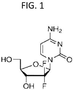
FIG. 1 is the molecular structure of gemcitabine.
FIG.2 is a drawing depicting the molecular structure of rottlerin.
FIG. 3 is a drawing depicting the molecular structure of embelin.
FIG. 4 is a drawing depicting the molecular structure of ellagic acid.
FIG. 5 is a drawing depicting the molecular structure of sulforaphane.
FIGS. 6A-6D are graphs showing the results of cell viability studies. In
particular, the effect of rottlerin on the growth of human pancreatic cancer
cells,
and cancer stem cells, is shown. Pancreatic cancer cells (A5PC-1, PANC-1 and
MIA PaCa-2) and pancreatic cancer stem cells (CSCs) were treated with
rottlerin
for 3 days, and cell viability was measured by XTT assay. Data represent mean
SD.
FIGS. 7A-7C are graphs showing the results of cell viability studies. In
particular, the effect of embelin on the growth of human pancreatic cancer
cells is
shown. Pancreatic cancer cells (AsPC-1, PANC-1 and MIA PaCa-2) were treated
with embelin for 3 days, and cell viability was measured by XTT assay. Data
represent mean SD.
FIGS. 8A-8B are graphs showing the results of cell viability studies. In
particular, the effect of ellagic acid on the growth of human pancreatic
cancer cells
4
CA 02831403 2013-09-25
WO 2012/159085
PCT/US2012/038705
is shown. Pancreatic cancer cells (A5PC-1 and MIA PaCa-2) were treated with
ellagic acid for 3 days, and cell viability was measured by XTT assay. Data
represent mean SD.
FIGS. 9A-9D are graphs showing the effect of embelin on prostate CSCs.
In particular, embelin is shown to inhibit spheroid and colony formation, and
induce caspase-3 and apoptosis. (A) Prostate CSCs were grown in CSC medium
and treated with embelin (0-6 M) for 7 days to obtain primary spheroids. At
the
end of incubation period, spheroids were collected, reseeded and treated with
embelin for another week to obtain secondary spheroids. Cell viability in
spheroids was measured by trypan blue assay at the end of 7 and 14 days. Data
represent mean SD. *, %, &, # and ** = significantly different from
controls, P
<0.05. (B) Prostate CSCs were seeded in soft agar and treated with embelin (0-
6
M) for 21 days. At the end of incubation period, numbers of colonies were
counted. *, % and & = significantly different from control, P < 0.05. (C and
D)
Activation of caspase-3 and induction of apoptosis is shown. Prostate CSCs
were
treated with embelin (0-6 M), and caspase-3 activity at 24 h and apoptosis at
48 h
were measured; as described *, #, % or & = significantly different from
control, P
<0.05.
FIGS. 10 is a graph showing the effect of embelin on prostate CSCs. In
particular, embelin is shown to inhibit the expression of Bc1-2, survivin and
XIAP
in prostate CSCs. Prostate CSCs were treated with embelin (0-6 M) for 24 h,
and
the expression of Bc1-2, survivin and XIAP was measured by the q-RT-PCR. Data
represent mean SD. * = significantly different from respective controls, P <
0.05. Data were normalized with GAPDH.
FIGS. 11 is a graph showing the regulation by embelin of Nanog and
Oct3/4 in prostate CSCs. Prostate CSCs were treated with embelin (0-6 M) for
24 h. The expression of Nanog and Oct3/4 was measured by qRT-PCR. Data
represent mean SD. * = significantly different from control, P < 0.05. Data
were
normalized with GAPDH.
FIGS. 12A-12D are graphs showing the regulation by embelin of the Shh
pathway in prostate CSCs. (A-C) Prostate CSCs were treated with embelin (0-6
M) for 24 h. The expression of Gli 1, G1i2, Patched-1, Patched-2 and SMO was
5
CA 02831403 2013-09-25
WO 2012/159085
PCT/US2012/038705
measured by qRT-PCR. Data represent mean SD. *, # and & = significantly
different from control, P < 0.05. Data were normalized with GAPDH. (D) Gli
transcriptional activity. Prostate CSCs were transduced with Gli-responsive
GIP/firefly luciferase viral particles (pGreen Fire I -Gli with EF1, System
Biosciences). After transduction, culture medium was replaced and CSCs were
treated with embelin (0-6 M) for 24 h. Gli-responsive reporter activity was
measured using a luciferase assay (Promega Corporation). Data represent mean
SD. *, # and & = significantly different from control, P < 0.05.
FIGS. 13A-13C are graphs showing the inhibition of invasion, migration
and EMT markers by embelin. (A and B) Prostate CSCs were treated with embelin
(0-6 M) for 24 h. Invasion and migration of CSCs were measured as we
described. *, # and & = significantly different from respective controls, P <
0.05.
(C) Expression of Snail and N-cadherin was measured by the qRT-PCR. Data
represent the mean S.D. * = significantly different from respective
controls, P <
0.05.
FIGS. 14A-14D are graphs showing that rottlerin inhibits spheroid and
colony formation, and induces caspase-3 and apoptosis. (A) Prostate CSCs were
grown in CSC medium and treated with rottlerin (0-2 M) for 7 days to obtain
primary spheroids. At the end of incubation period, spheroids were collected,
reseeded and treated with rottlerin for another week to obtain secondary
spheroids.
Cell viability in spheroids was measured by trypan blue assay at the end of 7
and
14 days. Data represent mean SD. *, % and & = significantly different from
controls, P < 0.05. (B) Prostate CSCs were seeded in soft agar and treated
with
rottlerin (0-2 M) for 21 days. At the end of incubation period, numbers of
colonies were counted. *, % and & = significantly different from control, P <
0.05.
(C and D) Activation of caspase-3 and induction of apoptosis. Prostate CSCs
were
treated with rottlerin (0-2 M) for 24 h, and caspase-3 activity and apoptosis
were
measured as we described. *, #, %, and & = significantly different from
control, P
<0.05.
FIG. 15 is a graph showing that rottlerin inhibits the expression of survivin,
XIAP, Bc1-2 and Bc1-XL in prostate CSCs. Prostate CSCs were treated with
rottlerin (0-2 1.1M) for 24 h, and the expression of survivin, XIAP, Bc1-2 and
Bcl-
6
CA 02831403 2013-09-25
WO 2012/159085
PCT/US2012/038705
XL, was measured by the q-RT-PCR. Data represent mean SD. * = significantly
different from respective controls, P < 0.05. Data were normalized with GAPDH.
FIG. 16 is a graph showing the regulation by rottlerin of cMyc, Nanog,
Oct3/4 and Sox-2 in prostate CSCs. Prostate CSCs were treated with rottlerin
(0-1
M) for 24 h. The expression of cMyc, Nanog, Oct3/4 and Sox-2 was measured
by qRT-PCR. Data represent mean SD. * and # = significantly different from
control, P < 0.05. Data were normalized with GAPDH.
FIGS. 17A-17E are graphs showing the regulation of Shh, Notch and TGFI3
pathways by rottlerin. Figure 17F shows the results of immunofluorescence
analysis of Glil and G1i2 expression in prostate CSCs (A-C) Prostate CSCs were
treated with rottlerin (0-2 uM) for 24 h. The expression of Patched-1, Patched-
2,
SMO, Glil and G1i2 was measured by qRT-PCR. Data represent mean SD. *, #,
and & = significantly different from control, P < 0.05. Data were normalized
with
GAPDH. (D) Prostate CSCs were treated with rottlerin (0-1 04) for 24 h. The
expression of Notchl, Notch3 and JAG1 was measured by qRT-PCR. (E)
TCF/LEF-1, Gli and Notch reporter activities. Prostate CSCs were transduced
with a mixture of TCF/LEF1-,Gli-, or Notch-responsive firefly luciferase
construct
and Renilla luciferase construct (40:1) along with lipofectamine. After
transduction, medium was changed and CSCs were treated with rottlerin (0-2 M)
for 24 h. Reporter activity was measured using a dual luciferase assay
(Promega
Corporation). * = significantly different from control, P < 0.05. (F)
Immunofluorescence analysis of Glil and G1i2 expression in prostate CSCs.
Green = Glil or G1i2; red = nucleus.
FIGS. 18A and 18B are photographs and a graph showing that rottlerin
inhibits cell viability in spheroids and colony formation by pancreatic CSCs.
(A)
Pancreatic CSCs were grown in six-well ultralow attachment plates (Corning
Inc.,
Corning, NY) at a density of 1,000 cells/ml in DMEM supplemented with 1% N2
Supplement (Invitrogen), 2% B27 Supplement (Invitrogen), 20 ng/ml human
platelet growth factor (Sigma-Aldrich), 100 ng/ml EGF (Invitrogen) and 1%
antibiotic-antimycotic (Invitrogen) at 37 C in a humidified atmosphere of 95%
air
and 5% CO2 and treated with rottlerin (0-2 M) for 7 days to obtain primary
spheroids. At the end of incubation period, spheroids were collected, reseeded
and
7
CA 02831403 2013-09-25
WO 2012/159085
PCT/US2012/038705
treated with rottlerin for another week to obtain secondary spheroids. (B)
Cell
viability in spheroids was measured by trypan blue assay at the end of 7 and
14
days from the above experiment. Data represent mean SD. *, #, and ** =
significantly different from controls, P <0.05.
FIG. 19 is a graph showing the regulation by rottlerin of cMyc, Nanog,
Oct-4, and Sox-2 in pancreatic CSCs. Pancreatic CSCs were treated with
rottlerin
(0-1 M) for 24 h. The expression of Nanog, Sox-2 and cMyc was measured by
qRT-PCR and normalized to GAPDH. Data represent mean SD. ** =
significantly different from control, P < 0.05.
FIGS. 20A and 20B are graphs showing the regulation by rottlerin of the
Shh pathway in pancreatic CSCs. (A) Pancreatic CSCs were treated with
rottlerin
(0-1 M) for 24 h. The expression of Patched-1, Smo and Gli-2 was measured by
qRT-PCR. Data represent mean SD. **, and ** = significantly different from
control, P < 0.05. (B) Pancreatic CSCs were transduced with Gli-responsive
GIP/firefly luciferase viral particles (pGreen Fire 1-Gli with EF1, System
Biosciences). After transduction, culture medium was replaced and CSCs were
treated with rottlerin (0-2 M) for 24 h. Gli-responsive reporter activity was
measured using a luciferase assay (Promega Corporation). Data represent mean
SD. *, @ and $ = significantly different from control, P < 0.05.
FIGS. 21A-21C are graphs showing that rottlerin induces apoptosis,
activates caspase-3/-7, and inhibits the expression of Bc1-2, XIAP and
Survivin in
pancreatic CSCs. (A and B) Induction of apoptosis and activation of caspase-3/-
7.
Pancreatic CSCs were treated with rottlerin (0-2 1_1M) for 48 h, and apoptosis
and
caspase-3/-7 activity were measured by XTT and colorometric assay,
respectively.
Data represent mean SD. *, # and @ = significantly different from control, P
<
0.05. (C) Pancreatic CSCs were treated with rottlerin (0-1 M) for 48 h, and
the
expression of Bc1-2, XIAP and Survivin was measured by qRT-PCR and
normalized to GAPDH. Data represent mean SD. **, and ## = significantly
different from control, P < 0.05.
FIGS. 22A and 22B are graphs, and Figure 22C are the results of an in vitro
Matrigel invasion assay, showing the regulation of EMT markers by rottlerin in
pancreatic CSCs. (A and B) Pancreatic CSCs were treated with rottlerin (0-1
1.1M)
8
CA 02831403 2013-09-25
WO 2012/159085
PCT/US2012/038705
for 24 h. The expression of Zeb-1 and Slug was measured by the qRT-PCR and
normalized to GAPDH. Data represent the mean S.D. * = significantly
different
from respective controls, P < 0.05. (C ) In vitro Matrigel invasion assay.
CSCs
were plated onto the Matrigel-coated membrane in the top chamber of the
transwell and treated with rottlerin (0-2 M) for 48 h. Cells invaded to the
lower
chamber were fixed with methanol, stained with crystal violet and photo
micrographed.
FIGS. 23A-23D are graphs showing that resveratrol, curcumin, honokiol,
and diallyl trisulphide inhibit cell viability in brain cancer stem cells.
Brain CSCs
were treated with resveratrol (0-20 M), curcumin (0-20 M), honokiol (0-20
M)
and diallyl trisulphide (0-10 M) for 3 days, and cell viability was measured
by
staining with trypan blue using Vi-CELL analyzer (Beckman Counter).
FIGS. 24A-24D are graphs showing that sulforaphane, rottlerin, EGCG,
and embelin inhibit cell viability in brain cancer stem cells. Brain CSCs were
treated with sulforaphane (0-20 M), rottlerin (0-1 M), EGCG (0-40 M) and
embelin (0-5 M) for 48 h, and cell viability was measured by staining with
trypan
blue using Vi-CELL analyzer (Beckman Counter).
FIGS. 25A-25D are graphs showing that resveratrol, curcumin, honokiol,
and diallyl trisulphide inhibit cell viability in prostate cancer stem cells.
Prostate
CSCs were treated with resveratrol (0-20 M), curcumin (0-20 M), honokiol (0-
20 M) and diallyl trisulphide (0-10 M) for 3 days, and cell viability was
measured by staining with trypan blue using Vi-CELL analyzer (Beckman
Counter).
FIGS. 26A-26D are graphs showing that sulforaphane, rottlerin, EGCG,
and embelin inhibit cell viability in prostate cancer stem cells. Prostate
CSCs were
treated with sulforaphane (0-20 M), rottlerin (0-5 M), EGCG (0-40 M) and
embelin (0-1 M) for 3 days, and cell viability was measured by staining with
trypan blue using Vi-CELL analyzer (Beckman Counter).
FIGS. 27A-27D are graphs showing that resveratrol, curcumin, honokiol,
and diallyl trisulphide inhibit cell viability in pancreatic cancer stem
cells.
Pancreatic CSCs were treated with resveratrol (0-20 M), curcumin (0-20 M),
honokiol (0-20 M) and diallyl trisulphide (0-20 M) for 3 days, and cell
viability
9
CA 02831403 2013-09-25
WO 2012/159085
PCT/US2012/038705
was measured by staining with trypan blue using Vi-CELL analyzer (Beckman
Counter).
PIGS. 28A-28D are graphs showing that sulforaphane, rottlerin, EGCG,
and embelin inhibit cell viability in pancreatic cancer stem cells. Pancreatic
CSCs
were treated with sulforaphane (0-20 p.M), rottlerin (0-2 M), EGCG (0-60 p.M)
and embelin (0-5 ttM) for 3 days, and cell viability was measured by staining
with
trypan blue using Vi-CELL analyzer (Beckman Counter).
FIGS. 29A-29D are graphs showing that sulforaphane, diallyl trisulphide,
resveratrol, and curcumin inhibit cell viability in breast cancer stem cells.
Breast
CSCs were seeded in 96-well plate and treated with sulforaphane, diallyl
trisulphide, resveratrol and curcumin for 3 days. At the end of incubation
period,
CSCs were harvested and cell viability was measured by XTT assay.
PIGS. 30A-30D are graphs showing that rottlerin, EGCG, embelin, and
honokiol inhibit cell viability in breast cancer stem cells. Breast CSCs were
seeded in 96-well plate and treated with rottlerin, EGCG, embelin, and
honokiol
for 3 days. At the end of incubation period, breast CSCs were harvested and
cell
viability was measured by XTT assay.
FIGS. 31A-31C are graphs showing that chromatin modulators inhibit cell
viability and promote apoptosis in pancreatic cancer stem cells. (A)
Pancreatic
CSCs were treated with SAHA (3 and 51.IM) and 5-Aza-dc (2 and 4 M) and cell
viability was measured at 48 h by staining with trypan blue using Vi-CELL
analyzer (Beckman Counter). (B) Pancreatic CSCs were untreated (a) or treated
with SAHA (b) or 5-Aza-dC (c) for 48 h, and apoptosis was measured by staining
with annexin-PI using Accuri Flow Cytometer. (C) Caspase-3/7 activity was
measured in pancreatic CSCs treated with SAHA (0.5 and 2 1.1M) or 5-Aza-dC (1
and 3 1.1M) for 25 h. Data represent mean SD. * and $ = significantly
different
from control, P < 0.05.
FIGS. 32A-32E are graphs showing that sulforaphane, rottlerin, and
embelin inhibit tumor growth in NOD/SCID/IL2R gamma mice. (A) Pancreatic
CSCs were orthotopically implanted in pancreas of NSG mice, and treated with
or
without sulforaphane 20 mg/kg, for 6 weeks, (B and C) Pancreatic CSCs were
xenografted sub-cutaneously in NSG mice, and treated with or without rottlerin
CA 02831403 2013-09-25
WO 2012/159085
PCT/US2012/038705
and embelin 20 mg/kg, for 6 weeks., (D and E) Prostate CSCs were xenografted
sub-cutaneously in NSG mice, and treated with or without rottlerin and embelin
20 mg/kg, for 6 weeks. At the end of the treatment, weights of tumors in
treated
mice were compared with control mice.
FIGS. 33A-33D are graphs showing that benzyl selenocyanate (BSC),
honokiol and phenyl isothiocyanate (PITC) inhibit cell viability in cancer
stem
cells. (A-B) Pancreatic CSCs were treated with BSC (0-20 M) and honokiol (0-
10
04); (C-D) Prostate CSCs were treated with PITC (0- 20 RIVI) and BSC (0-20
KM) for 3 days. Cell viability was measured by XTT assay.
FIGS. 34A-34D are graphs showing that PITC, BSC, sulforaphane and
honokiol inhibit cell viability in breast cancer stem cells. (A-D) Breast CSCs
were
treated with PITC(0-20 PM), BSC, (0-20 PM), sulforaphane (0- 20 RIVI) and
honokiol (0-10 04) for 3 days, and cell viability was measured by XTT assay.
FIG. 35 is a graph showing that rottlerin inhibits cell viability in breast
cancer stem cells. Breast CSCs were treated with rottlerin (0-1 04) for 3
days,
and cell viability was measured by XTT assay.
FIGS. 36A and 36B are graphs showing that sulforaphane and honokiol
inhibit cell viability in brain cancer stem cells. (A-B) Brain CSCs were
treated
with sulforaphane (0-20 PM), and honokiol (0-20 RIVI) for 3 days, and cell
viability
was measured by XTT assay.
FIGS. 37A-37C are graphs and photographs showing the effects of EGCG
on tumor spheroids and cell viability of pancreatic cancer stem cells (CSCs).
(A)
Pancreatic CSCs were seeded in suspension and treated with EGCG (0-60 1i1\4)
for
7 days. Pictures of spheroids formed in suspension were taken by a microscope.
(B) Pancreatic CSCs were seeded in suspension and treated with EGCG (0-60
[11\4)
for 7 days. At the end of incubation period, spheroids were collected, and
dissociated with Accutase (Innovative Cell Technologies, Inc.). For secondary
spheroids, cells were reseeded and treated with EGCG (0-60 [tM) for 7 days.
Cell
viability was measured by trypan blue assay. Data represent mean SD. *,
@,
or # = significantly different from respective controls, P < 0.05. (C) EGCG
inhibits colony formation by CSCs. Pancreatic CSCs were seeded in soft agar
and
treated with various doses of EGCG and incubated at 4 C for 21 days. At the
end
11
CA 02831403 2013-09-25
WO 2012/159085
PCT/US2012/038705
of incubation period, colonies were counted. Data represent mean SD. *, & or
#
= significantly different from respective controls, P < 0.05.
FIGS. 38A-38C are graphs showing the regulation of caspase-3/7 activity,
apoptosis and apoptosis-related proteins by EGCG on CSCs derived from human
primary pancreatic tumors. (A) Regulation of caspase-3/7 activity by EGCG.
CSCs were treated with EGCG (0-60 M) for 24 h, and caspase-3/7 activity was
measured as per manufacturer's instructions. Data represent mean SD. *, # or
% = significantly different from control, P < 0.05. (B) Regulation of
apoptosis by
EGCG. CSCs were treated with EGCG (0-60 tiM) for 48 h, and apoptosis was
measured by TUNEL assay. Data represent mean SD. *, # or % = significantly
different from control, P < 0.05. (C) Regulation of apoptosis-related
proteins.
Pancreatic CSCs were treated with EGCG (0-60 tiM) for 36 h. Real time PCR (q-
RT-PCR) was performed to examine the expression of Bc1-2, survivin, XIAP, and
GAPDH. Data represent mean SD. *, # or % = significantly different from
control, P < 0.05.
FIGS. 39A and 39B are graphs showing the regulation of pluripotency
maintaining transcription factors by EGCG in pancreatic cancer stem cells. (A)
Pancreatic CSCs were treated with EGCG (0-60 MM) for 36 h. At the end of
incubation period, cells were harvested and the expression of Nanog, Sox-2, c-
Myc
and Oct-4 was measured by the q-RT-PCR. Data represent mean SD. *, #, or %
= significantly different from respective controls, P < 0.05. (B) Nanog shRNA
enhances the inhibitory effects of EGCG on CSC's spheroid viability.
Pancreatic
CSCs were transduced with either scrambled shRNA or Nanog shRNA expressing
lentiviral vector (pLK0.1), and cell lysates were collected and western blot
analysis was performed using anti-Nanog antibody (data not shown).
CSC/scrambled and CSC/Nanog shRNA were seeded as described above and
treated with EGCG (0-60 tiM). After 7 days, spheroids were collected and cell
suspensions were prepared and viable cells were counted by trypan blue assay.
Data represent mean SD. *, &, @, #, ** or % = significantly different from
control, P <0.05.
FIGS. 40A-40C are graphs and photographs showing the inhibition of
components of sonic hedgehog pathway, Gli transcription and nuclear
12
CA 02831403 2013-09-25
WO 2012/159085
PCT/US2012/038705
translocation by EGCG. (A) Inhibition of components of sonic hedgehog pathway
and Gli transcription. Pancreatic CSCs were treated with EGCG (0-60 RM) for 36
h. The expression of Smothened (Smo), patched 1 (PTCH1), patched 2 (PTCH2),
was measured by q-RT-PCR. Data represent mean SD. *, #, or % = significantly
different from respective controls, P < 0.05. (B) Inhibition of Gli 1 and G1i2
expression and Gli transcription. Pancreatic CSCs were treated with EGCG (0-60
[iM) for 36 h. The expression of Gli 1 and G1i2 was measured by q-RT-PCR. Gli
reporter activity. CSCs were transduced with Gli-responsive GFP/firefly
luciferase
viral particles (pGreen Fire 1-Gli with EF1, System Biosciences). After
hunsduction, culture medium was replaced and CSCs were treated with EGCG (0-
60 M) for 24 h. Gli-responsive reporter activity was measured by luciferase
assay (Promega Corporation). Data represent mean SD. *, #, % or & =
significantly different from respective controls, P < 0.05. (C) EGCG inhibits
nuclear translocation of Gli 1 and G1i2. Pancreatic CSCs were treated with or
without EGCG (40 or 60 M) for 24 h. At the end of incubation period, CSCs
were fixed with paraformaldehyde, permeabilized with titron X100, and blocked
with 5% normal goat serum. Cells were then treated with either anti-Gli 1 or
anti-
G1i2 antibody, followed by secondary antibody plus DAPI. Stained cells were
mounted and visualized under a fluorescence microscope. Blue fluorescence of
DAPI was changed to red color for a better contrast.
FIGS. 41A-41D are graphs showing the regulation of epithelial
mesenchymal transition factors, migration, invasion and TCF/LEF activity by
EGCG in pancreatic CSCs. (A) Pancreatic CSCs were treated with EGCG (0-60
RM) for 48 h. At the end of incubation period, the expression of Snail, ZEB1
and
Slug was measured by q-RT-PCR. Data represent mean SD. * = significantly
different from respective controls, P < 0.05. (B) Transwell migration assay.
Pancreatic CSCs were plated in the top chamber of the transwell and treated
with
EGCG (0-60 M) for 24 h. Cells migrated to the lower chambered were fixed with
methanol, stained with crystal violet and counted. Data represent mean SD. *
#
or % = significantly different from respective controls, P < 0.05. (C)
Matrigel
invasion assay. CSCs were plated onto the Matrigel-coated membrane in the top
chamber of the transwell and treated with EGCG (0-60 [iM) for 48 h. Cells
13
CA 02831403 2013-09-25
WO 2012/159085
PCT/US2012/038705
invaded to the lower chambered were fixed with methanol, stained with crystal
violet and counted. Data represent mean SD. *, # or % = significantly
different
from respective controls, P <0.05. (D) Effects of EGCG on TCF-1/LEF activity.
Pancreatic CSCs were transduced with TCF/LEF responsive GFP/firefly luciferase
viral particles (pGreen Fire 1-Gli with EF1, System Biosciences). Transduced
CSCs were treated with EGCG (0-60) for 48 h and the GFP fluorescence was
measured. Data represent mean SD. *, # or ** = significantly different from
control, P < 0.05.
FIGS. 42A-42D are graphs showing quercetin synergizes with EGCG to
inhibit self-renewal capacity, invasion, migration, and TCF/LEF and Gli
transcriptional activities in pancreatic CSCs. (A) Effects of EGCG and
quercetin
on spheroid and colony formation. Upper Panel, uercetin synergizes with EGCG
to inhibit spheroid's cell viability. CSCs were seeded in suspension and
treated
with EGCG (0-60 pM) with or without quercetin (20 HM) for 7 days. At the end
of incubation period, all the spheroids were collected and resuspended. Cell
viability was measured by trypan blue assay. Data represent mean SD. *, #,
%,
**, ##, or %% = significantly different from control, P < 0.05. Lower panel,
uercetin synergizes with EGCG to inhibit colony foliiiation. Pancreatic CSCs
were
seeded in soft agar and treated with various doses of EGCG (0-60 [tM) with or
without quercetin (20 [iM) and incubated at 4 C for 21 days. At the end of
incubation period, colonies were counted. Data represent mean SD. *, #, %,
**,
##, or %% = significantly different from control, P < 0.05. (B) Effects of
EGCG
and quercetin on invasion and migration. Upper panel, Matrigel invasion assay.
CSCs were plated onto the Matrigel-coated membrane in the top chamber of the
transwell and treated with EGCG (0-60 [iM) with or without quercetin (20 HM)
for
48 hrs. Cells invaded to the lower chambered were fixed with methanol, stained
with crystal violet and counted. Data represent mean SD. *, #, %, **, ##, or
%% = significantly different from control, P < 0.05. Lower panel, Transwell
migration assay. Pancreatic CSCs were plated in the top chamber of the
transwell
and treated with EGCG (0-60 [tM) with or without quercetin (20 [tM) for 48
hrs.
Cells migrated to the lower chambered were fixed with methanol, stained with
crystal violet and counted. Data represent mean SD. *, #, %, **, ##, or %% =
14
CA 02831403 2013-09-25
WO 2012/159085
PCT/US2012/038705
significantly different from respective controls, P < 0.05. (C) Quercetin
synergizes
with EGCG to induce apoptosis. CSCs were seeded in suspension and treated with
EGCG (0-60 M) with or without quercetin (20 M) for 7 days. At the end of
incubation period, all the spheroids were collected. Apoptosis was measured by
TUNEL assay. Data represent mean SD. *, #, %, **, ##, or %% = significantly
different from control, P < 0.05. (D) Effects of EGCG and quercetin on TCF/LEF
and Gli transcriptional activities. Pancreatic CSCs were transduced with
either
lentivirus encoding TCF/LEF responsive GI-P and luciferase genes or Gli-
responsive GI-P and luciferase genes. Transduced CSCs were treated with EGCG
(40 M) with or without quercetin (20 M) for 48 hrs and the luciferase
activity
was measured. Data represent mean SD. *, #, or % = significantly different
from respective control, P < 0.05.
FIGS. 43A and 43B are graphs showing that quercetin synergizes with
sulforaphane (SFN) to inhibit self-renewal capacity of pancreatic cancer CSCs.
(A) Quercetin synergizes with SFN to inhibit spheroid cell viability.
Pancreatic
CSCs were seeded in suspension and treated with SFN (0-10 M) with or without
quercetin (20 M) for 7 days. At the end of incubation period, all the
spheroids
were collected and resuspended. Cell viability was measured by trypan blue
assay.
Data represent mean SD. *, &, @ or # * = significantly different from
control, P
< 0.05. (B) Quercetin synergizes with SFN to inhibit colony formation. SFN
inhibits colony formation by pancreatic CSCs. Pancreatic CSCs were seeded in
soft agar and treated with various doses of SFN and incubated at 4 C for 21
days.
At the end of incubation period, colonies were counted. Data represent mean
SD. *, &, @ or # = significantly different from respective controls, P < 0.05.
FIGS. 44A-44C are graphs showing the results of treating human cancer
stem cells (CSCs) and cancer cell lines from various organs with anthothecol
(0-20
M), sanguinarine (0-20 M), or mangostine (0-20 M) for 72 h, and measuring
cell viability. Structures of the compounds are shown on the right.
FIGS. 45A-45C are compound structures of phenyl-isothiocyanate (PITC)
(A), benzyl selenocyanate (BSC) (B), and benzyl isothiocyanate (BITC) (C).
CA 02831403 2013-09-25
WO 2012/159085
PCT/US2012/038705
DETAILED DESCRIPTION OF THE INVENTION
For the purposes of promoting an understanding of the principles of the
invention, reference will now be made to certain embodiments and specific
language will be used to describe the same. It will nevertheless be understood
that
no limitation of the scope of the invention is thereby intended, and
alterations and
modifications in the illustrated embodiments, and further applications of the
principles of the invention as illustrated therein are herein contemplated as
would
normally occur to one skilled in the art to which the invention relates.
Unless defined otherwise, all technical and scientific temis used herein have
the
same meaning as commonly understood by one of ordinary skill in the art to
which
this invention pertains.
For the purpose of interpreting this specification, the following definitions
will apply and whenever appropriate, terms used in the singular will also
include
the plural and vice versa. In the event that any definition set forth below
conflicts
with the usage of that word in any other document, including any document
incorporated herein by reference, the definition set forth below shall always
control
for purposes of interpreting this specification and its associated claims
unless a
contrary meaning is clearly intended (for example in the document where the
term
is originally used). The use of "or" means "and/or" unless stated otherwise.
The
use of the terms "a" and "an" and "the" and similar referents in the context
of
describing the invention (especially in the context of the following claims)
are to
be construed to cover both the singular and the plural, unless otherwise
indicated
herein or clearly contradicted by context. The terms "comprising," "having,"
"including," and "containing" are to be construed as open-ended terms (i.e.,
meaning "including, but not limited to,") unless otherwise noted. Recitation
of
ranges of values herein are merely intended to serve as a shorthand method of
referring individually to each separate value falling within the range, unless
otherwise indicated herein, and each separate value is incorporated into the
specification as if it were individually recited herein. All methods described
herein
can be performed in any suitable order unless otherwise indicated herein or
otherwise clearly contradicted by context. The use of any and all examples, or
exemplary language (e.g., "such as") provided herein, is intended merely to
better
16
CA 02831403 2013-09-25
WO 2012/159085
PCT/US2012/038705
illuminate the invention and does not pose a limitation on the scope of the
invention unless otherwise claimed. No language in the specification should be
construed as indicating any non-claimed element as essential to the practice
of the
invention.
One skilled in the art may refer to general reference texts for detailed
descriptions of known techniques discussed herein or equivalent techniques.
These
texts include Current Protocols in Molecular Biology (Ausubel et. al., eds.
John
Wiley & Sons, N.Y. and supplements thereto), Current Protocols in Immunology
(Coligan et al., eds., John Wiley St Sons, N.Y. and supplements thereto),
Current
Protocols in Pharmacology (Enna et al., eds. John Wiley & Sons, N.Y. and
supplements thereto) and Remington: The Science and Practice of Pharmacy
(Lippincott Williams & Wilicins, 2Vt edition (2005)), for example.
The present invention relates generally to compositions and methods for
treating cancer comprising administering to a subject in need thereof a
pharmaceutically effective dose of a stem cell inhibitor.
In some embodiments, providing a therapy or "treating" cancer refers to
indicia of success in the treatment, amelioration or prevention of cancer,
including
any objective or subjective parameter such as abatement, inhibiting
metastasis,
remission, diminishing of symptoms of making the disease, pathology or
condition
more tolerable to the patient, slowing the rate of degeneration or decline,
making
the final point of degeneration less debilitating, or improving a patient's
physical or
mental well-being. Those in need of treatment include those already with
cancer as
well as those prone to have cancer or in those in whom cancer is to be
prevented.
In general, a pharmaceutically effective dose is meant an amount that
produces the desired effect for which it is administered. The exact amount
will
depend on a variety of factors such as the purpose of the treatment,
composition or
dosage form, the selected mode of administration, the age and general
condition of
the individual being treated, the severity of the individual's condition, and
other
factors known to the prescribing physician and will be ascertainable by a
person
skilled in the art using known methods and techniques for determining
effective
doses. In some embodiments, a pharmaceutically effective dose results in a
cellular concentration of the drug of from about 1 nM to 30 jiM. In some
17
CA 02831403 2013-09-25
WO 2012/159085
PCT/US2012/038705
embodiments, a pharmaceutically effective dose results in a cellular
concentration
of the drug of from about 50 nM to about 10 M, from about 50 nM to about 1
M, from about 100 nM to about 1 [tM, or from about 100 nM to about 500 nM.
In some embodiments, a pharmaceutically effective dose includes between about
0.1 mg/kg/day to about 300 mg/kg/day. In some embodiments, a pharmaceutically
effective dose includes between about 1.0 p.g/kg/day to about 50 mg/kg/day.
The present invention also relates to methods of inhibiting the growth of
cancer stem cells comprising administering to a subject in need thereof a
pharmaceutically effective dose of a stem cell inhibitor.
The present invention also relates to methods of inhibiting the growth of
cancer stem cells comprising contacting cancer stem cells with an effective
dose of
a stem cell inhibitor.
In one embodiment, the present disclosure provides a method of enhancing
the biological effects of a chemotherapeutic drug on cancer cells comprising
administering to a subject in need thereof along with a chemotherapeutic drug
a
pharmaceutically effective dose of a stem cell inhibitor.
In one embodiment, the present invention relates to methods of treating
pancreatic cancer using stem cell inhibitors.
As described herein, there are certain natural products, including rottlerin,
embelin, ellagic acid, and sulforaphane, which can act as cancer stem cell
inhibitors and inhibit the growth of cancer stem cells and cancer cells. These
products have the advantages of being non-toxic and bioavailable and may
inhibit
the growth of pancreatic and other cancers and the growth of cancer stem
cells.
Without being bound by theory, in some embodiments it is believed that these
compounds inhibit oncogenic PI3/AKT and ERK pathways, and thus can be used
as cancer preventive agents. In some embodiments, sulforaphane inhibits the
growth of pancreatic cancer stem cells. In some embodiments, sulforaphane
blocks pancreatic cancer progression in an animal model, such as KrasG12D
mice.
In some embodiments, sulforaphane enhances the biological effects of
gemcitabine
and lapatinib on cancer stem cells. In some embodiments, sulforaphane enhances
the biological effects of gemcitabine and lapatinib on pancreatic cancer stem
cells.
18
CA 02831403 2013-09-25
WO 2012/159085
PCT/US2012/038705
Cancer stem cells (CSCs) have been proposed recently to be the cause
cancer initiation, progression and chemotherapy failure. CSCs also demonstrate
upregulation of signaling pathways such as sonic hedgehog (Shh), Wnt and
Notch.
Regulation of CSCs by non-toxic agents could be considered as a strategy for
the
treatment and/or prevention of cancer.
In one embodiment, the present invention provides a method of treating
cancer comprising administering to a subject in need a pharmaceutically
effective
dose of a stem cell inhibitor. In certain embodiments, the stem cell inhibitor
may
comprise rottlerin, embelin, ellagic acid, sulforaphane, resveratrol,
honokiol,
curcumin, diallyltrisulfide, benzyl isothiocyanate, quercetin,
epigallocatechin
gallate (EGCG), SAHA, m-Carboxycinnamic acid bis-hydroxamine, and/or MS-
275. In certain embodiments, the stem cell inhibitor may comprise epigenetic
regulators and agents that modify histones and DNA such as SAHA/vornostat, m-
Carboycinnamic acid bis-hydroxamine, MS-275, and demathylating agent such as
5- aza-2 ' -deoxycytidine.
Rottlerin is a polyphenolic compound derived from Mallotus philipinensis
(Euphorbiaceae). It is widely used as an inhibitor of PKC5 due to the
competition
between rottlerin and ATP, which plays a crucial role in apoptosis, cell
migration
and cytoskeleton remodeling. These cellular functions are important regulators
of
tumor progression and metastasis. In addition to inhibiting PKC5, rottlerin
targets
mitochondria to induce apoptosis. Rottlerin causes uncoupling of mitochondrial
respiration from oxidative phosphorylation and a collapse of mitochondrial
membrane potential in several cell types. Rottlerin has been shown to induce
apoptosis in various cancer cells, including prostate, colon, pancreatic and
lung
cancer cells, chronic leukemia, and multiple myeloma cells. Rottlerin has been
shown to inhibit cancer cell migration. Rottlerin has not previously been used
to
inhibit CSC self-renewal and tumor growth. Furthermore, there are no previous
studies demonstrating the regulation of CSCs by rottlerin, and whether
rottlerin can
inhibit sonic hedgehog, Wnt and Notch pathways.
Embelin is a polyphenolic compound derived from the fruit of Embelia
ribes Burm plant (Myrsinaceae). Embelin is a cell-permeable, non-peptide
inhibitor of X-linked inhibitor of apoptosis (XIAP); binds to the BIR3 domain,
19
CA 02831403 2013-09-25
WO 2012/159085
PCT/US2012/038705
preventing XIAP interaction with caspase-9 and Smac. It inhibits cell growth,
induces apoptosis and activates caspase-9 in cancer cells. Embelin possesses
wide
spectrum of biological activities with strong inhibition of nuclear factor
kappa B
and downstream antiapoptotic genes. These cellular functions are important
regulators of tumor progression and metastasis. Embelin has been shown to
induce
apoptosis in various cancer cells, including prostate, colon, pancreatic and
lung
cancer cells, chronic leukemia, and multiple myeloma cells. Embelin has not
previously been used to inhibit CSC self-renewal and tumor growth.
Furthermore,
there are no previous studies demonstrating the regulation of CSCs by embelin,
and whether embelin can inhibit Sonic hedgehog, Notch and Wnt pathways.
Ellagic acid is a compound derived from berries and nuts, it is a hydrolytic
product of ellagitannins.
Sulforaphane (SFN) is a compound found in cruciferous vegetables. It is
shown herein that sulforaphane inhibits the growth of human pancreatic cancer
cells and pancreatic cancer stem cells. Furthermore, SFN also inhibits the
growth
of pancreatic cancer progression in KrasG12D mice. In some embodiments of the
invention, quercetin can enhance the inhibitory effects of sulforaphane on
cancer
stem cells, such as pancreatic cancer stem cells.
In some embodiments, one or more of rottlerin, embelin, ellagic acid, and
sulforaphane can be used to kill cancer cells and inhibit cancer stem cell
growth by
targeting sonic hedgehog, Notch and Wnt pathways. Therefore, these compounds
may be used to target cancer stem cells and kill them. They are non-toxic and
bioavailable and, since these compounds are derived from plant/natural
sources,
they may be given to patients safely. In some embodiments, these compounds may
inhibit the self-renewal capacity of CSCs by inhibiting pluripotency
maintaining
factors and Notch, Wnt and Shh pathways. Thus, these compounds may be a
potent biologic inhibitor of cancer stem cells and can be used to treat and/or
prevent cancer. These compounds may also modulate the expression of genes and
pathways known to play roles in the carcinogenesis process and, therefore, may
be
used as agents for chemoprevention and/or therapy against cancer.
In some embodiments, the compounds may inhibit survival pathways such
as AKT and MAPK/ERK, which can be activated by oncogenic Kras. In some
CA 02831403 2013-09-25
WO 2012/159085
PCT/US2012/038705
embodiments, one or more of rottlerin, embelin and ellagic acid inhibit
pathways
downstream of Kras to treat or prevent cancer in pancreatic cancer subjects.
In some embodiments,sulforaphane enhances the biological effects of
gemcitabine and lapatinib on pancreatic cancer stem cells.
In some embodiments, these agents can be used in conjunction with other
cancer therapies. In some embodiments, one or more of the compounds are used
with other anticancer drugs, such as, for example gemcitabine and lapatinib,
irradiation to sensitize cancer stem cells, and/or surgical intervention.
Other
anticancer drugs that can be combined with the compounds as described herein
include, for example, Abraxane, Aldara, Alimta, Aprepitant, Arimidex,
Aromasin,
Arranon, Arsenic Trioxide, Avastin, Bevacizumab, Bexarotene, Bortezomib,
Cetuximab, Clofarabine, Clofarex, Clolar, Dacogen, Dasatinib, Ellence,
Eloxatin,
Emend, Erlotinib, Faslodex, Femara, Fulvestrant, Gefitinib, Gemtuzumab
Ozogamicin, Gemzar, Gleevec, Herceptin, Hycamtin, Imatinib Mesylate, Iressa,
Kepivance, Lenalidomide, Levulan, Methazolastone, Mylosar, Mylotarg,
Nanoparticle Paclitaxel, Nelarabine, Nexavar, Nolvadex, Oncaspar, Oxaliplatin,
Paclitaxel, Paclitaxel Albumin-stabilized Nanoparticle Formulation,
Palifermin,
Panitumumab, Pegaspargase, Pemetrexed Disodium, Platinol-AQ, Platinol,
Revlimid, Rituxan, Sclerosol Intrapleural Aerosol, Sorafenib Tosylate,
Sprycel,
Sunitinib Malate, Sutent, Synovir, Tamoxifen, Tarceva, Targretin, Taxol,
Taxotere,
Temodar, Temozolomide, Thalomid, Thalidomide, Topotecan Hydrochloride,
Trastuzumab, Trisenox, Vectibix, Velcade, Vidaza, Vorinostat, Xeloda,
Zoledronic
Acid, Zolinza, Zometa, doxorubicin, adriamycin, bleomycin, daunorubicin,
dactinomycin, epirubicin, idarubicin, mitoxantrone, valrubicin, hydroxyurea,
mitomycin, fluorouracil, 5-FU, methotrexate, floxuridine, interferon alpha-2b,
glutamic acid, plicamycin, 6-thioguanine, aminopterin, pemetrexed,
raltitrexed,
cladribine, clofarabine, fludarabine, mercaptopurine, pentostatin,
capecitabine,
cytarabine, carmustine, BCNU, lomustine, CCNU, cytosine arabinoside,
cyclophosphamide, estramustine, hydroxyurea, procarbazine, mitomycin,
busulfan,
medroxyprogesterone, estramustine phosphate sodium, ethinyl estradiol,
estradiol,
megestrol acetate, methyl testos terone, diethylstilbestrol
diphosphate,
chlorotrianisene, testolactone, mephalen, mechlorethamine, chlorambucil,
21
CA 02831403 2013-09-25
WO 2012/159085
PCT/US2012/038705
chlormethine, ifosfamide, bethamethasone sodium phosphate, dicarbazine,
asparaginase, mitotane, vincristine, vinblastine, etoposide, teniposide,
Topotecan,
IFN-gamma, irinotecan, campto, irinotecan analogs, caimustine, fotemustine,
lomustine, streptozocin, carboplatin, oxaliplatin, BBR3464, busulfan,
dacarbazine,
mechlorethamine, procarbazine, thioTEPA, uramustine, vindesine, vinorelbine,
alemtuzumab, tositumomab, methyl aminolevulinate, porfimer, verteporfin,
lapatinib, nilotinib, vandetanib, Z1)6474, alitretinoin, altretamine,
amsacrine,
anagrelide, denileukin diftitox, estramustine, hydroxycarbamide, masoprocol,
mitotane, tretinoin, or other anticancer drugs, including, for example,
antibiotic
derivatives, cytotoxic agents, angiogenesis inhibitors, hormones or hormone
derivatives, nitrogen mustards and derivatives, steroids and combinations, and
antimetbolites. Other chemotherapeutic drugs include Notch inhibitor, TGFbeta
inhibitor, TCF/LEF inhibitor, Nanog inhibitor, AKT inhibitor, FLT3 kinase
inhibitor, PI3 Kinase inhibitor, PI3 kinase / mTOR (dual inhibitor), PI3K/AKT
pathway inhibitor, Hedgehog pathway inhibitor, Gli inhibitor, Smoothened
inhibitor, JAK/STAT pathway inhibitor, Ras/MEK/ERK pathway inhibitor, and
BRAF inhibitor. In further particular aspects of the invention, an anticancer
drug
comprises two or more of the foregoing anticancer drugs.
Suitable cancers which can be treated by inhibiting cancer stem cells using
the compositions and methods of the present invention include cancers
classified
by site or by histological type. Cancers classified by site include cancer of
the oral
cavity and pharynx (lip, tongue, salivary gland, floor of mouth, gum and other
mouth, nasopharynx, tonsil, oropharynx, hypopharynx, other oral/pharynx);
cancers of the digestive system (esophagus; stomach; small intestine; colon
and
rectum; anus, anal canal, and anorectum; liver; intrahepatic bile duct;
gallbladder;
other biliary; pancreas; retroperitoneum; peritoneum, omentum, and mesentery;
other digestive); cancers of the respiratory system (nasal cavity, middle ear,
and
sinuses; larynx; lung and bronchus; pleura; trachea, mediastinum, and other
respiratory); cancers of the mesothelioma; bones and joints; and soft tissue,
including heart; skin cancers, including melanomas and other non-epithelial
skin
cancers; Kaposi's sarcoma and breast cancer; cancer of the female genital
system
(cervix uteri; corpus uteri; uterus, nos; ovary; vagina; vulva; and other
female
22
CA 02831403 2013-09-25
WO 2012/159085
PCT/US2012/038705
genital); cancers of the male genital system (prostate gland; testis; penis;
and other
male genital); cancers of the urinary system (urinary bladder; kidney and
renal
pelvis; ureter; and other urinary); cancers of the eye and orbit; cancers of
the brain
and nervous system (brain; and other nervous system); cancers of the endocrine
system (thyroid gland and other endocrine, including thymus); cancers of the
lymphomas (hodgkin's disease and non-hodgkin's lymphoma), multiple myeloma,
and leukemias (lymphocytic leukemia; myeloid leukemia; monocytic leukemia;
and other leukemias).
Other cancers, classified by histological type, that may be treated include,
but are not limited to, Neoplasm, malignant; Carcinoma, NOS; Carcinoma,
undifferentiated, NOS; Giant and spindle cell carcinoma; Small cell carcinoma,
NOS; Papillary carcinoma, NOS; Squamous cell carcinoma, NOS;
Lymphoepithelial carcinoma; Basal cell carcinoma, NOS; Pilomatrix carcinoma;
Transitional cell carcinoma, NOS; Papillary transitional cell carcinoma;
Adenocarcinoma, NOS; Gastrinoma, malignant; Cholangioc arc inoma;
Hepatocellular carcinoma, NOS; Combined hepatocellular carcinoma and
cholangiocarcinoma; Trabecular adenocarcinoma; Adenoid cystic carcinoma;
Adenocarcinoma in adenomatous polyp; Adenocarcinoma, familial polyposis coli;
Solid carcinoma, NOS; Carcinoid tumor, malignant; Branchiolo-alveolar
adenocarcinoma; Papillary adenocarcinoma, NOS; Chromophobe carcinoma;
Acidophil carcinoma; Oxyphilic adenocarcinoma; Basophil carcinoma; Clear cell
adenocarcinoma, NOS; Granular cell carcinoma; Follicular adenocarcinoma, NOS;
Papillary and follicular adenocarcinoma; Nonencapsulating sclerosing
carcinoma;
Adrenal cortical carcinoma; Endometroid carcinoma; Skin appendage carcinoma;
Apocrine adenocarcinoma; Sebaceous adenocarcinoma; Ceruminous
adenocarcinoma; Mucoepidermoid carcinoma; Cy stadenocarc inoma, NOS;
Papillary cystadenocarcinoma, NOS; Papillary serous cystadenocarcinoma;
Mucinous cystadenocarcinoma, NOS; Mucinous adenocarcinoma; Signet ring cell
carcinoma; Infiltrating duct carcinoma; Medullary carcinoma, NOS; Lobular
carcinoma; Inflammatory carcinoma; Paget's disease, mammary; Acinar cell
carcinoma; Adenosquamous carcinoma; Adenocarcinoma w/squamous metaplasia;
Thymoma, malignant; Ovarian stromal tumor, malignant; Thecoma, malignant;
23
CA 02831403 2013-09-25
WO 2012/159085
PCT/US2012/038705
Granulosa cell tumor, malignant; Androblastoma, malignant; Sertoli cell
carcinoma; Leydig cell tumor, malignant; Lipid cell tumor, malignant;
Paraganglioma, malignant; Extra-mammary paraganglioma, malignant;
Pheochromocytoma; Glomangiosarcoma; Malignant melanoma, NOS; Amelanotic
melanoma; Superficial spreading melanoma; Malignant melanoma in giant
pigmented nevus; Epithelioid cell melanoma; Blue nevus, malignant; Sarcoma,
NOS; Fibrosarcoma, NOS; Fibrous histiocytoma, malignant; Myxosarcoma;
Liposarcoma, NOS; Leiomyosarcoma, NOS; Rhabdomyosarcoma, NOS;
Embryonal rhabdomyosarcoma; Alveolar rhabdomyosarcoma; Stromal sarcoma,
NOS; Mixed tumor, malignant, NOS; Mullerian mixed tumor; Nephroblastoma;
Hepatoblastoma; Carcinosarcoma, NOS; Mesenchymoma, malignant; Brenner
tumor, malignant; Phyllodes tumor, malignant; Synovial sarcoma, NOS;
Mesothelioma, malignant; Dysgerminoma; Embryonal carcinoma, NOS; Teratoma,
malignant, NOS; Struma ovari, malignant; Choriocarcinoma; Mesonephroma,
malignant; Hemangiosarcoma; Hemangioendothelioma, malignant; Kaposi's
sarcoma; Hemangiopericytoma, malignant; Lymphangiosarcoma; Osteosarcoma,
NOS; Juxtacortical osteosarcoma; Chondrosarcoma, NOS; Chondroblastoma,
malignant; Mesenchymal chondrosarcoma; Giant cell tumor of bone; Ewing's
sarcoma; Odontogenic tumor, malignant; Ameloblastic odontosarcoma;
Ameloblastoma, malignant; Ameloblastic fibrosarcoma; Pinealoma, malignant;
Chordoma; Glioma, malignant; Ependymoma, NOS; Astrocytoma, NOS;
Protoplasmic astrocytoma; Fibrillary astrocytoma; Astroblastoma; Glioblastoma,
NOS; Oligodendroglioma, NOS; Oligodendroblastoma; Primitive
neuroectodermal; Cerebellar sarcoma, NOS; Ganglioneuroblastoma;
Neuroblastoma, NOS; Retinoblastoma, NOS; Olfactory neurogenic tumor;
Meningioma, malignant; Neurofibrosarcoma; Neurilemmoma, malignant; Granular
cell tumor, malignant; Malignant lymphoma, NOS; Hodgkin's disease, NOS;
Hodgkin's; paragranuloma, NOS; Malignant lymphoma, small lymphocytic;
Malignant lymphoma, large cell, diffuse; Malignant lymphoma, follicular, NOS;
Mycosis fungoides; Other specified non-Hodgkin's lymphomas; Malignant
histiocytosis; Multiple myeloma; Mast cell sarcoma; Immunoproliferative small
intestinal disease; Leukemia, NOS; Lymphoid leukemia, NOS; Plasma cell
24
CA 02831403 2013-09-25
WO 2012/159085
PCT/US2012/038705
leukemia; Erythroleukemia; Lymphosarcoma cell leukemia; Myeloid leukemia,
NOS; Basophilic leukemia; Eosinophilic leukemia; Monocytic leukemia, NOS;
Mast cell leukemia; Megakaryoblastic leukemia; Myeloid sarcoma; and Hairy cell
leukemia.
In some embodiments, the cancer to be treated and the cancer stem cells to
be inhibited are from cancers selected from the group consisting of breast
cancer,
prostrate cancer, brain cancer, lung cancer, mesothelioma, melanoma, multiple
myeloma, colon cancer, kidney cancer, ovarian cancer, pancreatic cancer,
leukemia, and lymphoma.
The "subject" of the cancer treatment methods and compositions according
to the invention includes, but is not limited to, a mammal, such as a human,
mouse,
rat, pig, cow, dog, cat, or horse. In one embodiment, the subject is a human
or
person.
In the compositions and methods of the invention, cancer stem cell
inhibitors can be administered by various routes of administration, including,
for
example, intraarterial administration, epicutaneous administration, eye drops,
intranasal administration, intragastric administration (e.g., gastric tube),
intracardiac administration, subcutaneous administration, intraosseous
infusion,
intrathecal administration, transmucosal administration, epidural
administration,
insufflation, oral administration (e.g., buccal or sublingual administration),
oral
ingestion, anal administration, inhalation administration (e.g., via aerosol),
intraperitoneal administration, intravenous
administration, transderm al
administration, intradermal administration, subdermal administration,
intramuscular administration, intrauterine administration, vaginal
administration,
administration into a body cavity, surgical administration (e.g., at the
location of a
tumor or internal injury), administration into the lumen or parenchyma of an
organ,
or other topical, enteral, mucosal, parenteral administration, or other method
or any
combination of the forgoing as would be known to one of ordinary skill in the
art
(see, for example, Remington's Pharmaceutical Sciences, 18th Ed. Mack Printing
Company, 1990, incorporated herein by reference).
Suitable compositions and dosage forms include tablets, capsules, caplets,
gel caps, troches, dispersions, suspensions, solutions, syrups, transdermal
patches,
CA 02831403 2013-09-25
WO 2012/159085
PCT/US2012/038705
gels, powders, magmas, lozenges, creams, pastes, plasters, lotions, discs,
suppositories, liquid sprays for nasal or oral administration, dry powder or
aerosolized formulations for inhalation, and the like.
Oral dosage forms are preferred for those therapeutic agents that are orally
active, and include tablets, capsules, caplets, solutions, suspensions and/or
syrups,
and may also comprise a plurality of granules, beads, powders or pellets that
may
or may not be encapsulated. Such dosage forms can be prepared using
conventional methods known to those in the field of pharmaceutical formulation
and described in the pertinent texts, e.g., in Remington: The Science and
Practice
of Pharmacy, 20th Edition, Gennaro, A. R., Ed. (Lippincott, Williams and
Wilkins,
2000).
Tablets and capsules represent the most convenient oral dosage forms, in
which case solid pharmaceutical carriers are employed. Tablets may be
manufactured using standard tablet processing procedures and equipment. One
method for forming tablets is by direct compression of a powdered, crystalline
or
granular composition containing the active agent(s), alone or in combination
with
one or more carriers, additives, or the like. As an alternative to direct
compression,
tablets can be prepared using wet-granulation or dry-granulation processes.
Tablets
may also be molded rather than compressed, starting with a moist or otherwise
tractable material; however, compression and granulation techniques are
preferred.
In addition to the active agent(s), tablets prepared for oral administration
will generally contain other materials such as binders, diluents, lubricants,
disintegrants, fillers, stabilizers, surfactants, coloring agents, and the
like. Binders
are used to impart cohesive qualities to a tablet, and thus ensure that the
tablet
remains intact after compression. Suitable binder materials include, but are
not
limited to, starch (including corn starch and pregelatinized starch), gelatin,
sugars
(including sucrose, glucose, dextrose and lactose), polyethylene glycol,
waxes, and
natural and synthetic gums, e.g., acacia sodium alginate,
polyvinylpyrrolidone,
cellulosic polymers (including hydroxypropyl cellulose, hydroxypropyl
methylcellulose, methyl cellulose, ethyl cellulose, hydroxyethyl cellulose,
and the
like), and Veegum. Diluents are typically necessary to increase bulk so that a
practical size tablet is ultimately provided. Suitable diluents include
dicalcium
26
CA 02831403 2013-09-25
WO 2012/159085
PCT/US2012/038705
phosphate, calcium sulfate, lactose, cellulose, kaolin, mannitol, sodium
chloride,
dry starch and powdered sugar. Lubricants are used to facilitate tablet
manufacture;
examples of suitable lubricants include, for example, magnesium stearate,
calcium
stearate, and stearic acid. Stearates, if present, preferably represent at no
more than
approximately 2 wt. % of the drug-containing core. Disintegrants are used to
facilitate disintegration of the tablet, and are generally starches, clays,
celluloses,
algins, gums or crosslinked polymers. Fillers include, for example, materials
such
as silicon dioxide, titanium dioxide, alumina, talc, kaolin, powdered
cellulose and
microcrystalline cellulose, as well as soluble materials such as mannitol,
urea,
sucrose, lactose, dextrose, sodium chloride and sorbitol. Stabilizers are used
to
inhibit or retard drug decomposition reactions that include, by way of
example,
oxidative reactions. Surfactants may be anionic, cationic, amphoteric or
nonionic
surface active agents.
The dosage form may also be a capsule, in which case the active agent-
containing composition may be encapsulated in the form of a liquid or solid
(including particulates such as granules, beads, powders or pellets). Suitable
capsules may be either hard or soft, and are generally made of gelatin,
starch, or a
cellulosic material, with gelatin capsules preferred. Two-piece hard gelatin
capsules are preferably sealed, such as with gelatin bands or the like. See,
for
example, Remington: The Science and Practice of Pharmacy, cited supra, which
describes materials and methods for preparing encapsulated pharmaceuticals. If
the
active agent-containing composition is present within the capsule in liquid
form, a
liquid carrier is necessary to dissolve the active agent(s). The carrier must
be
compatible with the capsule material and all components of the pharmaceutical
composition, and must be suitable for ingestion.
Solid dosage forms, whether tablets, capsules, caplets, or particulates, may,
if desired, be coated so as to provide for delayed release. Dosage forms with
delayed release coatings may be manufactured using standard coating procedures
and equipment. Such procedures are known to those skilled in the art and
described
in the pertinent texts, e.g., in Remington, supra. Generally, after
preparation of the
solid dosage form, a delayed release coating composition is applied using a
coating
pan, an airless spray technique, fluidized bed coating equipment, or the like.
27
CA 02831403 2013-09-25
WO 2012/159085
PCT/US2012/038705
Delayed release coating compositions comprise a polymeric material, e.g.,
cellulose butyrate phthalate, cellulose hydrogen phthalate, cellulose
proprionate
phthalate, polyvinyl acetate phthalate, cellulose acetate phthalate, cellulose
acetate
trimellitate, hydroxypropyl methylcellulose phthalate, hydroxypropyl
methylcellulose acetate, dioxypropyl methylcellulose succinate, carboxymethyl
ethylcellulose, hydroxypropyl methylcellulose acetate succinate, polymers and
copolymers formed from acrylic acid, methacrylic acid, and/or esters thereof.
Sustained release dosage forms provide for drug release over an extended
time period, and may or may not be delayed release. Generally, as will be
appreciated by those of ordinary skill in the art, sustained release dosage
forms are
formulated by dispersing a drug within a matrix of a gradually bioerodible
(hydrolyzable) material such as an insoluble plastic, a hydrophilic polymer,
or a
fatty compound, or by coating a solid, drug-containing dosage form with such a
material. Insoluble plastic matrices may be comprised of, for example,
polyvinyl
chloride or polyethylene. Hydrophilic polymers useful for providing a
sustained
release coating or matrix cellulosic polymers include, without limitation:
cellulosic
polymers such as hydroxypropyl cellulose, hydroxyethyl cellulose,
hydroxypropyl
methyl cellulose, methyl cellulose, ethyl cellulose, cellulose acetate,
cellulose
acetate phthalate, cellulose acetate trimellitate, hydroxypropylmethyl
cellulose
phthalate, hydroxypropylcellulose phthalate, cellulose hexahydrophthalate,
cellulose acetate hexahydrophthalate, and carboxymethylcellulose sodium;
acrylic
acid polymers and copolymers, preferably formed from acrylic acid, methacrylic
acid, acrylic acid alkyl esters, methacrylic acid alkyl esters, and the like,
e.g.
copolymers of acrylic acid, methacrylic acid, methyl acrylate, ethyl acrylate,
methyl methacrylate and/or ethyl methacrylate, with a terpolymer of ethyl
acrylate,
methyl methacrylate and trimethylammonioethyl methacrylate chloride (sold
under
the tradename Eudragit RS) preferred; vinyl polymers and copolymers such as
polyvinyl pyrrolidone, polyvinyl acetate, polyvinylacetate phthalate,
vinylacetate
crotonic acid copolymer, and ethylene-vinyl acetate copolymers; zein; and
shellac,
ammoniated shellac, shellac-acetyl alcohol, and shellac n-butyl stearate.
Fatty
compounds for use as a sustained release matrix material include, but are not
limited to, waxes generally (e.g., carnauba wax) and glyceryl tristearate.
28
CA 02831403 2013-09-25
WO 2012/159085
PCT/US2012/038705
Parenteral administration, if used, is generally characterized by injection,
including intramuscular, intraperitoneal, intravenous (IV) and subcutaneous
injection. Injectable formulations can be prepared in conventional forms,
either as
liquid solutions or suspensions, solid forms suitable for solution or
suspension in
liquid prior to injection, or as emulsions. In some embodiments, sterile
injectable
suspensions are formulated according to techniques known in the art using
suitable
dispersing or wetting agents and suspending agents. The sterile injectable
formulation may also be a sterile injectable solution or a suspension in a
nontoxic
parenterally acceptable diluent or solvent. Among the acceptable vehicles and
solvents that may be employed are water, Ringer's solution and isotonic sodium
chloride solution. In addition, sterile, fixed oils are conventionally
employed as a
solvent or suspending medium. In some embodiments, the formulation for
parenteral administration is a controlled release formulation, such as delayed
or
sustained release.
Any of the active agents may be administered in the form of a salt, ester,
amide, prodrug, active metabolite, derivative, or the like, provided that the
salt,
ester, amide, prodrug or derivative is suitable pharmacologically, i.e.,
effective in
the present method. Salts, esters, amides, prodrugs and other derivatives of
the
active agents may be prepared using standard procedures known to those skilled
in
the art of synthetic organic chemistry and described, for example, by J.
March,
Advanced Organic Chemistry: Reactions, Mechanisms and Structure, 4th Ed.
(New York: Wiley-Interscience, 1992). For example, acid addition salts are
prepared from the free base using conventional methodology, and involves
reaction
with a suitable acid. Suitable acids for preparing acid addition salts include
both
organic acids, e.g., acetic acid, propionic acid, glycolic acid, pyruvic acid,
oxalic
acid, malic acid, malonic acid, succinic acid, maleic acid, fumaric acid,
tartaric
acid, citric acid, benzoic acid, cinnamic acid, mandelic acid, methanesulfonic
acid,
ethanesulfonic acid, p-toluenesulfonic acid, salicylic acid, and the like, as
well as
inorganic acids, e.g., hydrochloric acid, hydrobromic acid, sulfuric acid,
nitric acid,
phosphoric acid, and the like. An acid addition salt may be reconverted to the
free
base by treatment with a suitable base. Particularly preferred acid addition
salts of
the active agents herein are salts prepared with organic acids. Conversely,
29
CA 02831403 2013-09-25
WO 2012/159085
PCT/US2012/038705
preparation of basic salts of acid moieties which may be present on an active
agent
are prepared in a similar manner using a pharmaceutically acceptable base such
as
sodium hydroxide, potassium hydroxide, ammonium hydroxide, calcium
hydroxide, trimethylamine, or the like. Preparation of esters involves
functionalization of hydroxyl and/or carboxyl groups that may be present
within
the molecular structure of the drug. The esters are typically acyl-substituted
derivatives of free alcohol groups, i.e., moieties that are derived from
carboxylic
acids of the formula RCOOH where R is alkyl, and preferably is lower alkyl.
Esters can be reconverted to the free acids, if desired, by using conventional
hydrogenolysis or hydrolysis procedures. Amides and prodrugs may also be
prepared using techniques known to those skilled in the art or described in
the
pertinent literature. For example, amides may be prepared from esters, using
suitable amine reactants, or they may be prepared from an anhydride or an acid
chloride by reaction with ammonia or a lower alkyl amine. Prodrugs are
typically
prepared by covalent attachment of a moiety, which results in a compound that
is
therapeutically inactive until modified by an individual's metabolic system.
Other derivatives and analogs of the active agents may be prepared using
standard techniques known to those skilled in the art of synthetic organic
chemistry, or may be deduced by reference to the pertinent literature. In
addition,
chiral active agents may be in isomerically pure form, or they may be
administered
as a racemic mixture of isomers.
To facilitate a better understanding of the present invention, the following
examples of certain aspects of some embodiments are given. In no way should
the
following examples be read to limit, or define, the entire scope of the
invention.
EXAMPLE 1
The effects of rottlerin, embelin, and ellagic acid on the growth of human
pancreatic cancer cells and cancer stem cells were studied. Pancreatic cancer
cells
AsPC-1, PANC-1, and MIA PaCa-2 and pancreatic cancer stem cells were treated
with rottlerin for 3 days and then cell viability was measured by XTT assay.
Pancreatic cancer cells AsPC-1, PANC-1, and MIA PaCa-2 were treated with
embelin for 3 days and cell viability was measured by XTT assay. Pancreatic
CA 02831403 2013-09-25
WO 2012/159085
PCT/US2012/038705
cancer cells AsPC-1 and MIA PaCa-2 were treated with ellagic acid for 3 days
and
cell viability was measured by XTT assay. The results of these studies are
illustrated in Figures 6-8.
EXAMPLE 2
Embelin inhibits growth and induces apoptosis in prostate CSCs.
The effects of embelin on growth of prostate CSCs was studied by
measuring cell viability and colony formation, as shown in Figures 9A and 9B.
Embelin inhibited the size of primary and secondary spheroids in suspension
(data
not shown), and cell viability in spheroids and colony formation in soft agar.
Since
embelin inhibited the self-renewal capacity of CSCs in vitro, effects of
embelin on
caspase-3 activity and apoptosis were examined, as shown in Figures 9C and 9D.
Embelin may induce caspase-3 activity and apoptosis. The data suggests that
embelin can inhibit self-renewal capacity of CSCs.
Embelin inhibits the expression of Bcl-2, Survivin and XIAP in prostate CSCs.
Since IAPs and Bc1-2 family members may play major roles in regulation
of cell survival and apoptosis, the effects of embelin on the expression of
Bc1-2,
survivin and XIAP were examined. Embelin inhibited the expression of Bc1-2,
survivin and XIAP, as shown in Figure 10. The data suggests that embelin can
regulate self-renewal capacity of prostate CSCs through inhibition of Bc1-2
and
IAPs.
Embelin inhibits the expression of Nanog and Oct3/4.
Since Nanog and 0ct3/4 may be highly expressed in CSCs, and may be
required for maintaining pluripotency, the effects of embelin on the
expression of
these genes in human prostate CSCs were examined, as shown in Figure 11.
Embelin inhibited the expression of Nanog and Oct3/4. The data suggests that
embelin inhibits the factors required for maintaining pluripotency in prostate
CSCs.
Embelin inhibits Shh signaling pathway.
The effects of embelin on Shh pathway by measuring the expression of Shh
receptors (Patched-1, Patched-2 and Smoothened) and effectors (Glil and G1i2)
by
qRT-PCR were examined. Embelin inhibited the expression of Glil, G1i2,
31
CA 02831403 2013-09-25
WO 2012/159085
PCT/US2012/038705
Patched-1, Patched-2, and smoothened (SMO), as shown in Figures 12A-C.
Embelin inhibited Ptch 1 and Ptch 2 because they are downstream targets of
Gli.
Since Gli transcription factor may mediate the effects of Shh which play
important
roles in maintaining stemness and tumorigenesis, the Gli transcriptional
activity
was measured, as shown in Figure 12D. As shown in Figure 12D, embelin
inhibited Gli transcriptional activity in a dose-dependent manner. The data
suggests that embelin can inhibit prostate CSC characteristics by inhibiting
Shh
pathway which has been shown to play an important role in maintaining
stemness.
Embelin inhibits markers of epithelial-mesenchymal transition (EMT) in human
prostate CSCs.
Recent studies revealed that there is a direct link between the EMT
program and the gain of epithelial stem cell properties. The effects of
embelin on
invasion, migration and the expression of mesenchymal marker N-cadherin and
EMT transcription factor Snail in prostate CSCs were examined, as shown in
Figure 13. Embelin inhibited CSC invasion, and migration, and also the
expression of Snail and N-cadherin. The data suggests that embelin can inhibit
or
reverse EMT which is required for early metastasis.
EXAMPLE 3
Rottlerin inhibits growth of prostate cancer stem cells.
The effects of rottlerin on growth of prostate CSCs by measuring cell
viability and colony formation were studied, as shown in Figures 14A and 14B.
Rottlerin inhibited the size of primary and secondary spheroids in suspension
(data
not shown), and cell viability in spheroids and colony formation in soft agar.
Since
rottlerin inhibited the self-renewal capacity of CSCs in vitro, the effects of
rottlerin
on caspase-3 activity and apoptosis were examined, as shown in Figures 14C and
14D. Rottlerin may induce caspase-3 activity and apoptosis. The data suggests
that rottlerin can inhibit self-renewal capacity of CSCs.
Rottlerin inhibits the expression of Survivin, XIAP, Bcl-2 and Bcl-XL in
prostate
CSCs.
Since IAPs and Bc1-2 family members may play major roles in regulation
of cell survival and apoptosis, the effects of rottlerin on the expression of
survivin,
32
CA 02831403 2013-09-25
WO 2012/159085
PCT/US2012/038705
XIAP, Bc1-2 and Bc1-XL were examined. Rottlerin inhibited the expression of
survivin, XIAP, Bc1-2 and Bc1-XL, as shown in Figure 15. The data suggests
that
rottlerin can inhibit survival and induce apoptosis in prostate CSCs through
inhibition of IAPs and anti-apoptotic Bc1-2 and Bc1-XL.
Rottlerin inhibits the expression of cMyc, Nanog, Oct3/4 and Sox-2.
Since cMyc, Nanog, Oct3/4 and Sox-2 may be highly expressed in CSCs,
and may be required for maintaining pluripotency, the effects of rottlerin on
the
expression of these genes in human prostate CSCs were examined, as shown in
Figure 16. Rottlerin inhibited the expression of cMyc, Nanog, Oct3/4 and Sox-
2.
The data suggests that rottlerin inhibits the factors required for maintaining
pluripotency in prostate CSCs.
Rottlerin inhibits Shh, Notch and TGFll signaling pathways.
The effects of rottlerin on the Shh pathway were examined by measuring
the expression of Shh receptors (Patched-1, Patched-2 and Smoothened) and
effectors (Glil and G1i2) by qRT-PCR. Rottlerin inhibited the expression of
Patched-1, Patched-2, SMO, Gli 1 and Gli2 were examined, as shown in Figures
17A-C. Rottlerin also inhibited the expression of Notchl, Notch3 and JAG1.
Shh,
Notch and TGFE3 signaling pathways interact together and play important roles
in
maintaining sternness and tumorigenesis, therefore the TCF/LEF1, Gli and Notch
reporter activities were measured, as shown in Figure 17E. Rottlerin inhibited
the
TCF/LEF1, Gli 1 and Notch responsive reporter activities in a dose-dependent
manner. Ptch 1 and 2 are the downstream targets of Gli transcription factor.
Rottlerin also inhibited the nuclear expression of constitutively active Gli 1
and
Gli2 as measured by LFC, as shown in Figure 17F. The data suggests that
rottlerin
can inhibit prostate CSC characteristics by inhibiting Shh, Notch and TGFr3
pathways which have been shown to interact together.
Rottlerin inhibits growth of pancreatic cancer stem cells.
The effects of rottlerin on growth of pancreatic CSCs isolated from human
pancreatic tumors by growing them in spheroids and measuring their cell
viability
in spheroids were examined, as shown in Figure 18. Rottlerin inhibited the
size of
primary and secondary spheroids in suspension, and cell viability of
spheroids, as
33
CA 02831403 2013-09-25
WO 2012/159085
PCT/US2012/038705
shown in Figures 18A and 18B. The data suggests that rottlerin is effective in
inhibiting the growth of pancreatic CSCs.
Rottlerin inhibits the expression of cMyc, Nanog, Oct-4 and Sox-2 in
pancreatic
CSCs.
Since transcription factors cMyc, Nanog, Sox-2, and Oct-4 may be highly
expressed in cancer stem cells and may be required for maintaining
pluripotency,
the effects of rottlerin on the expression of cMyc, Nanog, Sox-2, and Oct-4 in
human pancreatic CSCs were examined. Rottlerin inhibited the expression of
Nanog, Sox-2 and cMyc as measured by qRT-PCR, as shown in Figure 19. The
data suggests that rottlerin inhibits the factors required for maintaining
pluripotency in pancreatic CSCs.
Rottlerin inhibits hedgehog signaling pathway.
The Hedgehog (Hh) signaling pathway may be essential to the development
of tissues and organs. Aberrant activation of sonic hedgehog (Shh) signaling
pathway may play important roles in tumorigenesis and progression of several
tumors. Therefore, the effects of rottlerin on the expression of Shh receptors
(Patched-1, Smoothened) and effectors (G1i2) by qRT-PCR were examined.
Rottlerin inhibited the expression of Patched-1, Smo and G1i2, as shown in
Figure
20A. Since Gli transcription factor may mediate the effects of Shh which may
play
important roles in maintaining stemness and tumorigenesis, the Gli
transcriptional
activity was measured, as shown in Figure 20B. Rottlerin inhibited Gli
transcriptional activity in a dose-dependent manner. The data suggests that
rottlerin can regulate pancreatic carcinogenesis by inhibiting several
signaling
molecules of Shh pathway. Ptch 1 is the downstream target of Gli transcription
factor.
Rottlerin may activate caspase-3/-7, induce apoptosis, and inhibit the
expression of Bc1-2, XIAP and Survivin in pancreatic CSCs. The effects of
rottlerin on caspase-3/-7 activity, apoptosis, and expression of apoptosis
related
genes were examined, as shown in Figure 21. Rottlerin induced apoptosis and
caspase-3/-7 activity, as shown in Figures 21A and 21B. Since IAPs, Bc1-2
family
members may play major roles in regulation of apoptosis, the effects of
rottlerin on
the expression of Bc1-2, XIAP, and Survivin were studied. Rottlerin inhibited
the
34
CA 02831403 2013-09-25
WO 2012/159085
PCT/US2012/038705
expression of Bc1-2, XIAP, and Survivin , as shown in Figure 21C. The data
suggests that rottlerin induces apoptosis in pancreatic CSCs through
inhibition of
apoptosis-related genes (Bc1-2, XIAP and Survivin), and induction of caspase-
3/-7
activation respectively.
Rottlerin may inhibit epithelial-mesenchymal transition markers (EMT) and
cancer stem cell viability in spheroids, invasion in human pancreatic CSCs.
EMT
may play a crucial role in tumorigenesis and cancer progression. Recent
studies
revealed that there may be a direct link between the EMT program and the gain
of
epithelial stem cell properties. EMT may be sufficient to induce a population
with
stem cell characteristics from well-differentiated epithelial cells and cancer
cells.
The effects of rottlerin on the expression of EMT transcription factors in
pancreatic
CSCs were examined, as shown in Figure 22. Zeb-1 and Slug have been shown to
be upregulated during EMT. Rottlerin inhibited the expression of Zeb-1 and
Slug,
as shown in Figures 22A and 22B. The data suggests that rottlerin can regulate
EMT by inhibiting the expression of Zeb-1 and Slug in CSCs.
The effects of rottlerin on invasion were studied. Rottlerin inhibited the in
vitro invasion of pancreatic CSCs, as shown in Figure 22C. The data suggests
that
rottlerin can inhibit or reverse EMT by inhibiting /EB1 and Slug.
EXAMPLE 4
The effects of stem cell inhibitors on brain cancer stem cells, prostate
cancer stem cells, pancreatic cancer stem cells, and breast cancer stem cells
were
studied.
Brain CSCs were treated with resveratrol (0-20 M), curcumin (0-20 M)
honokiol (0-20 M), and diallyl trisulphide (0-10 M) for 3 days and cell
viability
was measured by staining with trypan blue using Vi-CELL analyzer. The results
of those studies are illustrated in Figure 23.
Brain CSCs were treated with sulforaphane (0-20 M), rottlerin (0-1 M),
EGCG (0-40 M), and embelin (0-5 M) for 48 hours and cell viability was
measured by staining with trypan blue using Vi-CELL analyzer. The results of
those studies are illustrated in Figure 24.
CA 02831403 2013-09-25
WO 2012/159085
PCT/US2012/038705
Prostate CSCs were treated with resveratrol (0-20 04), curcumin (0-20
M), honokiol (0-20 M), and diallyl trisulphide (0-10 M) for 3 days and cell
viability was measured by staining with trypan blue using Vi-CELL analyzer.
The
results of those studies are illustrated in Figure 25.
Prostate CSCs were treated with sulforaphane (0-20 M), rottlerin (0-5
M), EGCG (0-40 M), and embelin (0-1 M) for 3 days and cell viability was
measured by staining with trypan blue using Vi-CELL analyzer. The results of
those studies are illustrated in Figure 26.
Pancreatic CSCs were treated with resveratrol (0-20 M), curcumin (0-20
M), honokiol (0-20 M), and diallyl trisulphide (0-20 M) for 3 days and cell
viability was measured by staining with trypan blue using Vi-CELL analyzer.
The
results of those studies are illustrated in Figure 27.
Pancreatic CSCs were treated with sulforaphane (0-20 M), rottlerin (0-2
M), EGCG (0-60 M), and embelin (0-5 M) for 3 days and cell viability was
measured by staining with trypan blue using Vi-CELL analyzer. The results of
those studies are illustrated in Figure 28.
Breast CSCs were seeded in 96-well plate and treated with sulforaphane,
diallyl trisulphide, resveratrol, and curcumin for 3 days and cell viability
was
measured by XTT assay. The results of those studies are illustrated in Figure
29.
Breast CSCs were seeded in 96-well plate and treated with Rottlerin,
EGCG, embelin, and honokiol for 3 days and cell viability was measured by XTT
assay. The results of those studies are illustrated in Figure 30.
EXAMPLE 5
The effects of chromatin modulators on pancreatic cancers stem cells were
studied.
Pancreatic CSCs were treated with SAHA and Vorinostat (3 and 5 M) and
5-Aza-2'-deoxycytidine (5-Aza-dC, 2 and 4 M) and cell viability was measured
at 48 hours by staining with trypan blue using Vi-CELL analyzer. The results
of
those studies are illustrated in Figure 31A.
Pancreatic CSCs were (a) untreated, (b) treated with SAHA, or (c) treated
with 5-Aza-dC for 48 hours and apoptosis was measured by staining with annexin-
36
CA 02831403 2013-09-25
WO 2012/159085
PCT/US2012/038705
PI using Accuri Flow Cytometer. The results of those studies are illustrated
in
Figure 31B.
Pancreatic CSCs were treated with SAHA (0.5 and 2 M) or 5-Aza-dC (1
and 3 'LIM) for 24 hours and caspase-3/7 activity was measured. The results of
those studies are illustrated in Figure 31C.
EXAMPLE 6
EGCG inhibits the formation of primary and secondary tumor spheroids and
colonies by pancreatic cancer stem cells.
The ability of cells to self-renew is one of the main characteristics of CSCs.
Therefore, it was examined whether EGCG inhibits the growth of CSCs isolated
from human primary pancreatic tumors by measuring sphere formation and cell
viability in those spheroids. CSCs were grown in pancreatic cancer stem cell
defined medium in suspension, and treated with EGCG. At the end of incubation
period, primary and secondary spheroids in each well were photographed. EGCG
inhibited the growth (size) of spheroids in suspension in a dose dependent
manner
(Fig. 37A). The spheroids from each treatment group were collected and
resuspended for counting cell viability. EGCG inhibited CSC's viability in
primary and secondary spheroids in a dose-dependent manner (Fig. 37B). These
data suggest that EGCG can be effective in inhibiting the growth of pancreatic
CSCs.
Since EGCG inhibited the growth of tumor spheroid and cell viability of
CSCs, the effects of EGCG on colony formation were examined (Fig. 37C).
Pancreatic CSCs were grown in agar, and treated with various doses of EGCG for
3 weeks. At the end of incubation period, numbers of colonies were counted.
EGCG inhibited the growth of colonies in a dose-dependent manner. These data
suggest that EGCG can be effective in inhibiting the self-renewal capacity of
pancreatic CSCs.
EGCG induces caspase-3/7 activity and apoptosis, and inhibits the expression
of
Bcl-2, survivin and XIAP in human pancreatic CSCs.
Since members of the IAP and Bc1-2 play important roles in cell survival
and apoptosis (Srivastava RK. TRAIL/Apo-2L: mechanisms and clinical
37
CA 02831403 2013-09-25
WO 2012/159085
PCT/US2012/038705
applications in cancer. Neoplasia 2001;3:535-46), the effects of EGCG on
caspase-
3/7 activity and apoptosis, and on the expression of Bc1-2, survivin and XIAP
in
pancreatic CSCs were examined (Fig. 38). EGCG induced caspase-3/7 activity
and apoptosis in pancreatic CSCs in a dose-dependent manner (Figs. 38A and B).
Furthermore, EGCG inhibited the expression of Bc1-2, survivin and XIAP in
pancreatic CSCs (Fig. 38C). These data suggest that EGCG can induce apoptosis
in CSCs by engaging cell-intrinsic pathway of apoptosis.
EGCG inhibits the expression of pluripotency maintaining transcription
factors,
and inhibition of Nanog enhances the inhibitory effects of EGCG on pancreatic
CSC's self-renewal.
Since Nanog, Sox-2, c-Myc and Oct-4 are required for maintaining
pluripotency in stem cells (Cavaleri F, Scholer HR. Nanog: a new recruit to
the
embryonic stem cell orchestra. Cell 2003;113:551-2; Kashyap V, Rezende NC,
Scotland KB, Shaffer SM, Persson JL, Gudas LJ, Mongan NP. Regulation of stem
cell pluripotency and differentiation involves a mutual regulatory circuit of
the
NANOG, OCT4, and SOX2 pluripotency transcription factors with polycomb
repressive complexes and stem cell microRNAs. Stem Cells Dev 2009;18:1093-
108), the effects of EGCG on the expression of these factors were examined. As
shown in Fig. 39A, EGCG inhibited the expression of Nanog, c-Myc and Oct-4 in
pancreatic CSCs. However, EGCG has no effect on the expression of Sox-2.
A high level of Nanog is a key regulator of embryonic stem cell (ESC) self-
renewal and puripotency. Jeter CR, Badeaux M, Choy G, Chandra D, Patrawala L,
Liu C, Calhoun-Davis T, Zaehres H, Daley GQ, Tang DG. Functional evidence
that the self-renewal gene NANOG regulates human tumor development. Stem
Cells 2009;27:993-1005. Nanog-deficient ES cells and embryos lose their
pluripotency. Mitsui K, Tokuzawa Y, Itoh H, Segawa K, Murakami M, Takahashi
K, Maruyama M, Maeda M, Yamanaka S. The homeoprotein Nanog is required for
maintenance of pluripotency in mouse epiblast and ES cells. Cell 2003;113:631-
42. Since Nanog is highly expressed in CSCs compared to normal cells (Bae KM,
Su Z, Frye C, McClellan S, Allan RW, Andrejewski JT, Kelley V, Jorgensen M,
Steindler DA, Vieweg J, Siemann DW. Expression of pluripotent stem cell
reprogramming factors by prostate tumor initiating cells. J Urol 2010;183:2045-
38
CA 02831403 2013-09-25
WO 2012/159085
PCT/US2012/038705
53), it was examined whether inhibition of Nanog by shRNA can enhance the
inhibitory effects of EGCG on cell viability in spheroids. Lentiviral mediated
transduction of Nanog shRNA inhibited Nanog protein expression (data not
shown). EGCG inhibited CSC's viability in spheroids transduced with Nanog-
scrambled shRNA in a dose-dependent manner (Fig. 39B). The inhibition of
Nanog by shRNA further enhanced the antiproliferative effects of EGCG on CSCs.
These data suggest that inhibition of Nanog may be an attractive target for
regulation of self-renewal capacity of CSCs, and EGCG inhibits the factors
required for maintaining pluripotency in CSCs.
EGCG inhibits Shh signaling pathway.
The effects of EGCG on the Shh pathway were examined by measuring the
expression of Shh receptors (Patched-1, Patched-2 and Smoothened) and
effectors
(Glil and G1i2) by qRT-PCR (Fig. 40). EGCG inhibited the expression of
smoothened (SMO), Patched-1, and Patched-2 (Fig. 40A). Similarly, EGCG
inhibited the expression of transcription factor Glil and G1i2 (Fig. 40B).
Since Gli
mediates the effects of Shh which play important roles in maintaining stemness
and tumorigenesis (Varjosalo M, Taipale J. Hedgehog: functions and mechanisms.
Genes Dev 2008;22:2454-72), the Gli transcriptional activity was measured by
luciferase assay. As shown in Fig. 40B, EGCG inhibited Gli transcriptional
activity in a dose-dependent manner. EGCG inhibited the expression of Ptch 1
and
Ptch 2 because they are downstream targets of Gli.
The effects of EGCG on nuclear expression of Glil and G1i2 were next
examined by immunohistochemistry (Fig 40C). EGCG inhibited the nuclear
expression of Glil and Gli2 proteins. These data suggest that EGCG can inhibit
pancreatic CSC characteristics by inhibiting Shh pathway which has been shown
to
play an important role in maintaining stemness and metastasis.
EGCG inhibits the expression of epithelial-mesenchymal transition (EMT)
markers, migration, invasion and TCF/LEF activity.
During cancer metastasis, the mobility and invasiveness of cancer cells
increase. To detach from neighboring cells and invade adjacent cell layers,
carcinoma cells must lose cell-cell adhesion and acquire motility. The highly
conserved EMT program has been implicated in dissemination of carcinoma cells
39
CA 02831403 2013-09-25
WO 2012/159085
PCT/US2012/038705
from primary epithelial tumors. Thiery JP, Acloque H, Huang RY, Nieto MA.
Epithelial-mesenchymal transitions in development and disease. Cell
2009;139:871-90. Tumor progression is frequently associated with the
downregulation of E-cadherin (Thiery JP, Acloque H, Huang RY, Nieto MA.
Epithelial-mesenchymal transitions in development and disease. Cell
2009;139:871-90), and upregulation of vimentin and several transcription
factors
including Snail, ZEB1 and Slug. Iwatsuki M, Mimori K, Yokobori T, Ishi H,
Beppu T, Nakamori S, Baba H, Mori M. Epithelial-mesenchymal transition in
cancer development and its clinical significance. Cancer Sci 2010;101:293-9.
Cancer stem cells undergoing metastasis usually express EMT markers. The
regulation of EMT markers by EGCG was therefore examined. As expected,
EGCG inhibited the expression of Snail, 7EB1 and Slug as measured by q-RT-
PCR (Fig. 41A).
Since CSCs appear to play a significant role in early metastasis (Mueller
MT, Hermann PC, Heeschen C. Cancer stem cells as new therapeutic target to
prevent tumour progression and metastasis. Front Biosci (Elite Ed) 2010; 2:602-
13), the effects of EGCG on migration and invasion of CSCs were measured
(Figs.
40B and 40C). EGCG inhibited cell migration and invasion of CSCs. These data
suggest that EGCG can inhibit early metastasis of pancreatic CSCs.
Wnt/13-catenin signaling involves target gene activation by a complex of 0-
catenin with a T-cell factor (TCF) family member. Increased expression of 13-
catenin has been associated with enhanced transcriptional activation of
TCF/LEF,
invasion and migration by CSCs. The effects of EGCG on TCF/LEF
transcriptional activity were therefore examined by luciferase assay (Fig.
41D). As
expected, EGCG inhibited TCF/LEF activity in pancreatic CSCs. These data
suggest that inhibition of EMT markers by EGCG could inhibit early metastasis
of
CSCs.
Quercetin enhances the effects of EGCG on spheroid and colony formation,
apoptosis, invasion, migration, and the transcriptional activities of TCF/LEF
and Gli in pancreatic CSCs.
That quercetin can enhance the inhibitory effects of sulforaphane on CSC's
characteristics was recently demonstrated. Srivastava RK, Tang SN, Zhu W,
CA 02831403 2013-09-25
WO 2012/159085
PCT/US2012/038705
Meeker D, Shankar S. Sulforaphane synergizes with quercetin to inhibit self-
renewal capacity of pancreatic cancer stem cells. Front Biosci (Elite Ed)
2011;
3:515-28; Tang SN, Singh C, Nall D, Meeker D, Shankar S, Srivastava RK. The
dietary bioflavonoid quercetin synergizes with epigallocathechin gallate
(EGCG)
to inhibit prostate cancer stem cell characteristics, invasion, migration and
epithelial-mesenchymal transition. J Mol Signal 2010; 5:14. It was therefore
examined whether quercetin enhances the inhibitory effects of EGCG on self-
renewal, migration and invasion of pancreatic CSCs (Fig. 42). EGCG inhibited
cell viability in spheroids, colony formation, migration and invasion by CSCs
in a
dose-dependent manner (Figs. 42A and 42B). Quercetin, although effective
alone,
further enhanced the inhibitory effects of EGCG on cell viability, colony
formation, migration and invasion. Furthermore, EGCG and quercetin alone
induced apoptosis (Fig. 42C). Interestingly, EGCG synergizes with quercetin to
induce apoptosis in pancreatic CSCs. These data suggest that EGCG can be used
with quercetin to inhibit pancreatic CSC characteristics.
Since enhanced levels of TCF/LEF and Gli transcriptional activities have
been associated with CSC characteristics, the expression of TCF/LEF and Gli
activities in pancreatic CSCs was measured (Fig. 42D). EGCG inhibited both
TCF/LEF and Gli transcriptional activities in pancreatic CSCs. These data
suggest
that EGCG synergizes with quercetin to inhibit self-renewal capacity of
pancreatic
CSCs by inhibiting TCF/LEF and Gli transcription factors.
EXAMPLE 7
Quercetin enhances the effects of sulforaphane on spheroid and colony
formation by pancreatic cancer stem cells.
Quercetin has been shown to enhance the effects of anticancer drugs and
sensitize cancer cells to chemotherapy and radiotherapy. It was therefore
examined whether quercetin enhances the effects of sulforaphane (SFN) on
spheroid and colony formation by pancreatic CSCs (Fig. 43). SFN inhibited the
cell viability and colony formation of pancreatic CSCs in a dose-dependent
manner. Quercetin, although effective alone, further enhanced the biological
41
CA 02831403 2013-09-25
WO 2012/159085
PCT/US2012/038705
effects of SFN on cell viability (in spheroids) and colony formation. These
data
suggest that quercetin can be used with SFN to selectively target pancreatic
CSCs.
Therefore, the present invention is well adapted to attain the ends and
advantages mentioned as well as those that are inherent therein. While
numerous
changes may be made by those skilled in the art, such changes are encompassed
within the spirit of this invention as illustrated, in part, by the appended
claims.
All references, including publications, patent applications, and patents,
cited herein are hereby incorporated by reference to the same extent as if
each
reference was individually and specifically indicated to be incorporated by
reference and was set forth in its entirety herein.
Preferred embodiments of this invention are described herein, including the
best mode known to the inventors for carrying out the invention. Variations of
those preferred embodiments may become apparent to those of ordinary skill in
the
art upon reading the foregoing description. The inventors expect skilled
artisans to
employ such variations as appropriate, and the inventors intend for the
invention to
be practiced otherwise than as specifically described herein. Accordingly,
this
invention includes all modifications and equivalents of the subject matter
recited in
the claims appended hereto as permitted by applicable law. Moreover, any
combination of the above-described elements in all possible variations thereof
is
encompassed by the invention unless otherwise indicated herein or otherwise
clearly contradicted by context.
42