Note: Descriptions are shown in the official language in which they were submitted.
CA 02945179 2016-10-06
WO 2015/175974 PCT/US2015/031145
METHODS AND COMPOSITIONS FOR TREATMENT OF
MACROPHAGE-RELATED DISORDERS
[0001] This application claims the benefit to U.S. Provisional Application No.
61/994,736, filed on
May 16, 2014, and U. S. Provisional Application No. 62/051,849, filed on
September 17, 2014,
each of which is incorporated herein by reference in its entirety.
BACKGROUND OF THE INVENTION
[0002] Macrophages are white blood cells produced by the division of
monocytes. Monocytes and
macrophages are phagocytes, and play a role in innate immunity (non-specific
immune defenses) as
well as helping to initiate adaptive immunity (specific defense mechanisms).
These cells
phagocytose (engulf and then digest) cellular debris and pathogens either as
stationary or as mobile
cells. When activated by pathogens or by other mechanisms, macrophages
stimulate and recruit
lymphocytes and other immune cells to respond to the insult. Activated
macrophages are involved
in the progression of a number of diseases and disorders. Activated
macrophages elicit massive
leukocyte infiltration and flood the surrounding tissue with inflammatory
mediators, pro-apoptotic
factors, and matrix degrading proteases. These actions can result in
inflammation that can
dismantle tissues to the point of inflicting serious injury. Tissue
destruction perpetrated by
macrophage-induced inflammation has been associated with the development of
degenerative
diseases, tumors, autoimmune disorders, and other conditions.
[0003] Oxidative agents such as chlorite can return macrophages to their
inactivated state. Chlorite
has been used to treat various diseases or conditions. For example, chlorite
has been used to treat
macrophage-related diseases such as amyotrophic lateral sclerosis (ALS) and
Alzheimer's disease
(AD). However, the effectiveness of the chlorite treatment on all patients
suffering from the
diseases can vary. The present invention provides methods for treating sub-
populations of patients
suffering from macrophage-related diseases and related conditions with
chlorite, as well as
monitoring the treatment with chlorite.
SUMMARY OF THE INVENTION
[0004] The present invention provides a method of treating a subject suffering
from a macrophage-
related disease. The method can comprise steps of: (a) selecting a subject
suffering from a
macrophage-related disease if said subject has an elevated plasma level of one
or more
inflammatory factors chosen from the group consisting of LPS, IL-6, IL-8, IL-
18, IFN-g, and CRP;
and (b) administering to the subject a therapeutically effective amount of a
pharmaceutical
composition comprising chlorite.
- 1 -
CA 02945179 2016-10-06
WO 2015/175974 PCT/US2015/031145
[0005] The present invention provides a method of treating a subject suffering
from a macrophage-
related disease. The method can comprise steps of: (a) selecting a subject
suffering from a
macrophage-related disease if said subject has an elevated plasma level of one
or more
inflammatory factors chosen from the group consisting of LPS, IL-6, IL-8, IL-
18, IFN-g, and CRP;
and (b) administering to the subject a therapeutically effective amount of a
pharmaceutical
composition comprising chlorite.
[0006] In one aspect, the one or more inflammatory factors is IL-18. The
plasma level of IL-18
prior to said administering can be at least about 60 pg/ml. The plasma level
of IL-18 in said subject
can decrease after said administering.
[0007] In another aspect, the subject can further have an elevated plasma
level of one or more
inflammatory factors selected form the group consisting of: LPS, IL-6, IL-8,
IFN-g, and CRP. In
some cases, the one or more inflammatory factors is LPS. In another case, the
subject can further
have an elevated plasma level of one or more inflammatory factors selected
from the group
consisting of IL-18, IL-6, IL-8, IFN-g, and CRP.
[0008] In some cases, the plasma level of LPS prior to said administering is
at least about 0.05, 0.1,
0.15, or 0.2 EU/ml. In some cases, the plasma level of LPS prior to said
administering is at least
about 0.05 EU/ml. In still yet another case, the plasma level of LPS can be
higher than the normal
level. The plasma level of LPS in said subject can decrease after said
administering. In some cases,
the plasma level of LPS in said subject can decrease to an undetectable level
after said
administering.
[0009] In some cases, the subject has elevated plasma levels of IL-6 and IFN-
g. In practicing any
of the methods as described herein, the plasma level of IL-6 can be at least
about 6 pg/ml. The
plasma level of IFN-g can be at least about 20 pg/ml. The plasma level of CRP
can be at least about
1000 ng/ml. The subject can have an elevated plasma level of at least two
inflammatory factors
chosen from the group consisting of LPS, IL-6, IL-8, IL-18, IFN-g and CRP.
[0010] In another aspect, the macrophage-related disease can be selected from
amyotrophic lateral
sclerosis (ALS), Alzheimer's disease (AD), Parkinson's disease (PD) and HIV-
associated
neurocognitive disorder (HAND). The macrophage-related disease can be
amyotrophic lateral
sclerosis (ALS). In some cases, the subject was diagnosed as having the
macrophage-related
disease less than 3 years prior to said administering. In some cases, said
subject does not show
disease progression for at least 6 months after said administering.
[0011] In one aspect, said chlorite can be administered in an amount of at
least about 1 mg or at
least about 2 mg/kg body weight. Said composition can be administered
intravenously. Said
- 2 -
CA 02945179 2016-10-06
WO 2015/175974 PCT/US2015/031145
composition can be administered at least twice, three times or five times per
month. Said
composition can be administered for at least 2, 3, 4, 5 or 6 months.
[0012] In practicing any of the methods as described herein, the chlorite can
be greater than 95%,
99% or 99.5% pure. The composition comprising chlorite can further comprise a
pH adjusting
agent. The composition can be a liquid that exhibits 25% less pH drift
compared to an identical
composition without said pH adjusting agent. The pH adjusting agent can be a
phosphate buffer.
[0013] In some cases, said chlorite is sodium chlorite. In some cases, the
chlorite is in a form of
WF10.
[0014] Present invention also provides a method of monitoring the inflammation
progress a
macrophage-related disease in a subject. The method can comprise the steps of:
(a) administering to
the subject a pharmaceutical composition comprising chlorite; (b) measuring
the plasma level of at
least one monocyte activation marker selected from the group consisting of HLA-
DR and CD 16;
(c) comparing the measured plasma level of said monocyte activation marker to
a plasma level of
said monocyte activation marker in the subject prior to said administering
step; and (d) continuing
to administer the pharmaceutical composition to the patient if the plasma
level of said monocyte
activation marker has decreased as compared to the plasma level of said
monocyte activation
marker prior to said administering. In some cases, the plasma level of said
monocyte activation
marker is higher than normal level prior to said administering. In some cases,
the plasma level of
said monocyte activation marker decreases after said administering.
[0015] The plasma level of at least one monocyte activation marker can be
measured 24 hours prior
to said administering or 24 hours after said administering. The monocyte
activation marker can be
HLA-DR. In some cases, the subject has plasma level of HLA-DR higher than
normal level prior to
said administering. In some cases, said subject has decreased HLA-DR plasma
level after said
administering. Said method can further comprise measuring the plasma level of
CD14. In some
cases, the plasma level of CD14 in said subject can be higher than normal
level prior to said
administering. In some cases, the plasma level of CD14 decreases after said
administering.
[0016] The monocyte activation marker can be CD16. In some cases, the plasma
level of CD16 is
higher than normal level prior to said administering. In some cases, the
plasma level of CD16
decreases after said administering.
[0017] The plasma level of monocyte activation marker can be correlated with
the rate of
progression of said monocyte-related disease. In some cases, the elevated
plasma level of HLA-DR
and CD16 increase the rate of progression of said macrophage-related disease.
Administering said
composition can decrease the progression of said macrophage-related disease.
In some cases, the
administering said composition decreases the progression of said macrophage-
related disease by at
-3 -
CA 02945179 2016-10-06
WO 2015/175974 PCT/US2015/031145
least 1.0 unit/month using the ALSFRS-R scoring scale. In some cases, the
progression is
decreased by at least 0.5 unit/month using the ALSFRS-R scoring scale. In some
cases, the subject
suffering from a macrophage-related disease has progression rate of at least
1.0 unit/month using
the ALSFRS-R scoring scale.
[0018] In another aspect, the macrophage-related disease can be selected from
amyotrophic lateral
sclerosis (ALS), Alzheimer's disease (AD), Parkinson's disease (PD) and HIV-
associated
neurocognitive disorder (HAND). The macrophage-related disease can be
amyotrophic lateral
sclerosis (ALS).
INCORPORATION BY REFERENCE
[0019] All publications, patents, and patent applications mentioned in this
specification are herein
incorporated by reference to the same extent as if each individual
publication, patent or patent
application was specifically and individually indicated to be incorporated by
reference. In case of
any inconsistency between the incorporated by reference publications and the
instant specification,
the instant specification will control.
BRIEF DESCRIPTION OF THE DRAWINGS
[0020] The novel features of the invention are set forth with particularity in
the appended claims.
A better understanding of the features and advantages of the present invention
will be obtained by
reference to the following detailed description that sets forth illustrative
embodiments, in which the
principles of the invention are utilized, and the accompanying drawings of
which:
[0021] FIG.1 shows the overall design of the clinical trial.
[0022] FIG.2 depicts a diagram of the clinical study flow and patient
disposition to evaluate the
effects of chlorite in treating ALS.
[0023] FIG.3A shows the ALSFRS-R slope after six months of treatment without
(left) and with
(right) historical controls.
[0024] FIG.3B shows the mean change from baseline in ALSFRS-R score at Week 25
without
(left) and with (right) historical controls.
[0025] FIG.3C shows the ALSFRS-R slope after six months of treatment in
patients with baseline
wrCRP greater than or equal to the baseline median wrCRP.
[0026] FIG.4 shows the working mechanism of inflammation and ALS. LPS induces
macrophage
activation and production of NF-kB regulated factors. Plasma LPS would
disappear after
macrophage function turning back to normal.
[0027] FIG.5 shows the working mechanism of chlorite in treating microphage-
related diseases.
- 4 -
CA 02945179 2016-10-06
WO 2015/175974 PCT/US2015/031145
[0028] FIG.6 shows the ALSFRS-R score over the course of 6 months of treatment
in responders
and non-responders. "Responders" are the sub-population of the subjects that
respond positively to
the sodium chlorite treatment. "Non-responders" are the sub-population of the
subjects that do not
respond positively in terms of the ALSFRS-R score to the sodium chlorite
treatment.
[0029] FIG.7 shows the percentage of patients who were stable or improved on
change from
baseline ALSFRS-R score after six months of treatment.
[0030] FIG.8 is a chart showing the difference in the normalized baseline
level of the inflammation
plasma factors in the responders vs. non-responders. Responders have elevated
plasma
inflammation markers at baseline.
[0031] FIG.9 is a chart showing the difference in the normalized baseline
level of the inflammation
plasma factors in the responder, placebo group and non-responders. Placebo
group shows an
intermediate level of inflammation consistent.
[0032] FIG.10 is a ROC curve for comparing the area under the curve for each
marker's ability to
predict responders.
[0033] FIG.11 is a table showing the baseline level of the inflammatory plasma
factors in the
responders vs. non-responders treated with 2 mg/kg of sodium chlorite.
[0034] FIG.12 shows the plasma level some inflammatory factors at baseline and
week 25 for
responders, non-responders and placebo non-progressors. The "placebo non-
progressor" refers to a
sub-population of the placebo group that does not show disease progression in
the duration of the
study.
[0035] FIG.13 shows the plasma level IL-18, CRP, IL-8, wrCRP, INF-g and IL-6
at baseline and
Week 25 for responders and placebo non-progressors.
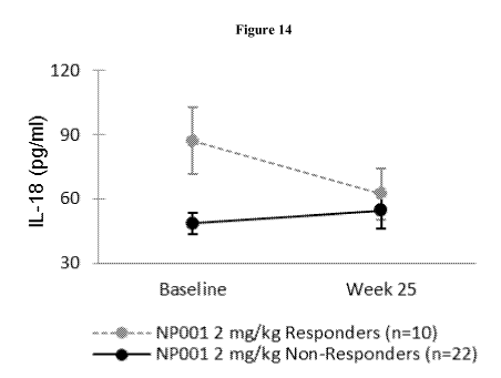
[0036] FIG.14 shows mean plasma IL-18 levels in high dose "responders" vs.
"non-responders" at
baseline and following 6-month treatment period (Week 25). Error bars
represent standard
deviation.
[0037] FIG.15 shows the IL-18 levels at baseline and Week 25 in responders,
non-responders and
placebos.
[0038] FIG.16 is a box and whisker plot of the distribution of the log of IL-
18, showing that the
IL-18 levels at baseline can differentiate responders, and non-responders.
[0039] FIG.17 is a table showing the baseline inflammation factor plasma
baseline value
interrelationships.
[0040] FIG.18 shows mean plasma LPS in all patients treated with 1 mg/kg or 2
mg/kg
chlorite/NP001 at baseline and following 6 month treatment period (Week 25).
Error bars represent
standard deviation. Limit of detection (LOD) for LPS = 0.05.
-5 -
CA 02945179 2016-10-06
WO 2015/175974 PCT/US2015/031145
[0041] FIG.19 shows mean LPS in placebo "responders" and "non-responders" at
baseline and
following 6 month treatment period (Week 25). Error bars represent standard
deviation. Limit of
detection (LOD) for LPS = 0.05.
[0042] FIG.20 shows the plasma level IL-18 at baseline and Week 25 for each
subject participating
in the study.
[0043] FIG.21 indicates a cut-off threshold value of the plasma level of IL-18
at baseline.
[0044] FIG.22 shows LPS positive and negative patients at baseline and ALS
disease progression
rate.
[0045] FIG.23 indicates ALS LPS negative patients have higher baseline ALSFRS-
R scores.
[0046] FIG.24 shows ALS LPS negative placebo patients become LPS positive
within 6 months.
[0047] FIG.25 shows decrease in ALSFRS-R score in ALS LPS negative patients
within 6 months.
[0048] FIG. 26 shows the relationship between baseline monocyte inflammatory
activation-related
markers and the historic rate of ALS disease progression, assessed by average
monthly change on
ALSFRS-R the disease progression rate (ALSFRS-R Score loss per month) in ALS.
FIG 26A
shows levels of baseline monocyte activation defined by CD14 co-expression of
HLA-DR was
directly related to the rate of ALS disease progression (r = 0.4310, p =
0.0138; n = 32). FIG 26B
depicts positive correlation was observed between baseline levels of CD
expression on the CD
bright subset of monocytes and disease progression rate in ALS (r = 0.4499, p
= 0.0098; n = 32).
[0049] FIG. 27 shows NP001 treatment changes CD14 monocyte expression of HLA-
DR as a
function of the degree of monocyte HLA-DR expression at baseline.
[0050] FIG. 28 shows the comparison of NP001 treatment response between ALS
patients with
elevated levels of baseline monocyte HLA-DR and those with lower range of
baseline monocyte
HLA-DR.
[0051] FIG. 29 illustrates the greatest change in monocyte levels of HLA-DR in
ALS patients with
the highest rate of disease progression.
[0052] FIG. 30 shows NP001 induced changes from baseline on CD16 levels
expressed on a CD16
bright subset of monocytes in a dose-dependent manner.
[0053] FIG. 31 shows the comparison of CD16 expression on monocyte CD16 bright
subset in
patients receiving 1.6mg/kg dose NP001 relative to healthy controls.
DETAILED DESCRIPTION OF THE INVENTION
[0054] The terminology used herein is for the purpose of describing particular
embodiments only
and is not intended to be limiting of the invention. As used herein, the
singular forms "a", "an" and
"the" are intended to include the plural forms as well, unless the context
clearly indicates
otherwise. Furthermore, to the extent that the terms "including", "includes",
"having", "has",
- 6 -
CA 02945179 2016-10-06
WO 2015/175974 PCT/US2015/031145
"with", or variants thereof are used in either the detailed description and/or
the claims, such terms
are intended to be inclusive in a manner similar to the term "comprising".
[0055] The term "about" or "approximately" means within an acceptable error
range for the
particular value as determined by one of ordinary skill in the art, which will
depend in part on how
the value is measured or determined, i.e., the limitations of the measurement
system. For example,
"about" can mean within 1 or more than 1 standard deviation, per the practice
in the art.
Alternatively, "about" can mean a range of up to 20%, up to 10%, up to 5%, or
up to 1% of a given
value. Alternatively, particularly with respect to biological systems or
processes, the term can
mean within an order of magnitude, preferably within 5-fold, and more
preferably within 2-fold, of
a value. Where particular values are described in the application and claims,
unless otherwise
stated the term "about" meaning within an acceptable error range for the
particular value should be
assumed.
[0056] "Treatment", "treating", "palliating" and "ameliorating", as used
herein, are used
interchangeably. These terms refer to an approach for obtaining beneficial or
desired results
including but not limited to therapeutic benefit and/or a prophylactic
benefit. By therapeutic
benefit is meant eradication or amelioration of the underlying disorder being
treated. Also, a
therapeutic benefit is achieved with the eradication or amelioration of one or
more of the
physiological symptoms associated with the underlying disorder such that an
improvement is
observed in the patient, notwithstanding that the patient may still be
afflicted with the underlying
disorder. For prophylactic benefit, the compositions may be administered to a
patient at risk of
developing a particular disease, or to a patient reporting one or more of the
physiological symptoms
of a disease, even though a diagnosis of this disease may not have been made.
[0057] As used herein, "agent" refers to a biological, pharmaceutical, or
chemical compound or
other moiety. Non-limiting examples include simple or complex organic or
inorganic molecule, a
peptide, a protein, an oligonucleotide, an antibody, an antibody derivative,
antibody fragment, a
vitamin derivative, a carbohydrate, a toxin, or a chemotherapeutic compound.
Various compounds
can be synthesized, for example, small molecules and oligomers (e.g.,
oligopeptides and
oligonucleotides), and synthetic organic compounds based on various core
structures. In addition,
various natural sources can provide compounds for screening, such as plant or
animal extracts, and
the like. A skilled artisan can readily recognize that there is no limit as to
the structural nature of
the agents of the present invention.
[0058] Generally, the term "concurrent administration", "co-administration",
or "administration in
conjunction with" in reference to two or more subjects of administration for
administration to a
subject body, such as components, agents, substances, materials, compositions,
and/or the like,
- 7 -
CA 02945179 2016-10-06
WO 2015/175974 PCT/US2015/031145
refers to administration performed using dose(s) and time interval(s) such
that the subjects of
administration are present together within the subject body, or at a site of
action in the subject
body, over a time interval in less than de minimus quantities. The time
interval may be any suitable
time interval, such as an appropriate interval of minutes, hours, days, or
weeks, for example. The
subjects of administration may be administered together, such as parts of a
single composition, for
example, or otherwise. The subjects of administration may be administered
substantially
simultaneously (such as within less than or equal to about 5 minutes, about 3
minutes, or about 1
minute, of one another, for example) or within a short time of one another
(such as within less than
or equal to about 1 hour, 30 minutes, or 10 minutes, or within more than about
5 minutes up to
about 1 hour, of one another, for example). The subjects of administration so
administered may be
considered to have been administered at substantially the same time. One of
ordinary skill in the art
will be able to determine appropriate dose(s) and time interval(s) for
administration of subjects of
administration to a subject body so that same will be present at more than de
minimus levels within
the subject body and/or at effective concentrations within the subject body.
When the subjects of
administration are concurrently administered to a subject body, any such
subject of administration
may be in an effective amount that is less than an effective amount that might
be used were it
administered alone.
[0059] The term "effective amount", "therapeutic amount" or "therapeutic
effective amount" which
is further described herein, encompasses both this lesser effective amount and
the usual effective
amount, and indeed, any amount that is effective to elicit a particular
condition, effect, and/or
response. As such, a dose of any such subject of concurrent administration may
be less than that
which might be used were it administered alone. One or more effect (s) of any
such subject (s) of
administration may be additive or synergistic. Any such subject(s) of
administration may be
administered more than one time. The effective amount may vary depending upon
the intended
application (in vitro or in vivo), or the subject and disease condition being
treated, e.g., the weight
and age of the subject, the severity of the disease condition, the manner of
administration and the
like, which can readily be determined by one of ordinary skill in the art. The
term also applies to a
dose that will induce a particular response in target cells, e.g., reduction
of proliferation or down-
regulation of activity of a target protein. The specific dose will vary
depending on the particular
compounds chosen, the dosing regimen to be followed, whether it is
administered in combination
with other compounds, timing of administration, the tissue to which it is
administered, and the
physical delivery system in which it is carried.
[0060] A "therapeutic effect," as used herein, encompasses a therapeutic
benefit and/or a
prophylactic benefit as described above. A prophylactic effect includes
delaying or eliminating the
- 8 -
CA 02945179 2016-10-06
WO 2015/175974 PCT/US2015/031145
appearance of a disease or condition, delaying or eliminating the onset of
symptoms of a disease or
condition, slowing, halting, or reversing the progression of a disease or
condition, or any
combination thereof
[0061] The term "pharmaceutically acceptable salt" refers to salts derived
from a variety of organic
and inorganic counter ions well known in the art. Pharmaceutically acceptable
acid addition salts
can be formed with inorganic acids and organic acids. Inorganic acids from
which salts can be
derived include, for example, hydrochloric acid, hydrobromic acid, sulfuric
acid, nitric acid,
phosphoric acid, and the like. Organic acids from which salts can be derived
include, for example,
acetic acid, propionic acid, glycolic acid, pyruvic acid, oxalic acid, maleic
acid, malonic acid,
succinic acid, fumaric acid, tartaric acid, citric acid, benzoic acid,
cinnamic acid, mandelic acid,
methanesulfonic acid, ethanesulfonic acid, p toluenesulfonic acid, salicylic
acid, and the like.
Pharmaceutically acceptable base addition salts can be formed with inorganic
and organic bases.
Inorganic bases from which salts can be derived include, for example, sodium,
potassium, lithium,
ammonium, calcium, magnesium, iron, zinc, copper, manganese, aluminum, and the
like. Organic
bases from which salts can be derived include, for example, primary,
secondary, and tertiary
amines, substituted amines including naturally occurring substituted amines,
cyclic amines, basic
ion exchange resins, and the like, specifically such as isopropylamine,
trimethylamine,
diethylamine, triethylamine, tripropylamine, and ethanolamine. In some
embodiments, the
pharmaceutically acceptable base addition salt is chosen from ammonium,
potassium, sodium,
calcium, and magnesium salts.
[0062] "Pharmaceutically acceptable carrier" or "pharmaceutically acceptable
excipient" includes
any and all solvents, dispersion media, coatings, antibacterial and antifungal
agents, isotonic and
absorption delaying agents and the like. The use of such media and agents for
pharmaceutically
active substances is well known in the art. Except insofar as any conventional
media or agent is
incompatible with the active ingredient, its use in the therapeutic
compositions of the invention is
contemplated. Supplementary active ingredients can also be incorporated into
the compositions.
[0063] "Subject" refers to an animal, such as a mammal, for example a human.
The methods
described herein can be useful in both human therapeutics, pre-clinical, and
veterinary applications.
In some embodiments, the subject is a mammal, and in some embodiments, the
subject is human.
[0064] The term "in vivo" refers to an event that takes place in a subject's
body.
A. Oxidative Aunts
[0065] In one aspect, the present invention provides a method of treating a
subject suffering a
macrophage-related disease, said method comprising administering to a subject
in need thereof an
effective amount of an oxidative agent. In another aspect, the present
invention provides a method
- 9 -
CA 02945179 2016-10-06
WO 2015/175974 PCT/US2015/031145
of monitoring a treatment with an oxidative agent to a subject suffering from
a macrophage related
disease. The oxidative agent can be chlorite or compositions comprising
chlorite.
I. Chlorite and Other Oxidative Agents
[0066] Substances that have the ability to oxidize other substances are
typically referred to as
oxidative and are known as oxidizing agents, oxidants, or oxidizers, which are
used
interchangeably herein. An oxidizing agent (also called an oxidant, oxidizer)
can be defined as
either: a chemical compound that readily transfers oxygen atoms, or a
substance that gains
electrons in a redox chemical reaction. In both cases, the oxidizing agent
becomes reduced in the
process. Various common oxidizers contain oxygen (e.g., KC104) and can be
considered as storage
forms of oxygen. Alternatively, the term "oxidizing agent" also includes any
time where formal
charge is increased (losing electrons), and applies to substances that contain
no oxygen, typically
halogens comprising fluorine, (F); chlorine, (C1); bromine, (Br); iodine, (I);
and astatine, (At), and
substances rich in these elements.
[0067] Common oxidizing or oxidative agents that can be used in the methods of
the present
invention include but are not limited to potassium nitrate (KNO3),
hypochlorite and other
hypohalite compounds, iodine and other halogens, chlorite, chlorate,
perchlorate, and other
analogous halogen compounds, permanganate salts, ammonium cerium(IV) nitrate
and related
cerium(IV) compounds, hexavalent chromium compounds such as chromic and
dichromic acids
and chromium trioxide, pyridinium chlorochromate (PCC), and
chromate/dichromate compounds;
peroxide compounds, Tollens' reagent, sulfoxides, persulfuric acid, ozone,
osmium tetroxide
(0s04), nitric acid, and nitrous oxide (N20). The oxidative agent can be non-
toxic to monocytes or
macrophages at physiologically effective concentrations.
[0068] The oxidative agents of the current invention can be compounds that
contain both readily-
transferrable oxygen and halogen atoms, including but not limited to
hypochlorite and other
hypohalite compounds, chlorite, chlorate, perchlorate and other analogous
halogen compounds, and
pyridinium chlorochromate (PCC). As used herein, such compounds are referred
to as activated-
oxygen activated-halogen compounds.
[0069] Alternatively, the oxidative agent may be a substance that contains no
oxygen, typically
halogens comprising fluorine, (F); chlorine, (C1); bromine, (Br); iodine, (I);
and astatine, (At). As
used herein, such compounds are referred to non-oxygen activated-halogen
compounds.
[0070] Many oxidative compounds have demonstrated protective and anti-
inflammatory activities,
likely due to induction of endogenous defense pathways. For example,
metabolites of the stress
induced enzyme heme oxygenase 1 (H0-1) such as carbon monoxide (CO) and
biliverdin exert
potent anti-inflammatory effects (Otterbein L E et al. Nat. Med. 6 (2000) 422-
428). The catalytic
- 10 -
CA 02945179 2016-10-06
WO 2015/175974 PCT/US2015/031145
products of HO-1 including the oxidants CO, Fe2+, and biliverdin are capable
of down-regulating
inflammatory reactions. Similar cell-protective properties have been described
for the redox-active
molecule thioredoxin (Hirota K. et al. J. Biol. Chem. 274 (1999) 27891-27897).
The use of chlorite
to treat various diseases and conditions is described in US Patent No.
4,725,437; US Patent No.
4,851,222; McGrath et al., Development of WF10, a novel macrophage-regulating
agent, Curr Opin
Investig Drugs, 3(3):365-73 (March 2002); US Patent No. 6,086,922; US Patent
No. 7,105,183; US
Patent No. 8,029,826; US Patent No. 8,501,244; US Patent No. 8,231,856; US
Patent No.
8,252,789; US Patent No. 8,067,035; and US Patent Application No. 13/388,411,
all of which are
incorporated herein by reference in their entirety.
[0071] Disclosed herein are compositions and methods for treatment of a
subject suffering from a
macrophage related disease using chlorite. The chlorite ion is C102-. A
chlorite (compound) is a
compound that contains this group, with chlorine in oxidation state +3.
Chlorites are also known as
salts of chlorous acid. Chlorine can assume oxidation states of -1, +1, +3,
+5, or +7 within the
corresponding anions Cl-, C10-, C102-, C103- or C104- known commonly and
respectively as
chloride, hypochlorite, chlorite, chlorate, and perchlorate.
II. Tetrachlorodecaoxide (TCDO) and WF10
[0072] The present invention also provides methods using one or more chlorite
containing agents.
The source of chlorite ions for administration of chlorite according to the
present invention can be
provided in a variety of forms. For example, chlorite can be administered as a
chlorite salt, for
example, alkali metal salt, e.g. sodium chlorite, potassium chlorite, and the
like, or a mixture of
chlorite salts, where the chlorite salts are preferably pharmaceutically
acceptable. In addition or
alternatively, chlorite can be administered as a matrix of chlorite ions,
e.g., described in U.S. Pat.
No. 4,507,285. In one embodiment, the chlorite ions as provided in a
composition having the
general formula:
C102- x n02
wherein "n" can be a value of about 0.1-0.25. Such agents can have an 02 band
at 1562 cm-1 in the
Raman spectrum and an 0-0 interval of 123 pm. Production of such agents is
known in the art, see
e.g., U.S. Pat. No. 4,507,285.
[0073] In one embodiment, the method of treatment involves administration of a
liquid
composition comprising an aqueous solution of a product known as
"tetrachlorodecaoxygen anion
complex", commonly known as TCDO. Production of TCDO is well known, see e.g.,
Example 1 of
U.S. Pat. No. 4,507,285. In some embodiments, the chlorite containing agents
that can be used in
the methods of the present invention for treating diabetes or related
disorders include but are not
limited to chlorite salt, such as alkali metal salt, sodium chlorite,
potassium chlorite, and the like, a
- 11 -
CA 02945179 2016-10-06
WO 2015/175974 PCT/US2015/031145
matrix of chlorite salts, a matrix of chlorite ions, e.g., compositions having
the general formula
C102xn02, where "n" can be a value or about 0.1-0.25. One example is TCDO. One
of the aqueous
TCDO formulations is WF10. WF10 is an aqueous formulation of the drug OXO-
K993. Oxoferin is
a topical formulation of the same drug and is registered and marketed as a
wound healing agent in
Europe and Asia. WF10 is a sterile, pyrogen-free, aqueous 10% (w/v) solution
of OXO-K993 with
no additional inactive ingredients and is intended for intravenous infusion.
TCDO is analytically
characterized as a solution containing 4.25% chlorite, 1.9% chloride, 1.5%
chlorate, 0.7% sulfate,
and sodium as the cation. The active principle is defined by the chlorite ion
content. In one
embodiment, WF10 solution contains about 63 mmo1/1 of chlorite.
[0074] Tetrachlorodecaoxide (TCDO) is a chlorite-containing drug used for the
dressing of
wounds, immunomodulation and as radiation protective agent. Due to its
oxidizing properties,
TCDO can destroy most pathogens although it is not regarded as antibiotic. But
the main reason for
its use for dressing of wounds is not its bactericidal activity. This drug is
regarded as
immunomodulating, that is, it acts by stimulating the immune system of the
body.
Tetrachlorodecaoxide combines with the heme part of hemoglobin, myoglobin and
peroxidase,
forming a TCDO-hemo complex. This in turn activates the macrophages and
accelerates the
process of phagocytosis which engulfs most of the pathogens and cell debris
present on the surface
of the wound, thus cleaning the wound surface and helping in the regenerative
process.
Tetrachlorodecaoxide is also mitogenic and chemotactic. The mitogenic impulse
gives rise to two
factors, MDGF (Macrophage derived growth factor) and WAF (Wound angiogenesis
factor). The
MDGF deposits fibroblasts and synthesizes collagen fibers, which fill the gap
in the wounds, the
WAF helps in the formation of new capillaries which further enhances the
healing process. The
chemotactic impulse acts on the myocyte (muscle cell) and causes it to
contract, thereby bringing
the wound edges closer and reducing the wound surface. Simultaneous influence
of all these factors
accelerates the wound healing with minimal scarring.
[0075] WF10 is a 1:10 dilution of tetrachlorodecaoxide (TCDO) formulated for
intravenous
injection. WF10 specifically targets macrophages. WF10 potentially modulates
disease-related up-
regulation of immune responses both in vitro and in vivo. Thus immune response
is influenced in a
way that inappropriate inflammatory reactions are downregulated
(Arzneimittelforschung. 2001;
51(7):554-62. Schempp H, et al). WF10 is currently being studied for treatment
of late-stage HIV
disease, as well as recurrent prostate cancer, late post-radiation cystitis,
autoimmune disease and
chronic active hepatitis C disease. WF10 is approved for use in Thailand under
the name
IMMUNOKINE in patients with post-radiation chronic inflammatory disease
including cystitis,
proctitis and mucositis.
- 12 -
CA 02945179 2016-10-06
WO 2015/175974 PCT/US2015/031145
[0076] In vivo studies have investigated the effects of WF10 on monocytes,
macrophages and
lymphocytes, on humoral and cellular immunity, and on response to local or
total body irradiation
(reviewed by McGrath M S et al. Current Opinion in Investigational Drugs 2002
3(3)). WF10
increased the number of macrophages infiltrating a skin blister in a human
wound healing model
(Hansel M et al. Skin Pharmacol 1988 1:64). In rats, WF10 increased the
proportion of
granulocytes, peripheral blood monocytes (PBMCs) and large granular
lymphocytes (LGLs), and
stimulated erythropoiesis after total body X-irradiation (Ivankovic S et al.
OX0 Study Report 1988
March; Ivankovic S et al. Radiat Res 1988 115: 115-123). In mice, WF10
stimulated regeneration
of hematopoietic stem cells receiving sublethal doses of J-irradiation (Mason
K A et al. Radiat Res
1993 136: 229-235). In other studies, WF10 displayed direct antitumor effects
against radiation-
induced, hemical-induced and metastatic malignant and benign tumors (Kempf S R
et al.
International Symposium on Tissue Repair 1990 Thailand; Milas L. OX0 Study
Report 1991
September; Kempf S R et al. Radiat Res 1994 139: 226-231). WF10 altered
proportions of T-helper
and suppressor/cytotoxic cells in spleen and thymus and increased both the
humoral and cellular
immune responses (Gillissen G et al. OX0 Study Report 1993).
[0077] Without being bound by theory, it has been suggested that WF10 causes
marked inhibition
of inducible genes related to T-cell proliferation and cause reproducible up-
regulation of
inflammatory gene expression in macrophages in vitro, which is thought to
contribute to the higher
rate of apoptosis in activated macrophages. These data, coupled with an
earlier report of WF10
inhibition of T-cell activation (McGrath M S et al. Transplant Proc. 1998 30:
4200-4202), show
that WF10 causes profound changes in T-cell function through regulation of
macrophage
activation. The WF 10 oxygen/chlorite matrix is stable until interaction with
heme-associated iron,
whereupon it is converted to an active chlorite molecule through a Michealis-
Menten reaction and
intermediate production of a reactive compound I. Chlorite is the active form
of the drug thought to
mediate the immunological effects in macrophages.
[0078] A dose-ranging clinical study was conducted from 1993 to 1994 in 44 HIV-
positive patients
with <500 CD4+ T cells/mm (Raffanti S P et al. Infection 1998 26: 201-206).
The study established
the maximum tolerated dose as 0.5 ml/kg/day of WF10, when administered in four
5-day cycles,
with each cycle followed by 16 days of without treatment. No significant
adverse events or clinical
laboratory toxicity were observed at this dosage. Plasma CD8+ T-cell counts
increased in a dose-
dependent manner over four cycles of WF10 administration. This study
demonstrated that WF10 at
a dose of 0.5 ml/kg was associated with a sustained immunological response,
i.e., sustained
elevation of CD8+ T cell numbers, consistent with the proposed mechanism of
action. Furthermore,
a single-center, phase I/II study, was conducted in 1997 to evaluate safety
and the effects of WF10
- 13 -
CA 02945179 2016-10-06
WO 2015/175974 PCT/US2015/031145
on the kinetics of red blood cell (RBC) survival, selective immunological
markers of HIV disease,
macrophage activation and viral kinetics (Hemdier B et al. Keystone Symposia
on Molecular and
Cellular Biology. 1998). Changes in immunological parameters of cells from
HIV+ patients in
response to WF10 treatment are summarized in Table 1 in McGrath M S et al.
Current Opinion in
Investigational Drugs 2002 3(3), including an increase in CD3+CD4+ cells, an
increase in CD3+
CD8+ cells, an increase in CD3+ CD4+ CD38- cells, an increase in CD3+ CD8+
CD38- cells, an
increase in CD3+ CD8+ CD28- cells, a decrease in CD3+ CD8+ CD28+ cells, a
decrease in CD3+
CD4+ CD38+ cells, a decrease in all CD14+ cells, and a decrease in CD20+ HLR-
DR+ cells. The
results suggested that WF10 reduced antigen presentation while concurrently
inducing
phagocytosis in macrophages with impaired function. WF10 had no effect on HIV
load over the
course of the trial. No significant differences were detected between the WF10
and placebo group
in hematological and blood chemistry values, including parameters specifically
associated with
hemolysis.
[0079] As appropriate, agents that provide a source of chlorite ions can be
administered in a free
base or free acid form, i.e., as the free compound and not as a salt. In some
embodiments, the
chlorite formulation contains about 150 ILIM chlorite.
[0080] Additionally, any pharmaceutically acceptable salt(s) of the
compound(s) can also be used.
Pharmaceutically acceptable salts are those salts which retain the biological
activity of the free
compounds and which are not biologically or otherwise undesirable. As
appropriate, stereoisomers
of the compounds disclosed can also be used in the invention, including
diastereomers and
enantiomers, as well as mixtures of stereoisomers, including but not limited
to racemic mixtures.
Unless stereochemistry is explicitly indicated in a structure, the structure
is intended to embrace all
possible stereoisomers of the compound depicted.
[0081] The oxidative compound or chlorite as described herein can be WF10.
WF10 is a chlorite-
based compound. After interaction with heme proteins, the chlorite matrix of
WF10 acquires
oxidizing and chlorinating properties (Schempp H. et al. 1999). It has been
suggested that WF10
exerts potent immunomodulatory effects most likely through generating
physiologic oxidative
compounds namely chloramines. Chloramines have been reported to exert cell-
protective and anti-
inflammatory activities (Choray M. et al. Amino Acids 23 (2002) 407-413).
[0082] Pro-oxidative substances can also have a direct effect on
transcriptional activities of the
NFAT species of transcription factors. The nuclear translocation of NFAT
requires their
dephosphorylation by the calcium/calmodulin dependent serine/threonine
phosphatase calcineurin.
The phosphatase activity of calcineurin is redox sensitive. WF10 is able to
inhibit antigen receptor
driven lymphocyte proliferation. Expression of NFAT regulated genes is
strongly suppressed by
- 14 -
CA 02945179 2016-10-06
WO 2015/175974 PCT/US2015/031145
WF10, and the nuclear translocation of NFATc is inhibited. The WF10 associated
inhibition of
NFAT regulated genes in activated T cells, in concert with the induction of
several monocyte
associated pro-inflammatory genes, suggest activation of the innate myeloid
functions concomitant
with the inactivation of adaptive proliferative lymphocyte response. This
approach represents a
novel method of targeting redox-regulation for the treatment of inflammatory
disorders. In some
embodiments, the macrophage related diseases that can be treated using the
methods of the present
invention are inflammatory diseases.
III. Chlorite Purity and pH
[0083] Methods of formulating chlorite have been described in US Patent Pub.
No. 20070145328,
filed Dec. 21, 2006 and entitled "Chlorite Formulations, and Methods of
Preparation and Use
Thereof," which is incorporated herein by reference in its entirety. Such
formulations are suitable
for various modes of administration, including but not limited to non-topical,
parenteral, systemic,
or intravenous administration.
[0084] Described in present invention are compositions and methods using
chlorite formulated in
aqueous solution in which the chlorite is greater than 95% pure. In some
cases, the chlorite can be
greater than 97%, 99%, 99.5% or 99.9% pure. In some cases, the chlorite can be
at least 95%,
97%, 99%, 99.5% or 99.9% pure. As used herein, the "purity" of chlorite in a
sample is calculated
as the percent weight of chlorite salt to the total weight of the sample. In
determining the purity of
chlorite in a solution, the weight of the solvent (e.g., water in an aqueous
solution) is not included.
Purity may be evaluated using ion chromatography and an ion detector, by
calibrated integration of
the respective peaks; for example, chlorite, chloride, chlorate, phosphate and
sulfate in the
compound or formulation. For example, chlorite is commercially available as
sodium chlorite,
technical grade, at a purity of 80% (catalog No. 244155 Sigma-Aldrich).
[0085] Alternatively, crystalline sodium chlorite is provided in a purity
greater than 95%, greater
than 96%, greater than 97%, greater than 98%, greater than 99%, greater than
99.5% or greater than
99.9%. Solid pharmaceutical formulations comprising crystalline sodium
chlorite in a purity greater
than 95%, greater than 96%, greater than 97%, greater than 98%, greater than
99%, greater than
99.5% or greater than 99.9% in addition to one or more pharmaceutical
excipients are also
encompassed.
[0086] The chlorite formulations for use with the present invention can
comprise low amounts of
chlorate, sulfate or chloride. As used herein, a formulation is "substantially
free" of a molecule if
the molecule comprises no more than 1 part in 1000 per weight of non-solvent
molecules in the
formulation. In certain embodiments, the weight ratio of chlorite to chlorate
is greater than 100:1.5,
greater than 100:0.5, greater than 100:1, or greater than 100:0.1. In one
embodiment, the
- 15 -
CA 02945179 2016-10-06
WO 2015/175974 PCT/US2015/031145
composition is substantially free of chlorate. In another embodiment, the
weight ratio of chlorite to
chloride is greater than 100:45.5 or greater than 100:8.5. In one embodiment
the composition is
substantially free of chloride. In a further embodiment, the weight ratio of
chlorite to sulfate is
greater than 100:16.4 or greater than 100:1.6. In one embodiment the
composition is substantially
free of sulfate.
[0087] The pH of a chlorite formulation for use with the present invention can
be adjusted to
between about 7 and about 11.5. In some embodiments, the pH of a chlorite
formulation is lowered
to between about 7 and about 11.5 using a pH adjusting compound that does not
expose the
formulation to high local acidity. In some embodiments, the pH adjusting
compound is any one or
more of monosodium phosphate, disodium phosphate, or acetic acid.
[0088] Also described herein are methods of preparing chlorite formulations
and pharmaceutical
formulations, including but not limited to the chlorite formulations
specifically described herein.
Also described herein are kits and methods of administration of the
formulations and
pharmaceutical formulations described herein. Various exemplary aspects and
variations of the
invention are described in the "Brief Summary of the Invention," as well as
elsewhere herein,
including but not limited to the Examples. It is also understood that the
invention includes
embodiments comprising, consisting essentially of, and/or consisting of one or
more elements as
described herein.
[0089] In some embodiments, the invention makes use of aqueous formulations
comprising
chlorite. In some embodiments, the chlorite formulation comprises an aqueous
solvent, and
optionally one or more other solvents for chlorite. In some embodiments, the
formulations
comprise chlorite and an aqueous solvent for chlorite, and have a pH of about
7 to about 11.5.
[0090] Solvents or combinations of solvents for use in the formulations
described herein can be
determined by a variety of methods known in the art. One non-limiting example
includes (1)
theoretically estimating solvent solubility parameter value(s) and choosing
the one(s) that match
with chlorite, using standard equations in the field; and (2) experimentally
determining the
saturation solubility of chlorite in the solvent(s), and (3) choosing one or
more that exhibits the
desired solubility, and (4) selecting a solvent or solvents that do not
diminish the activity of
chlorite, or that do not or only minimally react with chlorite. In some
embodiments, the liquid
formulations described herein comprise a plurality of solvents.
[0091] In some embodiments, the chlorite formulations comprise an aqueous
solvent. In some
variations, water is the principal solvent in the aqueous formulations. In
some variations, water is at
least about 50% by volume of the solvent component of an aqueous formulation.
In some
variations, water is at least about 50% by volume of the aqueous formulation.
In some variations,
- 16 -
CA 02945179 2016-10-06
WO 2015/175974 PCT/US2015/031145
water is any of between about 50 to about 60, between about 60 to about 70,
between about 70 to
about 80, between about 80 to about 90, between about 90 to about 99, at least
about 50, at least
about 60, at least about 70, at least about 80, at least about 90, or at least
about 95, about 50, about
60, about 70, about 80, about 90, or about 95 percent by volume of the solvent
component. In some
variations, water is any of between about 50 to about 60, between about 60 to
about 70, between
about 70 to about 80, between about 80 to about 90, between about 90 to about
99, at least about
50, at least about 60, at least about 70, at least about 80, at least about
90, or at least about 95,
percent by volume of the aqueous formulation. In some variations, water is at
least about 95% by
volume of the aqueous formulation. In some variations, water is between about
80 to about 90% by
volume of the aqueous formulation. In some variations, water is between about
90 to about 99% by
volume of the aqueous formulation.
[0092] The formulations may have differing concentration of chlorite. In some
embodiments, the
concentration of chlorite in the formulation is high, and then is diluted to a
less concentrated form
prior to administration. In some embodiments, a formulation described herein
is diluted about, at
least about or less than about 2.5x, about 5x, about 7.5x, about 10x, about
20x, about 25x, about
50x, about 100x, about 200x, about 250x, about 300x, about 500x, or about
1000x. In some
embodiments, a formulation described herein is diluted between about 2x and
about 10x, between
about 10x and about 50x, between about 50x and about 100x, between about 100x
and about 500x,
or between about 500x and about 1000x. In some embodiments, a formulation as
described herein
is diluted between about 2x and about 10x. In some embodiments, a formulation
as described
herein is diluted between about 10x and about 50x. In some embodiments, a
formulation as
described herein is diluted about 7.5x. In some embodiments, a formulation as
described herein is
diluted about 25x. In some embodiments, a formulation as described herein is
diluted about 200x.
[0093] In some embodiments, the concentration of chlorite in the formulations
described herein is
between about 1 ILIM and about 1.5 M. In another embodiments, the
concentration of chlorite in the
formulations described herein is between any of about 1 M and about 1.5 M;
between about 1 ILIM
and about 100 mM; between about between about 10 ILIM and about 100 mM;
between about 0.1
mM and about 10 mM; between about 0.1 mM and about 500 mM; between about 0.1
mM and
about 200 mM; between about 1 mM and about 100 mM; between about 0.1 mM and
about 5 mM;
between about 50 mM and about 100 mM; between about 55 mM and about 70 mM;
between about
60 mM and about 65 mM; between about 100 mM and about 500 mM; between about
200 mM and
about 400 mM; between about 300 mM and about 700 mM; about 1 mM; about 1.5 mM;
about 2
mM; about 2.5 mM; about 3 mM; about 3.5 mM; about 4 mM; about 5 mM; about 10
mM; about
20 mM; about 30 mM; about 40 mM; about 50 mM; about 60 mM; about 62 mM; about
65 mM;
- 17 -
CA 02945179 2016-10-06
WO 2015/175974 PCT/US2015/031145
about 70 mM; about 80 mM; about 90 mM; about 100 mM; at least about 0.1 mM; at
least about 1
mM; at least about 2 mM; at least about 5 mM; at least about 10 mM; at least
about 20 mM; at least
about 30 mM; at least about 40 mM; at least about 50 mM; at least about 60 mM;
at least about 70
mM; at least about 80 mM; at least about 90 mM; or at least about 100 mM. In
preferred
embodiments, the concentration of chlorite in the formulations described
herein is about or at least
about 60 mM.
[0094] In some embodiments, the concentration of chlorate in the formulations
described herein is
between about 50 mM and about 100 mM. In some embodiments, the concentration
of chlorate in
the formulations described herein is between about 55 mM and about 75 mM. In
some
embodiments, the concentration of chlorate in the formulations described
herein is between about
0.1 mM and about 10 mM. In some embodiments, the concentration of chlorate in
the formulations
described herein is between about 1 mM and about 5 mM.
[0095] In some embodiments, the chlorite formulation has a pH no greater than
about 12Ø In some
embodiments, the pH of the formulation is any of no greater than about 11.5,
about 11.0, about
10.5, about 10.0, about 9.5, about 9.0, about 8.5, about 8.0, about 7.5, about
7.0, about 6.5, or about
6Ø In some embodiments, the pH of the formulation is no greater than about
11.5. In some
embodiments, the pH of the formulation is no greater than about 10.5. In some
embodiments, the
pH of the formulation is no greater than about 8.5. In some embodiments, the
pH of the formulation
is no greater than about 7.5. In some embodiments, the pH of the formulation
is between any one or
more of about 7 and about 12; between about 7 and about 11.5; between about 7
and about 10.5;
between about 7 and about 10; between about 7 and about 9.5; between about 7
and about 9.0;
between about 7 and about 8.5; between about 7 and about 8.0; between about 7
and about 7.5;
between about 7.5 and about 8; between about 7.5 and about 8.5; between about
7 and about 8;
between about 8 and about 9; between about 7.0 and about 8.5; between about 8
and about 8.5;
between about 8.5 and about 9; between about 7.1 and about 7.7; between about
7.2 and about 7.6;
between about 7.3 and about 7.4; about 7.0; about 7.1; about 7.2; about 7.3;
about 7.4; about 7.5;
about 7.6; about 7.7; about 7.8; about 7.9; about 8.0; about 8.1; about 8.2;
about 8.3; about 8.4;
about 8.5; about 8.6; about 8.7; about 8.8; or about 8.9. In some embodiments,
the chlorite
formulation has a pH of about 7.0 to about 9Ø In some embodiments, the
chlorite formulation has
a pH of about 7.0 to about 8.5. In some embodiments, the chlorite formulation
has a pH of about
6.0 to about 8.5. In some embodiments, the chlorite formulation has a pH of
about 7.0 to about 8Ø
In some embodiments, the chlorite formulation has a pH of about 7.4. The
chlorite formulation can
have a pH that is at a physiological level.
- 18 -
CA 02945179 2016-10-06
WO 2015/175974 PCT/US2015/031145
[0096] In some embodiments, the chlorite formulations have a pH as described
above, and are
formulated for any one or more of parenteral, systemic, or intravenous
administration. In some
embodiments, the chlorite formulations have a pH as described above, and have
a percentage
chlorite purity as described herein.
[0097] In some embodiments, the formulations described herein have a pH as
described above, and
have a concentration of chlorite as described herein. In some embodiments, the
aqueous
formulations described herein have a pH between about 7 and about 11.5, or
between about 7.0 and
about 10, or between about 7.0 and about 9.0, or between about 7.0 and about
8.5, or between about
7.1 and about 7.7, and have a concentration of chlorite between about 1 and
about 100 mM. In
some embodiments, the aqueous formulations described herein have a pH between
about 7 and
about 11.5, or between about 7.0 and about 10, or between about 7.0 and about
9.0, or between
about 7.0 and about 8.5, or between about 7.1 and about 7.7, and have a
concentration of chlorite
between about 1 and about 5 mM. In some embodiments, the aqueous formulations
described
herein have a pH between about 7 and about 11.5, or between about 7.0 and
about 10, or between
about 7.0 and about 9.0, or between about 7.0 and about 8.5, or between about
7.1 and about 7.7,
and have a concentration of chlorite between about 50 and about 80 mM.
[0098] In some embodiments, the aqueous formulations described herein have a
pH between about
7 and about 11.5, or between about 7.0 and about 10, or between about 7.0 and
about 9.0, or
between about 7.0 and about 8.5, or between about 7.1 and about 7.7, wherein
the pH was adjusted
with a pH adjusting agent that is any one or more of a phosphate, or acetic
acid.
[0099] In some embodiments, the formulations described herein are stable with
respect to one or
more of pH or chlorite degradation over a period of any of at least about 1
day, at least about 2
days, at least about 3 days, at least about 4 days, at least about 5 days, at
least about 6 days, at least
about 1 week, at least about 2 weeks, at least about 3 weeks, at least about 4
weeks, at least about 5
weeks, at least about 6 weeks, at least about 7 weeks, at least about 8 weeks,
at least about 1 month,
at least about 2 months, at least about 3 months, at least about 4 months, at
least about 5 months, or
at least about 6 months. In some embodiments, the formulations described
herein are stable with
respect to one or more of pH or chlorite degradation over a period of any of
at least about 1 week.
In some embodiments, the formulations are stable with respect to one or more
of pH or chlorite
degradation over a period of any of at least about 1 month. In some
embodiments, the formulations
described herein are stable with respect to one or more of pH or chlorite
degradation at one or more
of room temperature, refrigerated conditions, or approximately 4 degree C. In
some embodiments,
the formulations described herein are stable with respect to one or more of pH
or chlorite
degradation under conditions of diminished light or storage in a container
that limits the amount of
- 19 -
CA 02945179 2016-10-06
WO 2015/175974 PCT/US2015/031145
light to which the formulation is subjected. In some embodiments, the
formulations described
herein are stable with respect to one or more of pH or chlorite degradation
when stored in the dark.
Examples of stable pH, as used herein, means that the pH of the formulation
changes by less than
any of about 0.1, about 0.2, about 0.3, about 0.4, about 0.5, about 0.6, about
0.7, about 0.8, about
0.9, or about 1 relative to the pH of the formulation as initially prepared.
In some embodiments, the
pH of the formulation changes by less than about 0.2 relative to the pH of the
formulation as
initially prepared. The pH may be measured using, for example, a pH meter.
Examples of stable
chlorite formulations include those in which less than any of about 0.1%, less
than about 0.2%, less
than about 0.3%, less than about 0.4%, less than about 0.5%, less than about
0.6%, less than about
0.7%, less than about 0.8%, less than about 0.9%, less than about 1%, less
than about 2%, less than
about 3%, less than about 4%, less than about 5%, less than about 6%, less
than about 7%, less than
about 8%, less than about 9%, or less than about 10% of the chlorite degrades
into a non-chlorite
ion relative to the amount of chlorite present in the formulation as initially
prepared. In some
embodiments, less than about 2% of the chlorite degrades into a non-chlorite
compound relative to
the amount of chlorite present in the formulation as initially prepared. In
some embodiments, less
than about 0.5% of the chlorite degrades into a non-chlorite compound relative
to the amount of
chlorite present in the formulation as initially prepared. The presence of non-
chlorite elements may
be measured, for example, using gas chromatography (GC), mass spectrometry, or
other methods
known by those of skill in the art.
[00100] In some embodiments, the chlorite formulations described herein
comprise no greater than
about 5% by weight of deleterious non-chlorite elements of other commercially
available
formulations. In some embodiments, the chlorite formulations described herein
comprise any of no
greater than about 4%, about 3%, about 2%, about 1%, about 0.5%, about 0.3%,
about 0.25%,about
0.2%, about 0.1%, about 0.05%, or about 0.02%, by weight of deleterious non-
chlorite elements of
other commercially available formulations. In some embodiments, the chlorite
formulations
described herein comprise any of no greater than about 4% by weight of
deleterious non-chlorite
elements of other commercially available formulations. In some embodiments,
the chlorite
formulations described herein comprise any of no greater than about 2% by
weight of deleterious
non-chlorite elements of other commercially available formulations. In some
embodiments, the
chlorite formulations described herein comprise any of no greater than about
0.5% by weight of
deleterious non-chlorite elements of other commercially available
formulations. In some
embodiments, the chlorite formulations described herein comprise any of no
greater than about
0.05% by weight of deleterious non-chlorite elements of other commercially
available
formulations. In some embodiments, the chlorite formulations described herein
are substantially
- 20 -
CA 02945179 2016-10-06
WO 2015/175974 PCT/US2015/031145
free of the deleterious non-chlorite elements of other commercially available
formulations. Non-
limiting examples of methods of detection of non-chlorite components include
HPLC; SPCS, for
example using a Novosep A2 column with 3.6 mM Sodium Carbonate as a mobile
phase, 5 ,
250x4.0 mm, flow rate 0.8 mL/min; DS-Plus Suppressor, for example using a
Novosep A2 column
with 3.6 mM Sodium Carbonate as a mobile phase, 5 , 250x4.0 mm, flow rate 0 8
mL/min; an
Allsep A-2 Anion column using 2.1mM NaHCO3/1.6 mM Na2CO3 as a mobile
phase,
100x4.6 mm, flow rate 2.0 mL/min; an anion HC column using 2.8 mM
NaHCO3:2.2 mM
Na2CO3 in 10% Methanol as a mobile phase, 150x4.6 mm, flow rate 1.4 mL/min; or
an Allsep A-2
Anion column using 2.1 mM NaHCO3/1.6 mM Na2CO3 as a mobile phase, 5 , 100x4.6
mm, flow
rate 1.0 mL/min. See, for example, the Alltech Associates, Inc. Grace Davison
line of products and
product information for details. In some embodiments, formulations described
herein comprise no
greater than about 10%, about 20%, about 30%, about 40%, about 50%, about 60%,
about 70%,
about 75%, about 80%, about 85%, about 90%, or about 95% of the amount of a
member of the
group consisting of, or alternatively any one or more of, chlorate ions and
sulfate ions present in an
equal weight/volume percent of chlorite formulated as WF10 or a dilution
thereof That is, in some
embodiments, when a non-WF10 formulation as described herein comprises a
certain percent w/v
of chlorite, such formulation has no greater than about the stated percentage
of the amount of one
or more of the specified non-chlorite components in WF10 or a dilution
thereof, wherein the WF10
or dilution thereof comprises the same percent w/v of chlorite as is found in
the non-WF10
formulation with which it is being compared. In some embodiments, the
formulations described
herein comprise no greater than about 75% of the amount of a member of the
group consisting of,
or alternatively any one or more of, chlorate ions and sulfate ions present in
an equal
weight/volume percent of chlorite formulated as WF10. In some embodiments, the
formulations
described herein comprise no greater than about 85% of the amount of a member
of the group
consisting of, or alternatively any one or more of, chlorate ions and sulfate
ions present in an equal
weight/volume percent of chlorite formulated as WF10. In some embodiments, the
formulations
described herein comprise no greater than about 50% of the amount of a member
of the group
consisting of, or alternatively any one or more of, chlorate ions and sulfate
ions present in an equal
weight/volume percent of chlorite formulated as WF10.
[00101] It can be understood from the product insert of WF10 that WF10
reportedly includes a
ratio of chlorite to chlorate of 100:35.7 (4.25% to 1.5%), a ratio of chlorite
to chloride of 100:45.5
(4.25% to 1.9%) and a chlorite to sulfate ratio of 100:16.4 (4.25% to 0.7%).
[00102] Examples of deleterious non-chlorite components include non-chlorite
components that
cause an adverse reaction when administered to physiological systems. In some
variations, a
- 21 -
CA 02945179 2016-10-06
WO 2015/175974 PCT/US2015/031145
deleterious non chlorite component is associated with one or more indicia of
toxicity in one or more
of in vitro or in vivo assays known in the art, or are associated with one or
indicia of toxicity when
administered to a physiological system, including but not limited to a
subject, including but not
limited to a human subject. Deleterious non chlorite components include but
are not limited to
sulfate, chlorine dioxide, chlorate, and borate. In some embodiments, the
chlorite formulations
described herein are substantially free of the deleterious non-chlorite
elements of WF10. In some
variations, the chlorite formulations described herein are substantially free
of sulfate and chlorate
ions.
[00103] In some embodiments, the chlorite formulations described herein
contain less than about
1.9% of chloride ions. In some embodiments, the chlorite formulation contains
any of less than
about 1.9%, less than about 1.8%; less than about 1.5%; less than about 1.0%;
less than about
0.5%; less than about 0.3%; less than about 0.1%; less than about 0.05%; less
than about 0.01%;
less than about 0.001%; between about 0.001 to about 0.1%; between about 0.1
to about 0.5%;
between about 0.5 to about 1.0%; between about 1.0 to about 1.5%; or between
about 1.5 to about
1.8%by weight of chloride ions. In some embodiments, the chlorite formulation
contains less than
about 0.5% by weight of chloride ions. In some embodiments, the chlorite
formulation contains less
than about 0.24% by weight of chloride ions. In some embodiments, the chlorite
formulation
contains less than about 0.2% by weight of chloride ions. In some embodiments,
the chlorite
formulation contains less than about 0.1% by weight of chloride ions. In some
embodiments, the
chlorite formulation is substantially free of chloride ions. In some
embodiments, the level of
chloride ions is below the level of detection using HPLC.
[00104] In some embodiments, the chlorite formulation contains less than about
1.5% of chlorate
ions. In some embodiments, the chlorite formulation contains any of less than
about 1.4%, less than
about 1.3%; less than about 1.0%; less than about 0.5%; less than about 0.3%;
less than about
0.1%; less than about 0.01%; less than about 0.001%; between about 0.001 to
about 0.1%; between
about 0.001 to about 0.01%; between about 0.01 to about 0.1%; between about
0.1 to about 0.5%;
between about 0.5 to about 1.0%; or between about 1.0 to about 1.4% of
chlorate ions. In some
embodiments, the chlorite formulation is substantially free of chlorate ions.
In some embodiments,
the chlorite formulation contains less than about 0.5% by weight of chlorate
ions. In some
variations, the chlorite formulation is substantially free of chlorate ions.
In some embodiments, the
chlorite formulation contains less than about 0.19% by weight of chlorate
ions. In some
embodiments, the chlorite formulation contains less than about 0.1% by weight
of chlorate ions. In
some embodiments, the level of chlorate ions is below the level of detection
using HPLC.
- 22 -
CA 02945179 2016-10-06
WO 2015/175974 PCT/US2015/031145
[00105] In some embodiments, the chlorite formulation contains less than about
0.7% of sulfate
ions. In some embodiments, the chlorite formulation contains any of less than
about 0.65%; less
than about 0.6%; less than about 0.5%; less than about 0.4%; less than about
0.3%; less than about
0.2%; less than about 0.1%; less than about 0.08%; less than about 0.07%; less
than about 0.06%;
less than about 0.05%; less than about 0.005%; less than about 0.0005%;
between about 0.001 to
about 0.1%; between about 0.01 to about 0.1%; between about 0.01 to about
0.5%; between about
0.06 to about 0.08%; or between about 0.5 to about 0.65% of sulfate ions. In
some embodiments,
the chlorite formulation contains between about 0.5 to about 0.65% of sulfate
ions. In some
embodiments, the chlorite formulation is substantially free of sulfate ions.
In some embodiments,
the chlorite formulation contains less than about 0.5% by weight of sulfate
ions. In some
embodiments, the chlorite formulation is substantially free of sulfate ions.
In some embodiments,
the chlorite formulation contains less than about 0.08% by weight of sulfate
ions. In some
embodiments, the level of sulfate ions is below the level of detection using
HPLC.
[00106] In some embodiments, the chlorite formulations described herein
comprise phosphate
ions. In some embodiments, the chlorite formulations described herein comprise
sodium ions. In
some embodiments, a chlorite formulation comprises chlorite, an aqueous
solvent, sodium, and
phosphate ions. In some variations, the aqueous solvent consists essentially
of water. In some
embodiments, a chlorite formulation consists essentially of chlorite, water,
sodium, and phosphate,
and is substantially free of chlorate. In some embodiments, a chlorite
formulation consists
essentially of chlorite, water, sodium, and phosphate, and is substantially
free of chlorate, and
further comprises a pharmaceutically acceptable diluent. In some embodiments,
sodium and
phosphate are provided in whole or in part as monosodium phosphate or disodium
phosphate. In
some embodiments, the pharmaceutically acceptable diluent is a saline
solution.
[00107] In some embodiments, the chlorite formulations described herein
comprise no greater than
about 10% by weight of by products or impurities present in commercially
available technical
grade chlorite. Non-limiting examples of by-products or impurities present in
commercially
available technical grade chlorite include chlorate, sulfate, chlorine
dioxide, chloride, sodium
bicarbonate, and sodium carbonate. In some embodiments, the chlorite
formulations described
herein comprise no greater than about any of 15%, about 12%, about 10%, about
9%, about 8%,
about 7%, about 6%, about 5%, about 4%, about 3%, about 2%, about 1%, about
0.5%, about 0.3%,
about 0.1%, between about 0.1 to about 5%; between about 5 to about 10%; or
between about 10 to
about 15% by weight of one or more degradation products or impurities present
in commercially
available technical grade chlorite, including but not limited to one or more
of chlorate or sulfate. In
some embodiments, the chlorite formulations described herein comprise no
greater than about 0.5%
- 23 -
CA 02945179 2016-10-06
WO 2015/175974 PCT/US2015/031145
by weight of degradation products or impurities present in commercially
available technical grade
chlorite, including but not limited to one or more of chlorate or sulfate. In
some embodiments, the
chlorite formulations described herein comprise no greater than about 5% by
weight of degradation
products or impurities present in commercially available technical grade
chlorite, including but not
limited to one or more of chlorate or sulfate. In some embodiments, the
chlorite formulations
described herein are substantially free of the degradation products or
impurities present in
commercially available technical grade chlorite, including but not limited to
chlorate or sulfate.
[00108] In some embodiments, the formulations described herein are less toxic
to a subject than
previously reported chlorite formulations at the same concentration of
chlorite, when administered
by at least one of the routes of administration described herein, including
but not limited to by non-
topical, systemic, parenteral, or intravenous administration. In some
embodiments, the toxicity of a
chlorite formulation is analyzed for toxicity using an in vivo or in vitro
toxicity assay, including
well-known toxicity assays. In some embodiments, the chlorite formulation is
analyzed for toxicity
using a non-specific in vitro toxicity assay.
[00109] In another variation, toxicity is measured according to various
response indicia of toxicity
in a subject after administration of the chlorite formulations described
herein, as compared to
administration of other commercially available chlorite formulations. In some
variations, toxicity is
measured relative to systemic administration of chlorite formulated as WF10.
In another variation,
toxicity is measured relative to intravenous administration of chlorite
formulated as WF10 to a
subject. In some variations, toxicity is measured after administration to a
mammalian subject,
including but not limited to a human subject. In some variations, toxicity is
measured as one or
more of irritation to the surface to which the chlorite formulation is
exposed, including but not
limited to the gastrointestinal tract, nausea, vomiting, diarrhea, abdominal
pain, hemolysis,
methemoglobinemia, cyanosis, anuria, coma, convulsions, liver damage, kidney
damage, loss of
appetite, or weight loss. In some variations, toxicity is measured as one or
more of asthenia,
injection site pain, headache, rhinitis, or diarrhea. See, e.g., McGrath M S,
"Development of WF10,
A Novel Macrophage-Regulating Agent," Curr Opin Investig Drugs, 3(3):365-73
(March 2002),
which is incorporated by reference in its entirety. In another variation,
toxicity is measured as
anemia. See, e.g., Kempf et al., "Comparative Study on the Effects of Chlorite
Oxygen Reaction
Product TCDO (Tetrachlorodecaoxygen) and Sodium Chlorite Solution (NaC102)
With Equimolar
Chlorite Content on Bone Marrow and Peripheral Blood of BDIX Rats," Drugs
Under
Experimental and Clinical Research, 19(4):165-1 (1993). In some variations,
toxicity is measured
as asthenia. In some variations, toxicity is measured as injection site
reaction. In some variations,
toxicity is measured as injection site pain.
- 24 -
CA 02945179 2016-10-06
WO 2015/175974 PCT/US2015/031145
IV. Methods of Adjusting the pH of Formulations Sensitive to pH
[00110] Various methods can be used to adjust the pH of formulations and
pharmaceutical
formulations comprising chlorite. It is intended that the methods described
herein can be used to
produce the formulations or pharmaceutical formulations described herein for
use with the present
invention. However, the formulations and pharmaceutical formulations described
herein may also
be produced by other methods, and the formulations and pharmaceutical
formulations described
herein are not limited to those produced by the methods described herein.
[00111] Some compounds or formulations are sensitive to high local acidity or
alkalinity,
requiring proper methods to adjust the pH of such compounds or formulations.
Preferred pH
adjusting agent(s) or pH adjusting compound(s) are weak acids or weak bases
having a pKa of
about 4 to about 9, a pKa of about 5 to about 9, or a pKa of about 5 to about
8, or a pKa of about 6
to about 7.5. Examples include, but are not limited to a phosphate buffer
having a pKa of about 4 to
about 9 as well known in the field, for example, monobasic phosphates, or
monosodium phosphate
and/or disodium phosphate and lower alkanoic acids, for example, acetic acid
or propionic acid. In
some embodiments, the pH of a formulation sensitive to acidity is lowered to
between about 7 and
about 11.5 using a pH adjusting compound that does not expose the formulation
to acidity,
including but not limited to a high local acidity in the area around the pH
adjusting compound. In
some embodiments, the pH of a formulation sensitive to acidity is lowered to
between about 7 and
about 10 using a pH adjusting compound that does not expose the formulation to
acidity, including
but not limited to a high local acidity in the area around the pH adjusting
compound. In some
embodiments, the pH of a formulation sensitive to acidity is lowered to
between about 7 and about
9.5 using a pH adjusting compound that does not expose the formulation to
acidity, including but
not limited to a high local acidity in the area around the pH adjusting
compound. In some
embodiments, the pH of a formulation sensitive to acidity is lowered to
between about 7 and about
9.0 using a pH adjusting compound that does not expose the formulation to
acidity, including but
not limited to a high local acidity in the area around the pH adjusting
compound. In some
embodiments, the pH of a formulation sensitive to acidity is lowered to
between about 7 and about
8.5 using a pH adjusting compound that does not expose the formulation to
acidity, including but
not limited to a high local acidity in the area around the pH adjusting
compound. In some
embodiments, the pH of a formulation sensitive to acidity is lowered to
between about 7.1 and
about 7.7 using a pH adjusting compound that does not expose the formulation
to acidity, including
but not limited to a high local acidity in the area around the pH adjusting
compound.
[00112] "High local acidity," as used herein, refers to the pKa of one or more
molecules local to a
chlorite molecule, as opposed to the overall acidity of a solution as would be
measured, for
- 25 -
CA 02945179 2016-10-06
WO 2015/175974 PCT/US2015/031145
example, using a pH meter. To determine whether a pH-adjusting agent will
subject chlorite to high
local acidity, the pKa of the pH adjusting agent can be identified using, for
example, the CRC
Handbook of Chemistry and Physics (86th Edition, David R. Lide ed., CRC Press,
2005).
[00113] Lowering the pH of chlorite formulations has been challenging because
many pH
adjusting agents expose compounds or formulations to high acidity in the local
area of the
molecules of the pH-adjusting compound. In the presence of high local acidity,
some amount of
non-chlorite compounds are generated, e.g., chlorate and/or chlorine dioxide.
See, e.g., Ullmann's
Encyclopedia of Industrial Chemistry, Vol. A6, Ed. Wolfgang Gerhartz, 5th Ed.
(1986), which is
incorporated herein by reference in its entirety. Such degradation products
may not be desired in
formulations for parenteral or systemic administration to physiological
systems, e.g., because they
are not inactive in physiological systems. Some such degradation products
result in toxicity,
including but not limited to the toxicities, including but not limited to non-
specific toxicity,
described herein.
[00114] Unless the context makes clear, the pH of any of the formulations or
pharmaceutical
formulations described herein may be adjusted using the methods described
herein.
[00115] In some variations, the activity of a therapeutic agent, including but
not limited to chlorite,
is diminished by exposure to high local acidity. "Diminished activity," as
used herein, refers to an
activity of a therapeutic agent that is qualitatively or quantitatively
inferior to that of the therapeutic
agent prior to the exposure to high local acidity. As one example, a changed
activity that is
qualitatively or quantitatively inferior to that of the therapeutic agent
prior to the exposure to high
local acidity would be a lesser efficacy of wound healing, or a lesser
efficacy in treating one or
more of the diseases or conditions described herein. In some variations, the
changed activity is any
of at least about 3%, at least about 5%, at least about 10%, at least about
15%, at least about 20%,
or at least about 25% lower than the activity of the therapeutic agent prior
to the exposure to high
local acidity. In some variations, the changed activity is at least about 5%
lower than the activity of
the therapeutic agent prior to the exposure to high local acidity.
[00116] In some embodiments, the pH of a chlorite formulation is adjusted to
any one or more of
the pH levels described in the formulations section or elsewhere herein. In
some embodiments, the
pH of a chlorite formulation described between about 7 and about 11.5. In some
embodiments, the
method comprises lowering the pH of a formulation comprising chlorite to any
of between about
between about 7 and about 11; between about 7 and about 10.5; between about 7
and about 10;
between about 7 and about 9.5; between about 7 and about 9; between about 7
and about 8.5;
between about 7 and about 8.0; between about 7 and about 7.5; between about
7.5 and about 8;
between about 7.5 and about 8.5; between about 7 and about 8; between about
7.1 and about 7.7;
- 26 -
CA 02945179 2016-10-06
WO 2015/175974 PCT/US2015/031145
between about 7.2 and about 7.6; between about 7.3 and about 7.5; between
about 8 and about 9;
between about 8 and about 8.5; between about 8.5 and about 9; about 7.0; about
7.1; about 7.2;
about 7.3; about 7.4; about 7.5; about 7.6; about 7.7; about 7.8; about 7.9;
about 8.0; about 8.1;
about 8.2; about 8.3; about 8.4; about 8.5; about 8.6; about 8.7; about 8.8;
or about 8.9 using a pH
adjusting agent that does not expose the chlorite to a high local acidity. In
some embodiments, the
method comprises lowering the pH of a formulation comprising chlorite to
between about 7 and
about 8.5. In some embodiments, the method comprises lowering the pH of a
formulation
comprising chlorite to between about 7 and about 8Ø In some embodiments, the
method comprises
lowering the pH of a formulation comprising chlorite to between about 7.1 and
about 7.7. In some
embodiments, the method comprises lowering the pH of a formulation comprising
chlorite to about
7.4.
[00117] In one non-limiting example, the pH of a mixture comprising chlorite
is adjusted using a
pH adjusting agent that does not subject the chlorite to a local pH of below 7
when exposed to the
mixture comprising chlorite. In some embodiments, the pH adjusting agent is
monosodium
phosphate, disodium phosphate, or a mixture thereof. In some embodiments,
monosodium
phosphate and/or disodium phosphate is used as a solid or in solution. In some
embodiments, the
pH adjusting agent is acetic acid.
[00118] In some embodiments, the pH of chlorite is adjusted by adding chlorite
or an aqueous
mixture comprising chlorite to a solution containing buffer. In some
embodiments, the pH of
chlorite is adjusted by adding chlorite or an aqueous mixture comprising
chlorite to a solution of a
phosphate buffer.
[00119] In some variations, one or more pH-adjusting agents are used to adjust
the pH of a chlorite
solution or mixture, and the resulting solution or mixture is analyzed for the
presence of
degradation products of chlorite, including but not limited to degradation
products generated by
high local acidity. In some variations, pH-adjusting agents such as acetic
acid, monosodium
phosphate, and/or disodium phosphate are used to adjust the pH of a chlorite
solution or mixture,
and the resulting solution or mixture is analyzed for the presence of chlorate
or chlorine dioxide.
[00120] In some embodiments, the resulting solution or mixture is analyzed for
degradation
products using well known analytical methods such as HPLC, mass spectrometry,
etc. In some
embodiments, the resulting solution or mixture is analyzed for degradation
products using a toxicity
assay, including well-known toxicity assays. In some embodiments, the
resulting solution or
mixture is analyzed for impurities using a non-specific toxicity assay.
[00121] In some embodiments, the pH of a chlorite formulation is adjusted
after a chlorite
purification step. In some embodiments, the pH of a chlorite formulation is
adjusted to between
- 27 -
CA 02945179 2016-10-06
WO 2015/175974 PCT/US2015/031145
about 7 and about 11.5 without the generation of chlorite degradation products
that are a result of
high local acidity. In some embodiments, the pH of a chlorite formulation is
adjusted to between
about 7 and about 8.0 without the generation of chlorite degradation products
that are a result of
high local acidity. In some embodiments, the pH of the chlorite formulation is
adjusted to any of
between about 7 and about 11; between about 7 and about 10.5; between about 7
and about 10;
between about 7 and about 9.5; between about 7 and about 9; between about 7
and about 8.5;
between about 7 and about 8; between about 7 and about 7.5; between about 7.5
and about 8;
between about 7.5 and about 8.5; between about 7 and about 8; between about 8
and about 9;
between about 8 and about 8.5; or between about 8.5 and about 9 without the
generation of chlorite
degradation products that are a result of high local acidity.
V. Pharmaceutical Formulations
[00122] Unless the context clearly indicates otherwise, any of the
formulations described herein
may be used in any of the pharmaceutical formulations described herein. In a
preferred
embodiment, the pharmaceutical composition can comprise: (a) chlorite; and (b)
a pharmaceutically
acceptable excipient. The pharmaceutical composition can further comprise a pH
adjusting agent.
In some embodiments, the pH adjusting agent comprises monosodium phosphate
and/or disodium
phosphate. The pH adjusting agent can comprise a phosphate buffer. The pH of
the composition
can be between about 7.1 and about 7.7, e.g., 7.4. The formulations can have
low levels of harmful
chlorate, e.g., the weight ratio of chlorite:chlorate can be greater than
100:1.5, or substantially free
of chlorate. Such formulations can be formulated to be administered
intravenously.
[00123] The pharmaceutical formulations described herein can be suitable for
administration to a
subject. By "suitable for administration to a subject" is meant that the
pharmaceutical formulation,
when obtained from a newly opened bottle and administered via the desired
route, causes no greater
than a clinically acceptable level of deleterious side effects.
[00124] The formulations or pharmaceutical formulations described herein can
further comprise a
saline solution. A saline solution, as used herein, refers to a
physiologically acceptable solution
with a physiologically acceptable level of sodium chloride. In some
embodiments, the saline
solution is isotonic.
[00125] The chlorite formulations for use with the present invention can be
pharmaceutically
acceptable chlorite formulations comprising one or more pharmaceutically
acceptable excipients.
Excipients, as used herein, refer to any non-chlorite, non-water, or non-
saline element of a
pharmaceutical formulation. Excipients include but are not limited to
carriers, adjuvants, diluents,
stabilizers, wetting agents, emulsifiers, buffers, preservatives, flavorings,
inactive ingredients, gel
formulations, erodible and non-erodible polymers, microspheres, liposomes,
etc., including
- 28 -
CA 02945179 2016-10-06
WO 2015/175974 PCT/US2015/031145
combinations of the foregoing, known to skilled artisans and described further
herein. In some
embodiments, the percent by weight of the excipient per the total volume of
the formulation or
pharmaceutical formulation is no greater than any of about 10%, about 9%,
about 8%, about 7%,
about 6%, about 5%, about 4%, about 3%, about 2%, about 1%, about 0.5%, about
0.4%, about
0.3%, about 0.2%, about 0.1%, or about 0.05%. In some embodiments, the percent
by weight of the
excipient per the total volume of the formulation or pharmaceutical
formulation is no greater than
about 1%. In some embodiments, the percent by weight of the excipient per the
total volume of the
formulation or pharmaceutical formulation is no greater than about 3%.
[00126] Below is a non-limiting and non-exhaustive list of excipients that are
commonly used in
the pharmaceutical arts. These excipients are commonly used in various types
of formulations,
including those formulated for intravenous, oral, intramuscular, or parenteral
administration. Given
the reactivity of chlorite, it is likely that some of the excipients listed
below are inappropriate for a
given pharmaceutical formulation. Whether or not a particular excipient is
inappropriate for a given
pharmaceutical formulation may depend upon the amount of the excipient being
added to the
pharmaceutical formulation. Before adding one or more of any excipient,
including but not limited
to the excipients described herein, to a pharmaceutical formulation of
chlorite, it is important to
consider the reactivity of the excipient with chlorite. Some organic molecules
that are commonly
used as excipients react with chlorite in such a way that the excipient is
changed, including but not
limited to a change that results in increased toxicity of the pharmaceutical
formulation prior to
exposure of the excipient to chlorite. In some embodiments, the pharmaceutical
formulations
described herein comprise one or more pharmaceutically acceptable excipients
that do not react
with chlorite. Preferably, the pharmaceutical formulations described herein
comprise one or more
pharmaceutically acceptable excipients that do not diminish the therapeutic
effect of the
pharmaceutical formulation relative to prior to exposure to the excipient.
[00127] The chlorite formulations described herein can comprise one or more
pharmaceutically
acceptable excipients that do not generate one or more of the deleterious non-
chlorite elements of
other commercially available chlorite formulations. In some embodiments, the
chlorite
formulations described herein comprise an excipient, and are substantially
free of one or more of
the deleterious non-chlorite elements of other commercially available chlorite
formulations. The
chlorite formulations described herein can comprise an excipient, and can be
substantially free of
one or more of the degradation products or impurities of other commercially
available chlorite
formulations as described herein.
[00128] The chlorite formulation can comprise a stabilizer. Stabilizers
include but are not limited
to agents that will do any of (1) improve the compatibility of excipients with
a container, including
- 29 -
CA 02945179 2016-10-06
WO 2015/175974 PCT/US2015/031145
a glass bottle or an encapsulating materials such as gelatin, (2) improve the
stability of chlorite
(e.g., prevent degradation), (3) improve formulation stability, or
combinations thereof Stabilizers
may be selected from, for example, fatty acids, fatty alcohols, alcohols, long
chain fatty acid esters,
long chain ethers, hydrophilic derivatives of fatty acids, polyvinyl
pyrrolidones, polyvinyl ethers,
polyvinyl alcohols, hydrocarbons, hydrophobic polymers, moisture-absorbing
polymers, and
combinations thereof Amide analogues of stabilizers can also be used. The
chosen stabilizer may
change the hydrophobicity of the formulation (e.g., oleic acid, waxes), or
improve the mixing of
various components in the formulation (e.g., ethanol), control the moisture
level in the formula
(e.g., PVP or polyvinyl pyrrolidone), control the mobility of the phase
(substances with melting
points higher than room temperature such as long chain fatty acids, alcohols,
esters, ethers, amides
etc. or mixtures thereof; waxes), and/or improve the compatibility of the
formula with
encapsulating materials (e.g., oleic acid or wax). Some of these stabilizers
may be used as
solvents/co-solvents (e.g., ethanol). Stabilizers may be present in sufficient
amount to inhibit
chlorite's degradation.
[00129] The formulations described herein may contain one or more of a gelling
agent or a release
modifying agent.
[00130] The formulations described herein may contain one or more adjuvants
appropriate for the
indicated route of administration. Again, prior to the addition of any
excipient to the formulations
described herein, the reactivity of chlorite should be considered with respect
to whether the
resulting pharmaceutical formulation will be appropriate for administration
via the desired route of
administration. Adjuvants with which the therapeutic agent may be admixed with
include but are
not limited to lactose, sucrose, starch powder, cellulose esters of alkanoic
acids, stearic acid, talc,
magnesium stearate, magnesium oxide, sodium and calcium salts of phosphoric
and sulphuric
acids, acacia, gelatin, sodium alginate, polyvinylpyrrolidine, and/or
polyvinyl alcohol. When a
solubilized formulation is required the therapeutic agent may be in a solvent
including but not
limited to polyethylene glycol of various molecular weights, propylene glycol
of various molecular
weights, carboxymethyl cellulose colloidal solutions, methanol, ethanol, DMSO,
corn oil, peanut
oil, cottonseed oil, sesame oil, tragacanth gum, and/or various buffers. Other
adjuvants and modes
of administration are well known in the pharmaceutical art and may be used in
the practice of the
methods and formulations described herein. The carrier or diluent may include
time delay material,
such as glyceryl monostearate or glyceryl distearate alone or with a wax, or
other materials well
known in the art. The formulations for use as described herein may also
include gel formulations,
erodible and non-erodible polymers, microspheres, and liposomes.
- 30 -
CA 02945179 2016-10-06
WO 2015/175974 PCT/US2015/031145
[00131] Additives and diluents normally utilized in the pharmaceutical arts
can optionally be
added to the pharmaceutical composition and the liquid formulation. These
include thickening,
granulating, dispersing, flavoring, sweetening, coloring, and stabilizing
agents, including pH
stabilizers, other excipients, anti-oxidants (e.g., tocopherol, BHA, BHT,
TBHQ, tocopherol acetate,
ascorbyl palmitate, ascorbic acid propyl gallate, and the like), preservatives
(e.g., parabens), and the
like. Exemplary preservatives include, but are not limited to, benzylalcohol,
ethylalcohol,
benzalkonium chloride, phenol, chlorobutanol, and the like. Some antioxidants
provide oxygen or
peroxide inhibiting agents and may be used in the formulations described
herein, including but not
limited to, butylated hydroxytoluene, butylhydroxyanisole, propyl gallate,
ascorbic acid palmitate,
a-tocopherol, and the like. Thickening agents, such as lecithin,
hydroxypropylcellulose, aluminum
stearate, and the like, may be used if desired, for example to improve one or
more qualities of the
formulation, such as the texture.
[00132] In some variations, the chlorite formulations for use with the
invention are sterile.
Sterilization can be by any method that is compatible with chlorite. In some
embodiments,
sterilization is via a method that does not generate a substantial amount of a
degradation product of
chlorite. In some embodiments, sterilization is via a method that does not
cause a structural change
in chlorite. In some embodiments, the formulations described herein are
sterile pharmaceutical
formulations for parenteral or intravenous administration. In some
embodiments, the chlorite
formulations described herein are sterile filtered, for example, through a
sterile 0.22 micron filter.
[00133] The formulations or pharmaceutical formulations can be sterile-
filterable. In some
embodiments, the chlorite formulations described herein are formulated for
administration by one
or more of the routes of administration described herein. A formulation that
is "formulated for
administration" by a specified route of administration, as used herein, is a
formulation that does not
include pharmaceutical excipients that are considered inappropriate for the
route of administration
by those of skill in the relevant art. As one example, a formulation that is
suitable for intravenous
administration would not include a toothpaste excipient or carrier intended
for topical
administration, where the excipient or carrier is considered inappropriate for
the specified route of
administration by those of skill in the relevant art.
[00134] Chlorite-containing agent in any form disclosed herein can be provided
in any suitable
formulation, which can be selected according to the desired route of
administration as disclosed
herein. In one embodiment, the formulation of the drug product comprises
purified sodium chlorite
which may include a certain amount of water content, buffer such as sodium
phosphate dibasic, and
sterile water for injection (USP) as a vehicle. In one embodiment, the amount
of purified sodium
chlorite is about 5.6 mg/mL (including a batch factor to reflect the water
content of the batch), the
- 31 -
CA 02945179 2016-10-06
WO 2015/175974 PCT/US2015/031145
amount of sodium phosphate dibasic is about 0.107 mg/mL, and sterile water to
bring the volume
up to 1 mL. In certain embodiments, a formulation according to the invention
consists essentially of
purified sodium chlorite, buffer, and sterile water for injection (USP) as the
vehicle. In certain
embodiments, the formulated drug product is stable for up to 3 months at
25° C./60%
relative humidity and/or 40° C./75% relative humidity conditions.
[00135] U.S. Pat. No. 4,725,437 describes an aqueous solution of a chemically
stabilized chlorite
matrix suitable for intravenous administration in a dosed amount of about
6.2x10⁻⁶ mole of C102 to
9.3x10⁻⁵ mole of C102 per kg of body weight in humans and non-human animals.
The solution
contains the chlorite matrix in a concentration of about 12 to 72 micromol of
C102 per ml. Further
chlorite formulations are described in U.S. Pat. Nos. 4,507,825, and
4,725,437, which are herein
incorporated by reference in their entireties.
[00136] The present invention also provides methods of treating diseases or
complications
comprising administering an effective amount of TCDO in a subject.
Formulations of TCDO are
provided in this application. In one example, the TCDO formulation is WF10.
WF10 is also known
as Oxoferin® and is available commercially. In another example, the
chlorite formulation
contains chlorite. Other formulations of TCDO or chlorite are encompassed
within the scope of the
present invention. Alternatively, in some embodiments TCDO and/or WF10 can be
excluded in
part or in whole.
[00137] Chlorite-containing compositions, such as TCDO, can be formulated for
parenteral or
enteral administration, generally parenteral administration. Accordingly,
formulations of chlorite,
or chlorite-containing agents such as TCDO and WF10, are suitable for
parenteral, topical or
transdermal administration, usually intravenous, intramuscular, or
subcutaneous administration, and
may be suitable for administration by bolus injection, sustained release
(including controlled
release), infusion, and the like. More details on the route of administration
are disclosed herein
below. In some embodiments, the administration of the chlorite containing
agents is by infusion
e.g., by subcutaneous or intravenous infusion, or in the form of
suppositories.
VI. Administration and Dosing of Chlorite or Chlorite Containing Agents
[00138] Unless the context indicates otherwise, all of the formulations and
pharmaceutical
formulations described herein may be administered by any of systemic,
parenteral (e.g.,
intramuscular, intraperitoneal, intravenous, ICV, intracisternal injection or
infusion, subcutaneous
injection, or implant), by inhalation spray, nebulized or aerosolized using
aerosol propellants, nasal,
vaginal, rectal, sublingual, urethral (e.g., urethral suppository), by
infusion, intraarterial, intrathecal,
intrabronchial, subcutaneous, intradermal, intravenous, intracervical,
intraabdominal, intracranial,
intrapulmonary, intrathoracic, intratracheal, nasal routes, oral
administration that delivers the
- 32 -
CA 02945179 2016-10-06
WO 2015/175974 PCT/US2015/031145
therapeutic agent systemically, drug delivery device, or by a dermal patch
that delivers the
therapeutic agent systemically, transdermally or transbuccally. In some
variations, the formulation
is formulated for other than oral or transbuccal administration.
[00139] In some variations, the formulations described herein are not
administered topically.
[00140] In some embodiments, the formulations, pharmaceutical formulations,
and methods of
administration and treatment described herein are suitable for use in any
vertebrate, such as warm-
or cold-blooded animal. In some embodiments, the formulations, pharmaceutical
formulations, and
methods of administration and treatment described herein are suitable for use
in a mammal,
including in the veterinary context, including domestic pets (such as cats,
dogs, rabbits, birds,
horses, etc.) and agricultural animals (such as bovine, ovine, fowl, etc.). In
some variations, the
formulations, pharmaceutical formulations, and methods of administration and
treatment described
herein are suitable for use in primates, including but not limited to humans.
[00141] Chlorite formulations are generally dosed in vivo corresponding to the
body weight of the
subject. Due to the continuous breakdown of the active agent in the blood, the
agent is normally
administered at regular intervals. Those of skill in the art will readily
appreciate that actual dosages
and regimen will vary as a function of the agent, formulation, the severity of
the symptoms, the
susceptibility of the subject to treatment and/or side effects, and the like.
Dosages are readily and
routinely determined by those of skill in the art by a variety of means.
[00142] Exemplary doses of chlorite-containing formulations can vary between
about 0.1 ml/kg of
body weight to about 1.5 ml/kg of body weight, and at a concentration of about
40 to about 80
mmol C102- per liter, respectively. For example, the dose of chlorite-
containing formulation can
comprise about 0.5 ml/kg of body weight and usually about 60 mMol C102 per
liter, respectively.
In the case of TCDO, for example, WF10 is administered intravenously to
patients with diabetes or
a diabetes related disease or complication at a maximum dose of approximately
0.5 ml/kg of body
weight. Other suitable doses may be approximately 0.25 ml/kg of body weight.
[00143] The regimen of administration e.g. dose combined with frequency of
administration will
generally involve administration in an amount and at a frequency to provide a
desired effect, e.g.
administration of an amount effective to provide for improvement in one or
more symptoms of a
patient suffering from diabetes or a diabetes related disease or complication,
such as a
cardiovascular disease, a metabolic disease such as metabolic syndrome, and
macular degeneration
symptoms. For example, chlorite or a chlorite-containing agent can be
administered for 2, 3, 4, 5, 6,
7, 8, 9, 10 or more consecutive days, which administration period may be
reinitiated after 1, 2, 3 or
more weeks following the last dose.
- 33 -
CA 02945179 2016-10-06
WO 2015/175974 PCT/US2015/031145
[00144] The regimen of administration e.g. dose combined with frequency of
administration will
generally involve administration in an amount and at a frequency to provide a
desired effect, e.g.
administration of an amount effective to provide for improvement in one or
more symptoms of a
patient suffering from a macrophage-related disease or complication, such as
inflammation, lesion,
muscle degeneration, and obesity. Some examples of macrophage-related disease
can include but is
not limited to Alzheimer's disease (AD), Parkinson's disease (PD),
Huntington's disease (HD),
Multiple sclerosis, and Amyotrophic lateral sclerosis (ALS), cancer, and
chronic granulomatous
disease. For example, chlorite or a chlorite-containing agent can be
administered for 2, 3, 4, 5, 6, 7,
8, 9, 10 or more consecutive days, which administration period may be
reinitiated after 1, 2, 3 or
more weeks following the last dose.
[00145] Chlorite according to the invention can be administered on a daily
basis. In some
embodiments, chlorite is administered on a daily basis at a dose of about 0.2
mg/kg/day of chlorite
to about 3.3 mg/kg/day of chlorite. In some embodiments, chlorite is
administered on a daily basis
at a dose of about 0.2 mg/kg/day of chlorite per day, about 0.4 mg/kg/day of
chlorite per day, about
0.5 mg/kg/day of chlorite, about 0.6 mg/kg/day of chlorite, about 0.7
mg/kg/day of chlorite, about
0.8 mg/kg/day of chlorite, about 0.9 mg/kg/day of chlorite, about 1.0
mg/kg/day of chlorite, about
1.1 mg/kg/day of chlorite, about 1.2 mg/kg/day of chlorite, about 1.3
mg/kg/day of chlorite, about
1.4 mg/kg/day of chlorite, about 1.5 mg/kg/day of chlorite, about 1.6
mg/kg/day of chlorite, about
1.7 mg/kg/day of chlorite, about 1.8 mg/kg/day of chlorite, about 1.9
mg/kg/day of chlorite, about
2.0 mg/kg/day of chlorite, about 2.1 mg/kg/day of chlorite, about 2.2
mg/kg/day of chlorite, about
2.3 mg/kg/day of chlorite, about 2.4 mg/kg/day of chlorite, about 2.5
mg/kg/day of chlorite, about
2.6 mg/kg/day of chlorite, about 2.7 mg/kg/day of chlorite, about 2.8
mg/kg/day of chlorite, about
2.9 mg/kg/day of chlorite, about 3.0 mg/kg/day of chlorite, about 3.1
mg/kg/day of chlorite, about
3.2 mg/kg/day of chlorite, about 3.3 mg/kg/day of chlorite, about 3.4
mg/kg/day of chlorite, or
about 3.5 mg/kg/day of chlorite.
[00146] In some embodiments, the pharmaceutical composition used in the
methods of the
invention can be further administered in a cycle. An exemplary cycle consists
of: a) a first period of
time wherein the pharmaceutical composition is administered at a first dose
for a first number of
times; and b) a second period of time wherein the pharmaceutical composition
is administered at a
second dose for a second number of times. In some embodiments, the first
period of time is about
one week, the first number of times is about five, the second period of time
is about two weeks, and
the second number of times is zero. In other embodiments, the first period of
time is about one
week, the first number of times is about three, the second period of time is
about one week, and the
second number of times is zero. The first dose can be about 0.4 mg/kg/day of
chlorite to about 3.3
- 34 -
CA 02945179 2016-10-06
WO 2015/175974 PCT/US2015/031145
mg/kg/day of chlorite. For example, the first dose can be about 2.1 mg/kg/day
of chlorite. The cycle
can be performed multiple times, e.g., about 2, 3, 4, 5, 6, 7, 8, 9, 10 or 10
or more times. In some
embodiments, the cycle is performed about 2-4 times.
[00147] In some embodiments, the dosing schedule consists of periods of
administration
alternating with periods of non-administration. For example, chlorite might be
administered in a
three week cycle, comprising dosing chlorite up to 5 times in a week followed
by two weeks
without treatment. The cycle could be repeated as necessary to achieve the
desired result. In another
embodiment, chlorite is administered in a two week cycle, e.g., up to 3 times
in a week followed by
a week without administration. In some embodiments, a total of 2-4 cycles are
performed. In an
exemplary embodiment, the dosing regimen comprises administration of 2.1
mg/kg/day of chlorite
for a total of 2-4 three week cycles.
B. Macrophne Activation
[00148] In one aspect, the present invention provides a method of treating a
subject suffering from
a macrophage-related disease comprising administering an effective amount of
an oxidative agent
to a subject in need thereof The macrophage-related disease can be related to
activated
macrophage. The subject suffering from a macrophage-related disease may have a
plasma level of
one or more inflammation factors that is higher than a threshold level, its
normal level or its disease
level. The oxidative agent may include, but is not limited to, chlorite. The
term "normal level"
refers to the average concentration of a factor measured in subjects that are
not suffering from the
macrophage-related disease to be treated by the administering of the oxidative
agent. The term
"disease level" refers to the average concentration of a factor measured in
subjects that are
suffering from the macrophage-related disease to be treated by administering
of the oxidative agent.
[00149] Macrophages are released from the bone marrow as immature monocytes,
circulated in
the blood stream, and can eventually migrate into tissues to undergo final
differentiation into
resident macrophages. Resident macrophages include Kupffer cells in the liver,
alveolar
macrophages in the lung, and osteoclasts in the bone. Monocytes and
macrophages are phagocytes,
acting in innate immunity as well as to help adaptive immunity of vertebrate
animals. Their role is
to phagocytose (engulf and then digest) cellular debris and pathogens either
as stationary or mobile
cells, and to stimulate lymphocytes and other immune cells to respond to the
pathogen. They can be
identified by specific expression of a number of proteins including CD14, CD1
1 b, F4/80
(mice)/EMR1 (human), Lysozyme M, MAC-1/MAC-3, and CD68 by flow cytometry or
immunohistochemical staining (Khazen W, et al. 2005 FEBS Lett. 579 (25): 5631-
4). When a
monocyte enters damaged tissue through the endothelium of a blood vessel (a
process known as the
leukocyte extravasation), it undergoes a series of changes to become a
macrophage. Monocytes are
- 35 -
CA 02945179 2016-10-06
WO 2015/175974 PCT/US2015/031145
typically attracted to a damaged site by chemical substances through
chemotaxis, triggered by a
range of stimuli including damaged cells, pathogens and cytokines released by
macrophages
already at the site. At some sites such as the testis, macrophages have been
shown to populate the
organ through proliferation. Unlike short-lived neutrophils, macrophages
survive longer in the body
up to a maximum of several months.
[00150] Macrophages perform a multitude of functions essential for tissue
remodeling,
inflammation, and immunity, including but not limited to phagocytosis,
cytotoxicity, and secretion
of a variety of cytokines, growth factors, lysozymes, proteases, complement
components,
coagulation factors, and prostaglandins. One important role of the macrophage
is the removal of
necrotic cellular debris in the lungs. Removing dead cell material is
important in chronic
inflammation as the early stages of inflammation are dominated by neutrophil
granulocytes, which
are ingested by macrophages if they come of age. The removal of necrotic
tissue is to a greater
extent handled by fixed macrophages, which typically stay at strategic
locations such as the lungs,
liver, neural tissue, bone, spleen and connective tissue, where microbial
invasion or accumulation
of dust is likely to occur, ingesting foreign materials such as pathogens,
recruiting additional
macrophages if needed. Macrophages can express paracrine functions within
organs that are
specific to the function of that organ. In the testis for example, macrophages
have been shown to be
able to interact with Leydig cells by secreting 25-hydroxycholesterol, an
oxysterol that can be
converted to testosterone by neighboring Leydig cells. Also, testicular
macrophages may
participate in creating an immune privileged environment in the testis, and in
mediating infertility
during inflammation of the testis. A list of different types of macrophages in
tissues is shown in
Table 1.
Table 1: Different Types of Macrophages in Tissues
Name of cell Location
Dust cells/Alveolar macrophages pulmonary alveolus of lungs
Histiocytes connective tissue
Kupffer cells liver
Microglia neural tissue
Epithelioid cells granulomas
Osteoclasts bone
Sinusoidal lining cells spleen
Mesangial cells kidney
[00151] Macrophages as scavengers that remove dying cells and other debris
from the body. They
are a type of antigen presenting cells which play a crucial role in initiating
an immune response. As
- 36 -
CA 02945179 2016-10-06
WO 2015/175974 PCT/US2015/031145
secretory cells, monocytes and macrophages are vital to the regulation of
immune responses and
the development of inflammation as they produce monokines including enzymes,
complement
proteins, and regulatory factors such as interleukin-1. Macrophages also carry
receptors for
lymphokines for lymphocyte activation important for killing microbes and tumor
cells. After
digesting a pathogen, a macrophage presents the antigen on a MHC class II
molecule to the
corresponding helper T cell. Eventually the antigen presentation results in
the production of
antibodies that bind to the antigens of pathogens, leading to phagocytosis or
antibody-dependent
cell cytotoxicity by macrophages. The antigen presentation on the surface of
infected macrophages
(in the context of MHC class II) in a lymph node stimulates TH1 (type 1 helper
T cells) to
proliferate (mainly due to IL-12 secretion from the macrophage). When a B-cell
in the lymph node
recognizes the same unprocessed surface antigen on the microbe with its
surface bound antibody,
the antigen is endocytosed and processed. The processed antigen is then
presented in MHCII on the
surface of the B-cell. TH1 receptor that has proliferated recognizes the
antigen-MHCII complex
(with co-stimulatory factors- CD40 and CD4OL) and causes the B-cell to produce
antibodies that
help opsonisation of the antigen so that the pathogen can be better cleared by
macrophages.
[00152] Macrophages provide yet another line of defense against tumor cells
and somatic cells
infected with fungus or parasites. Once a T cell has recognized its particular
antigen on the surface
of an aberrant cell, the T cell becomes an activated effector cell producing
lymphokines including
families of interleukins, chemokines and interferons that further stimulate
and activate
macrophages. These activated macrophages can then engulf and digest affected
cells more
efficiently. The macrophage does not generate a response specific for an
antigen, but attacks the
cells present in the local area in which it was activated.
[00153] Macrophages also play a role in muscle regeneration. A previous study
has shown
macrophage influences on muscle repair of soleus muscle on mice (Tidball J G,
Wehling-Henricks
M, 2007, The Journal of Physiology 578: 327-336). Macrophage depletion also
reduces muscle
growth during a growth period.
I. Classically Activated Macrophages
[00154] In one aspect, the present invention provides a method of modulating
macrophage
accumulation or activation comprising administering an effective amount of an
oxidative agent
(e.g., chlorite). The oxidative agent can be chlorite or WF10. The oxidative
agent can modulate the
stimulation of macrophages via receptors expressed by macrophages including
but not limited to
interferon (IFN)-gamma receptor, CD14/LPS receptor, MHC II molecule, or
interleukin receptors
such as IL-4 and IL-13 receptors. In some embodiments, the oxidative agent
modulates the release
of chemokines by macrophages. In some embodiments, the oxidative agent
modulates the release
- 37 -
CA 02945179 2016-10-06
WO 2015/175974 PCT/US2015/031145
of pro-inflammatory cytokines such as IL-1, IL-6, IL-18, NF-g, CRP and TNF-
alpha, or anti-
inflammatory cytokines such as IL-10 and TGF-beta by macrophages. In some
embodiments, the
oxidative or immunomodulating agent modulates the release of proteolytic
enzymes by
macrophages. In some embodiments, the oxidative or immunomodulating agent
modulates the
release of extracellular matrix (ECM) related molecules by macrophages.
[00155] A model of two major macrophage classes has developed (Gordon, S.
(1999)
Fundamental Immunology, 4th Ed., Paul, W. E., ed., Lippincott-Raven
Publishers, Philidelphia, pp.
533-545; Stein, M. et al. (1992) J. Exp. Med. 176:287). Classically activated
macrophages typically
exhibit a Thl -like phenotype, promoting inflammation, extracellular matrix
(ECM) destruction, and
apoptosis, while alternatively activated macrophages typically display a Th2-
like phenotype,
promoting ECM construction, cell proliferation, and angiogenesis. Although
both phenotypes are
important components of both the innate and adaptive immune systems, the
classically activated
macrophage tends to elicit chronic inflammation and tissue injury whereas the
alternatively
activated macrophage tends to resolve inflammation and facilitate wound
healing (See reviews:
Duffield, J. S. (2003) Clin. Sci. 104:27; Gordon, S. (2003) Nat. Rev. Immunol
3:23; Ma, J. et al.
(2003) Cell. Mol. Life Sci. 60:2334; Mosser, D. M. (2003) J. Leukoc. Biol.
73:209).
[00156] Typically, differentiation of classically activated macrophages
requires a priming signal in
the form of IFN-gamma via the IFN-gamma R (Dalton, D. K. et al. (1993) Science
259:1739;
Huang, S. et al. (1993) Science 259:1742). When the primed macrophage
subsequently encounters
an appropriate stimulus, such as bacterial LPS, it becomes classically
activated. LPS is first bound
by soluble LBP and then by either soluble or membrane-bound CD14. CD14
delivers LPS to the
LPS recognition complex (Janeway, C. A. & R. Medzhitov (2002) Annu. Rev.
Immunol 20:197),
which consists of at least TLR410 and MD-2 (Nagai, Y. et al. (2002) Nat.
Immunol. 3:667).
Pathogens and pathogen components are subsequently taken up by phagocytosis
(Honey, K. & A.
Y. Rudensky (2003) Nat. Rev. Immunol. 3:472) and delivered to lysosomes where
they are exposed
to a variety of degradation enzymes including several cathepsin cysteine
proteases. Suitable
antigens are processed and loaded onto MHC class II molecules in late
endocytic compartments
and antigen/MHCII complexes as well as co-stimulatory B7 family members are
presented to T
cells (Harding, C. V. et al. (2003) Curr. Opin. Immunol 15:112).
[00157] These events are followed closely by a significant change in cellular
morphology and a
dramatic alteration in the secretory profile of the cell. A variety of
chemokines including IL-
8/CXCL8, IP-10/CXCL10, MIP-1 alpha/CCL3, MIP-1 beta/CCL4, and RANTES/CCL5, are
released as chemoattractants for neutrophils, immature dendritic cells,
natural killer cells, and
activated T cells (Luster, A. D. (2002) Curr. Opin. Immunol. 14:129). Further,
several pro-
- 38 -
CA 02945179 2016-10-06
WO 2015/175974 PCT/US2015/031145
inflammatory cytokines are released including IL-1 beta/IL-1F2, IL-6, and TNF-
alpha/TNFSF1A.
TNF-alpha also contributes to the pro-apoptotic activity of the classically
activated macrophage
(Boyle, J. J. et al. (2003) Arterioscler. Thromb. Vasc. Biol. 23:1553;
Duffield, J. S. et al. (2001)
Am. J. Pathol. 159:1397; Song, E. et al. (2000) Cell. Imunol. 204:19). TNF-
alpha is accompanied
by Fas Ligand/TNFSF6 secretion and NO release as a result of iNOS upregulation
(Hesse, M. et al.
(2001) J. Immunol. 167:6533; Thomassen, M. J. & M. S. Kavuru (2001) Int.
Immunopharmacol.
1:1479; Duffield, J. S. et al. (2000) J. Immunol 164:2110; Munder, M. et al.
(1998) J. Immunol
160:5347). In addition, the classically activated macrophage releases
proteolytic enzymes including
MMP-1, -2, -7, -9, and -12, which degrade collagen, elastin, fibronectin, and
other ECM
components (Chizzolini, C. et al. (2000) J. Immunol. 164:5952; Gibbs, D. F. et
al. (1999) Am. J.
Respir. Cell Mol. Biol. 20:1136; Gibbs, D. F. et al. (1999) Am. J. Respir.
Cell Mol. Biol. 20:1145).
[00158] Although the release of these molecules is important for host defense
and direction of the
adaptive immune system, when uncontrolled their release can levy significant
collateral damage on
the microenvironment. By eliciting massive leukocyte infiltration and flooding
the surrounding
tissue with inflammatory mediators, pro-apoptotic factors, and matrix
degrading proteases, the
classically activated macrophage is capable of dismantling tissues to the
point of inflicting serious
injury. Tissue destruction perpetrated by chronic inflammation has been
associated with the
development of tumors, type 1 autoimmune diseases, and glomerulonephritis
among other
pathologies (Gordon, S. (2003) Nat. Rev. Immunol. 3:23; Mosser, D. M. (2003)
J. Leukoc. Biol.
73 :209).
[00159] In some embodiments, the methods of the present invention comprise
administering an
oxidative compound, e.g., chlorite, for the treatment of a macrophage related
diseases. In some
embodiments, the present invention provides a method for treating a macrophage
related disease
with an oxidative agent by modulating at least one IFN-gamma receptor. In some
embodiments, the
present invention provides a method for treating a macrophage related disease
with an oxidative
agent by modulating LPS, modulating MHC II antigen presentation pathway,
modulating release of
chemokines including but not limited to IL-18, IL-6, CRP, and/or IFN-g.
II. Alternatively Activated Macrophages
[00160] Differentiation of alternatively activated macrophages does not
require any priming. IL-4
and/or IL-13 can act as sufficient stimuli (Stein, M. et al. (1992) J. Exp.
Med. 176:287; Doherty, T.
M. et al. (1993) J. Immunol. 151:7151). The binding of these factors to their
respective receptors is
followed by fluid-phase pinocytosis of soluble antigen (Brombacher, F. (2000)
BioEssays 22:646;
Montaner, L. J. et al. (1999) J. Immunol 162:4613; Conner, S. D. & S. L.
Schmid (2003) Nature
422:37). Soluble antigen is then loaded onto MHC class II molecules and
antigen/MHCII
- 39 -
CA 02945179 2016-10-06
WO 2015/175974 PCT/US2015/031145
complexes and co-stimulatory B7 family members are subsequently displayed to T
cells (Harding,
C. V. et al. (2003) Curr. Opin. Immunol 15:112).
[00161] Similar to the classically activated macrophage, the alternatively
activated macrophage
changes its cellular morphology and secretory pattern as a result of
appropriate stimulation.
Leukocytes are attracted by the macrophage via its release of chemokines
including MDC/CCL22
(Andrew, D. P. et al. (1998) J. Immunol 161:5027; Imai, T. et al. (1999) Int.
Immunol 11:81),
PARC/CCL18 (Kodelja, V. et al. (1998) J. Immunol. 160:1411; Goerdt, S. et al.
(1999)
Pathobiology 67:222) and TARC/CCL17. Inflammation is counteracted by the
release of factors
such as IL-lra/IL-1F3 (Mantovani, A. et al. (2001) Trends Immunol 22:328),
Yml, Ym2, RELMa
(Raes, G. et al. (2002) J. Leukoc. Biol. 71:597; Loke, P. et al. (2002) BMC
Immunol. 3:7), IL-10,
and TGF-beta. TGF-beta also functions indirectly to promote ECM building by
inducing nearby
fibroblasts to produce ECM components. The alternatively activated macrophage
itself secretes the
ECM components, Fibronectin and bIG-H3 (Gratchev, A. et al. (2001) Scand. J.
Immunol 53:386),
the ECM cross-linking enzyme, Trans-glutaminase (Haroon, Z. A. et al. (1999)
Lab. Invest.
79:1679), and Osteopontin, which is involved in cell adhesion to the ECM
(Murry, C. E. et al.
(1994) Am. J. Pathol. 145:1450).
[00162] In addition, alternatively activated macrophages upregulate the enzyme
Arginase I, which
is involved in proline as well as polyamine biosynthesis. Proline promotes ECM
construction while
polyamines are involved in cell proliferation (Hesse, M. et al. (2001) J.
Immunol. 167:6533). Other
factors secreted by the alternatively activated macrophage that promote cell
proliferation include
PDGF, IGF, and TGF-beta (Song, E. et al. (2000) Cell. Imunol. 204:19; Cao, B.
et al. (2000) Chin.
Med. J. 113:776). These factors, along with FGF basic, TGF-alpha, and VEGF,
also participate in
angiogenesis (Cao, B. et al. (2000) Chin. Med. J. 113:776; Sunderkotter, C. et
al. (1991) Pharmac.
Ther. 51:195).
[00163] The molecules secreted by the alternatively activated macrophage work
toward resolution
of inflammation and promotion of wound repair due to their anti-inflammatory,
fibrotic,
proliferative, and angiogenic activities. This macrophage is also especially
efficient at combating
parasitic infections such as Schistosomiasis. In addition to its beneficial
activities, the alternatively
activated macrophage has been implicated in several pathologies, the most
prominent of which are
allergy and asthma (Duffield, J. S. (2003) Clin. Sci. 104:27; Gordon, S.
(2003) Nat. Rev. Immunol.
3:23).
[00164] The present invention also encompasses methods of modulating
macrophage
accumulation or activation with an oxidative agent targeting a signaling
pathway including but not
limited to lipopolysaccharide (LPS), toll-like receptor (TLR), prostaglandin
E2 (PGE2), interferon
- 40 -
CA 02945179 2016-10-06
WO 2015/175974 PCT/US2015/031145
(IFN)-a, IFN-b, IFN-g, interleukin (IL)-1, IL-4, IL-6, sIL1Ra, IL-10, IL-12,
IL-12p40, IL-13, IL-
18, CRP, IP10, MHC (major histocompatibility complex) Class II molecules
(MHCII), TNF-a,
macrophage inflammatory protein 1 alpha (MIP1-a), IFN-g-inducing factor
(IGIF), macrophage-
stimulating protein (MSP), inter-cellular adhesion molecule 1(ICAM-1), colony
stimulating factor
1 (CSF-1R), L-arginine, and nitric oxide signaling pathways. The oxidative
agent of the present
invention may target or have an effect on any receptor, cytosolic or nuclear
intermediate signaling
molecule, or transcription factor involved in any one of the signaling
pathways disclosed herein.
Examples of important signaling molecules as part of one or more signaling
pathways that can be
modulated by the oxidative agent of the present invention include but are not
limited to TLR2,
TLR4, CAT2, ICSBP, IL1 -R, Tie-2, TRIF/IRF3, IFNR-I, IFNR-II, IRF1, IRF2, Raf-
1, MEK1,
MEK2, ERK1, ERK2, p38, MAPKK4, MAPKK6, PKC, JAK1, JAK2, STAT1, STAT3, Elkl,
JNK/SAPK, AP1, Pul, NFkB, NFAT, iNOS, USF1, ISGF3, SP1, Bc16, ATF2, c-Jun, and
COX-2.
Molecules important to macrophage activation or effects that can be modulated,
either directly or
indirectly, by the oxidative agent of the present invention include those that
belong to transcription
factors, cell surface receptors, cytokines, chemokines, cytokine or chemokine
receptors, growth
factors, interferons, interferon receptors, and adhesion molecules.
Specifically, the oxidative agent
of the present invention can modulate molecules including but not limited to
TLR-2, TLR-4, mkp-
1, COX-2, SOCS-3, Fc.gamma.R1, IFN-a, IFN-b, IFN-g, CRP, IL-4, IL-6, IL-18, IL-
1Ra, IGIF, IL-
b, MHCI, MHCII IAA, MHCII IAB, MHCII IEB, IP10, IL-10, cathepsin H, lysozyme,
CathB, stk,
TNF-a, IL-12p35, IL-12p40, MIP-1 a, ICAM-1, NOS, mig, Cat-2, CIITA, ICSBP,
CathL, CSF1R,
GM-CSF, IRF1, IRF-2, c-fos, VEGF, IL-8, bFGF, CSF-1, EGF, MMP-2, MMP-7, MMP-9,
MMP-
12, EMAPII, endothelin 2, HIF-1, HIF-2, CXCL8, TGF-b, PGE2, and/or MDF.
III. Monocytes
[00165] Monocytes are known as a type of white blood cell. Monocytes have two
main functions
in the immune system: (1) replenish resident macrophages and dendritic cells
under normal states;
and (2) in response to inflammation signals, monocytes can move quickly to
sites of infection in the
tissues and divide/differentiate into macrophages and dendritic cells to
elicit an immune response.
Monocytes are produced by the bone marrow from haematopoietic stem cell
precursors called
monoblasts. Monocytes circulate in the bloodstream for about one to three days
and then typically
move into tissues throughout the body. In the tissues monocytes mature into
different types of
macrophages at different anatomical locations. Monocytes which migrate from
the bloodstream to
other tissues will then differentiate into tissue resident macrophages or
dendritic cells.
Macrophages are responsible for protecting tissues from foreign substances but
are also suspected
to be the predominant cells involved in triggering atherosclerosis. They are
cells that possess a
- 41 -
CA 02945179 2016-10-06
WO 2015/175974 PCT/US2015/031145
large smooth nucleus, a large area of cytoplasm and many internal vesicles for
processing foreign
material.
[00166] There are two types of monocytes in human blood: a) the classical
monocyte, which is
characterized by high level expression of the CD14 cell surface receptor
(CD14++ monocyte) and
b) the non-classical, pro-inflammatory monocyte with low level expression of
CD14 and with
additional co-expression of the CD16 receptor (CD14+CD16+ monocyte). After
stimulation with
microbial products the CD14+CD16+ monocytes produce high amounts of pro-
inflammatory
cytokines such as tumor necrosis factor (TNF-a) and interleukin-12.
[00167] An increase or decrease in the number of CD14+CD16+ monocytes has been
indicated in
various diseases (Loems Ziegler-Heitbrock, Journal of Leukocyte Biology, Vol
81, 2007). These
CD14+CD16+ monocytes may play a role in giving rise to macrophages that
contribute to the
inflammation of a disease. CD14+CD16+ monocytes are involved in many
inflammatory diseases
including but not limited to rheumatoid arthritis, diabetes, hemodialysis,
atherosclerosis, Kawasaki
disease, as well as bacterial infections and viral infections, which are
disclosed in more details
herein below. In some embodiments, the present invention provides a method of
treating a
macrophage related disease comprising administering to a subject in need
thereof an effective
amount of an oxidative and/or immunomodulatory agent, wherein the agent
modulates or has an
effect on CD14+CD16+ monocytes. Monocytes are bone marrow derived precursors
of tissue
macrophages that are critical effectors of wound healing, clearance of
bacteria and cellular debris
and induction and resolution of inflammation. Macrophages that are associated
with classical
inflammation are termed M1 and those cells produce factors such as TNF-a, IL-1
and other pro-
inflammatory factors. Macrophages that are associated with reversal of
inflammation and
suppression of immune responses are termed M2. In the context of ALS
pathogenesis, the M2
macrophage phenotype within the spinal cord is associated with normal
function, whereas the
appearance of new M1 type macrophages within the spinal cord is associated
with disease
progression (Henkel et al., (2009) J Neuroimmune Pharmacol 4(4): 389-398).
[00168] Recent studies have shown that disease progression in the G93A strain
of ALS mice is
directly associated with migration of inflammatory monocytes into the spinal
cord (Butovsky et al.,
(2012) The Journal of Clinical Investigation, 122(9): 3063-3087). Preliminary
studies of NP001 in
the G93A SOD1 congenic strain of mice showed a significant survival
improvement in treated as
compared to control mice (McGrath et al., (2010) 21st internation al symposium
on ALS/MND,
Clinical Work in Progress 11-13). Inflammation associated disease progression
might be affected in
a manner similar to that seen in the ALS mouse model.
- 42 -
CA 02945179 2016-10-06
WO 2015/175974 PCT/US2015/031145
IV. Tumor-Associated Macrophages (TAM)
[00169] In some embodiments, the present invention provides a method of
modulating tumor
associated macrophages comprising administering an oxidative agent into a
subject. Macrophages
are prominent in the stromal compartment of virtually all types of malignancy.
Macrophages
respond to the presence of stimuli in different parts of tumors with the
release of a distinct
repertoire of growth factors, cytokines, chemokines, and enzymes that regulate
tumor growth,
angiogenesis, invasion, and/or metastasis. The distinct microenvironments
where tumor-associated
macrophages (TAM) act include: 1) areas of invasion where TAMs promote cancer
cell motility; 2)
stromal and perivascular areas where TAMs promote metastasis; and 3) avascular
and perinecrotic
areas where hypoxic TAMs stimulate angiogenesis (reviewed by Lewis C E et al.
Cancer Res. 2006
(66) 605-612). TAMs have a phenotype that are relatively immature,
characterized by low
expression of the differentiation-associated macrophage antigens,
carboxypeptidase M and CD51,
high constitutive expression of IL-1 and IL-6, and low expression of TNF-a.
[00170] TAM infiltration correlates positively with tumor cell proliferation
as measured by MIB-1
levels in breast carcinomas, Ki67 levels in endometrial carcinomas, or mitotic
index in renal cell
carcinoma (reviewed by Lewis C E et al. Cancer Res. 2006 (66) 605-612).
Various studies have
shown that TAMs express a number of factors that stimulate tumor cell
proliferation and survival,
including epidermal growth factor (EGF) (Goswami S. et al. Cancer Res 2005;
65; 5278-83; Lewis
C E et al. Lancet 1993; 342; 148-9), platelet-derived growth factor (PDGF),
TGF-hl, hepatocyte
growth factor, MMP-9, and basic fibroblast growth factor (bFGF). TAMs also
play an important
part in regulating angiogenesis. TAMs release a number of potent proangiogenic
cytokines and
growth factors, such as vascular endothelial growth factor (VEGF), TNF-a, IL-
8, and bFGF.
Additionally, they express a broad array of angiogenesis-modulating enzymes,
including MMP-2,
MMP-7, MMP-9, MMP-12, and cyclooxygenase-2 (COX-2) (Sunderkotter C. et al.
Pharmacol
Ther 1991; 51: 195-216; Klimp A H et al. Cancer Res. 2001; 61: 7305-9). TAMs
respond to tumor
hypoxia by upregulating the hypoxia-inducible transcription factors HIF-1 and
HIF-2.
Macrophages also upregulate VEGF and other proangiogenic factors in response
to hypoxia. For
example, macrophages synthesize elevated levels of MMP-7 when exposed to
hypoxia in vitro and
in avascular areas of human tumors. A cDNA array study has identified
upregulation of messages
encoding more than 30 other proangiogenic genes in primary macrophages exposed
to hypoxia,
including CXCL8, angiopoietin, COX-2 and other factors (White J R, et al.
Genomics 2004; 83: 1-
8).
[00171] TAMs have also been implicated in the regulation of metastasis. High
numbers of TAMs
in primary tumors have been correlated with early establishment of metastases
in a number of
- 43 -
CA 02945179 2016-10-06
WO 2015/175974 PCT/US2015/031145
tumor types (Hanada T et al. Int J. Urol 2000; 7: 263-9). TAMs play roles in
both the release of
metastatic cells from the primary tumor as well as the establishment of
secondary tumors at distant
sites.
[00172] TAMs also play a role in tumor immunosuppression. Unlike macrophages
from healthy
tissues, which are capable of presenting tumor-associated antigens, lysing
tumor cells, and
stimulating the antitumor functions of T cells and NK cells, TAMs in the tumor
microenvironment
lack these activities, leaving the host without the ability to mount an
effective antitumor immune
response. A number of studies have shown that tumor-derived molecules, like
cytokines, growth
factors, chemotactic molecules, and proteases, influence TAM functions (Elgert
K D et al. J Leukoc
Biol 1998; 64: 275-90). For example, tumor cells secrete proteins that can
inhibit the cytotoxic
activity of TAMs, e.g., IL-4, IL-6, IL-10, MDF, TGF-hl and PGE 2 (Ben-Baruch,
Semin Cancer
Biol 2005). Moreover, TGF-hl, IL-10, and PGE 2 may suppress the expression of
MHC class II
molecules by macrophages in the tumor microenvironment as well as distant
sites like the spleen
and peritoneum. This effect may limit the ability of TAMs to present tumor-
associated antigens to
T cells effectively in these areas. Another important aspect of TAM
involvement in antitumor
immune mechanisms is the ability of these cells to release immunostimulatory
cytokines. For
example, macrophage expression of IL-12, a cytokine known to stimulate both
the proliferation and
cytotoxicity of T cells and NK cells, is markedly suppressed in tumors,
possibly by exposure to IL-
10, PGE 2, and TGF-hl (Mitsuhashi M. et al. J Leuko Biol 2004; 76: 322-23).
Hypoxia in the
tumor microenvironment is likely to contribute suppressing the antitumor
activity of TAMs as it
stimulates the release of the potent immunosuppressive factors PGE 2 and IL-
10. They act on
TAMs to reduce their cytotoxicity activity toward tumor cells. Hypoxia also
inhibits the ability of
macrophages to phagocytose dead or dying cells and present antigens to T
cells. One mechanism by
which this may be achieved is by reduced surface expression of CD80, a
costimulatory molecule
needed for the full activation of T-cell responses to antigenic peptides.
[00173] Many signaling pathways are important to TAM functions. Exemplary
signaling pathways
regulating TAM function include but are not limited to NFkB pathway, TLR
pathways, specifically
TLR/IL-1R signaling, TLR2 and TLR4 signaling, the Tie-2/Ang-2 pathway, the
TRIF/TBK1/IRF3
pathway, and hypoxia-induced pathways. NFkB is one of the most crucial
transcription factors
regulating the inflammatory repertoire of macrophages, particularly their
expression of
proinflammatory cytokines, costimulatory molecules, and other activation
markers in response to
diverse environmental cues (e.g., stress signals, inflammatory cytokines,
pathogens, and hypoxia).
TLR/IL-1R signaling is an important upstream component of NFkB activation in
macrophages. In
inflammation-induced cancers, activation of TLR/IL-1R on stromal macrophages
may be triggered
- 44 -
CA 02945179 2016-10-06
WO 2015/175974 PCT/US2015/031145
by: 1) direct interaction with bacteria at sites of chronic infection (e.g.,
enteric bacteria in colitis-
associated colon cancer or H. pylori in gastric cancer) (Karin M et al. Cell
2006 124: 823-835); or
2) interaction with tumor-cell-derived proinflammatory cytokines like IL-1;
and/or 3) recognition
of components of necrotic tumor cell debris like HMGB1 (high mobility group
box 1) or S100
(reviewed by Biswas S K et al. J. Immunol. 2008 180: 2011-2017). TLR4
activation on human lung
cancer cells promotes production of the immunosuppressive cytokine TGF-.beta.
and the
proangiogenic factors VEGF and CXCL8 as well as conferring resistance to TNF-
.alpha.-induced
apoptosis and tumor cell survival (He W et al. Mol. Immunol 2007 44: 2850-
2859). A preferential
role of TLR2 activation in triggering an M2 (immunosuppressive)-like cytokine
profile (IL-12 low,
IL-10 high) in dendritic cells and macrophages through ERK/MAPK
phosphorylation has been
reported (Dillon S et al. J Immunol 2004 172: 4733-4743).
[00174] Tie-2-expressing monocytes (TEM) exist in human and murine tumors (De
Palma et al
2005 Cancer Cell 8: 211-226). Endothelial cells as well as tumor cells are
known to up-regulate
Ang-2, a ligand for Tie-2 in tumors. It has been suggested that tumor-derived
Ang-2 may facilitate
the recruitment of Tie-2 monocytes/macrophages into tumors (Murdoch C et al. J
Immunol 178:
7405-7411). Importantly, Ang-2 also significantly inhibits the release of
proinflammatory cytokines
like TNF-.alpha. and IL-12 by Tie-2 monocytes in vitro (Biswas S K et al. J.
Immunol. 2008 180:
2011-2017), an effect more pronounced in hypoxia. These findings suggest that
the Ang-2/Tie-2
axis may represent another potential mechanism for dampening the
antiangiogenic phenotype and
prompting the immunosuppressive phenotype of TAM, especially in hypoxic areas
of tumors.
[00175] Preferential activation of the TRIF-dependent IRF3/STAT1 pathway
(where TRIF is
TLR/IL-1R domain-containing adaptor inducing IFN-.beta., TBK is TANK-binding
kinase, and
IRF is IFN regulatory factor) has been demonstrated in TAM in murine
fibrosarcoma (Biswas S et
al. Blood 107: 2112-2122). This was evident from the constitutive activation
of STAT1 and the up-
regulation of type I IFN-inducible genes including CCL5, CXCL9, and CXCL10 in
the TAM under
basal and LPS-activated conditions (Biswas S K et al. J. Immunol 2008 180:
2011-2017). IL-10
transcription has also been shown to be regulated by the TRIF/IRF3 pathway via
TRAF3 and type I
IFNs (Chang E Y et al. J Immunol 178: 6705-6709). Taken together, TRIF pathway
members such
as TBK1 and IRF3 may play a role in mediating the effects of TAM and may
represent a potential
therapeutic target.
[00176] As mentioned hereinabove, hypoxia has profound effects on macrophage
functions
including their migration into tumors and patterns of gene expression,
especially those encoding
proangiogenic cytokines and enzymes. Hypoxia induces gene expression in these
cells through up-
regulation of the transcription factors hypoxia-inducible factors (HIF) 1 and
2 (HIF-1 and HIF-2).
- 45 -
CA 02945179 2016-10-06
WO 2015/175974 PCT/US2015/031145
Macrophages up-regulate both HIFs and subsequently a wide array of HIF target
genes in
hypoxic/necrotic areas of human tumors (Murdoch C et al. 2005 Int J Cancer,
117: 701-708). Most
importantly, hypoxia is a potent inducer of both VEGF and MMP7 in TAM, both of
which are
known to support tumor angiogenesis, invasion, and metastasis. In addition,
hypoxia up-regulates
the expression of M2 macrophage markers like IL-10, arginase, and PGE 2. It
also modulates
expression of proinflammatory genes like TNF-a, IL-1, migration inhibitory
factor (MIF), CCL3,
and COX2.
[00177] In some embodiments, the present invention provides a method of
treating cancer
comprising administering an oxidative agent. In some embodiments, macrophage
activation or
function is modulated by the oxidative or immunomodulatory agent of the
present invention such
that the antitumor activity is enhanced. In some embodiments, the oxidative
agent of the present
invention modulates one or more pathways involved in macrophage activation or
function, wherein
the pathways include but are not limited to the NFkB pathway, TLR pathway, Tie-
2/Ang-2
pathway, TRIF/TBK1/IRF3 pathway, hypoxia-induced pathway and any pathway
involving any
molecule disclosed herein.
C. Patient Selection and Monitorin2 of Treatment in Macropha2e-Re1ate Diseases
[00178] In one aspect, the present invention provides a method of treating a
subject suffering from
a macrophage-related disease comprising administering an effective amount of
an oxidative agent
to a subject in need thereof The present invention also provides identifying a
sub-population of
subjects that are suffering from the macrophage-related disease. The sub-
population of the subjects
may respond to the administration of oxidative agent more effectively than the
other sub-population
of the subjects suffering from the macrophage-related disease. The present
invention also provides
method of identifying a subject suffering the macrophage-related disease by
measuring the plasma
level of one or more inflammation factors. The subject suffering from a
macrophage-related
disease may have a plasma level of one or more inflammation factors that is
higher than a threshold
level, its normal level or its disease level. The oxidative agent may include,
but is not limited to,
chlorite.
[00179] Macrophage-related diseases can be heterogeneous. In some cases, the
macrophage-
related diseases can also be partially related to genetic mutations. Depending
on the level of
disease progression, or the cause of the disease, the effectiveness of the
subject responding to the
treatment of oxidative agent may vary. The present invention also provides a
method of monitoring
the treatment of a macrophage-related disease by administering an oxidative
agent. To prevent un-
neccessary treatment and better diagnosis for the sub-population of the
disease, correctly
- 46 -
CA 02945179 2016-10-06
WO 2015/175974 PCT/US2015/031145
identifying a subject suffering from a macrophage-related disease that can
respond positively to the
oxidative agent (e.g., chlorite) treatment is important.
I. Inflammation Factors Screening
[00180] In some cases, the subject suffering from a macrophage-related disease
that can respond
positively to the treatment of an oxidative agent administration can be
identified and subsequently
treated by measuring the level of one or more inflammation factors in the
plasma or bloodstream of
the subject. The subject can have a plasma level of the one or more
inflammation factors that is
higher than a threshold level, a normal level or a disease level of the one or
more inflammation
factors respectively. The subject that does not have a plasma level of the
inflammation factors that
is higher than the threshold level, the normal level or the disease level can
be monitored
continuously for the level of the one or more inflammation factors to
determine the right timing for
such treatment.
[00181] The inflammation factors can include, without limitation, IL-18, LPS,
IL-6, INF-g, CRP,
IL-8, wrCRP, and combinations thereof. In preferred embodiments, the subject
can be identified
and treated by measuring the plasma level of at least one inflammation factor,
e.g., IL-18, LPS, or
both. In preferred embodiments, the subject can be identified and treated by
measuring the plasma
level of IL-6 and INF-g.
[00182] The subject suffering from the macrophage-related disease that can
respond to the
treatment of oxidative agent administration positively may have a plasma level
of IL-18 that is
higher than a threshold level, a normal level or a disease level. The
threshold level can be about or
at least about or more than about 30, 40, 50, 60, 70, 80, 90, 100 pg/ml in the
plasma. The threshold
level can be about, at least about or more than about 60 pg/ml. The level
measured can be higher
than the threshold level, the normal level or the disease level by at least
10%, 15%, 20%, 25%,
30%, 35%, 40%, 45%, 50%, 60%, 70%, 80% 90%, 1 fold, 2 fold, 3 fold, 4 fold or
5 fold.
[00183] The plasma level of IL-18 can be used as a marker for screening a
subject suffering from a
macrophage-related disease. In some cases, comparison of the plasma levels of
IL-18 prior to and
after administering a composition comprising chlorite as disclosed herein can
indicate the efficacy
of said treatment of macrophage-related disease.
[00184] Provided herein are methods of treating a subject suffering from a
macrophage-related
disease, said method comprising a) selecting a subject suffering from a
macrophage-related disease
if said subject has an elevated plasma level of one or more inflammatory
factors chosen from the
group consisting of LPS, IL-6, IL-8, IL-18, IFN-g, and CRP; and b)
administering to the subject a
therapeutically effective amount of a pharmaceutical composition comprising
chlorite.
- 47 -
CA 02945179 2016-10-06
WO 2015/175974 PCT/US2015/031145
[00185] Further provided herein are methods of diagnosing a subject suffering
from a
macrophage-related disease as treatable with a pharmaceutical composition
comprising chlorite
comprising a) measuring a plasma level of one or more inflammatory factors
chosen from the
group consisting of LPS, IL-6, IL-8, IL-18, IFN-g, and CRP; and b) diagnosing
the subject as
suffering from a macrophage-related disease treatable with the pharmaceutical
composition
comprising chlorite if said subject has an elevated plasma level of the one or
more inflammatory
factors. A subject suffering from a macrophage-related disease may be
treatable with a
pharmaceutical composition comprising chlorite if the plasma level of IL-18 is
at least about 60
pg/mL. A subject suffering from a macrophage-related disease may be treatable
with a
pharmaceutical composition comprising chlorite if the plasma level of LPS is
at least about 0.05
EU/mL. A subject suffering from a macrophage-related disease may be treatable
with a
pharmaceutical composition comprising chlorite if the plasma level of IL-6 is
at least about 6
pg/mL. A subject suffering from a macrophage-related disease may be treatable
with a
pharmaceutical composition comprising chlorite if the plasma level of INF-g is
at least about 20
pg/mL. A subject suffering from a macrophage-related disease may be treatable
with a
pharmaceutical composition comprising chlorite if the plasma level of CRP is
at least about 1000
ng/mL.
[00186] Further provided herein are methods for selecting responders to
treatment with a
pharmaceutical composition comprising chlorite comprising: a) measuring a
plasma level of one or
more inflammatory factors chosen from the group consisting of LPS, IL-6, IL-8,
IL-18, IFN-g, and
CRP in a subject suffering from a macrophage-related disease; and b) selecting
the subject for
administration of the pharmaceutical composition of chlorite if said subject
has an elevated plasma
level of the one or more inflammatory factors.
[00187] The macrophage-related disease may be a neurodegenerative disease
including but is not
limited to Amyotrophic lateral sclerosis (ALS), Alzheimer's disease, and
Parkinson's disease, or
HIV-associated neurocognitive disorder (HAND). The macrophage-related disease
may also be a
neurodegenerative disease of infancy or childhood selected from the group
consisting of the
following: Achondroplasia and variants (DE), Acute cerebellar ataxia (SE),
Acute delayed measles
encephalitis (Lyon) (PE), Acute disseminated encephalomyelitis (LE), Acute
hemorrhagic
necrotizing leukoencephalitis (LE), Adrenoleukodystrophy and variants (LE),
Adrenomyeloneuropathy (LE), Aicardi syndrome of flexor spasms, callosal
agenesis, andoptic
hypoplasia (DE), Albinism with degenerative features and variants (DE),
Albright hereditary
osteodystrophy (CE), Alcoholic encephalopathy (DE), Alexander fibrinoid
leukodystrophy and
variants (LE), Alpers poliodystrophy (PE), Alpha-aminoadipic aciduria (DE),
Alpha-ketoadipic
- 48 -
CA 02945179 2016-10-06
WO 2015/175974 PCT/US2015/031145
aciduria (DE), Alpha-methyl-beta-hydroxybutyric aciduria (DE), Angleman happy
puppet
syndrome (DE), Arginemia (DE), Arginosuccinic aciduria (DE),
Aspartylglucosaminuria (DE),
Ataxia telangiectasia (CE), Autism with polioencephalopathy (PE), Balo
encephalitis periaxialis
concentrica (LE), Bassen-Kornzweig disease (SE), Behget syndrome (CE), Behr
optic-
spinocerebellar degeneration (SE), Biemond posterior column ataxia (SE), Bloch-
Sulzberger
disease (incontinentia pigmenti) (DE), Blue diaper syndrome (DE), Canavan
spongiform
leukodystrophy (LE), Carbamyl phosphate synthetase deficiency (DE), Carbon
monoxide
encephalopathy (CE), Carnitine deficiency (DE), Carnosinemia
(hypercarnosinemia) (DE), Central
pontine myelinolysis (LE), Cerebrohepatorenal syndrome (Zellweger disease)
(DE),
Cerebrotendinous xanthomatosis (DE), Charcot-Marie-Tooth disease and variants
(SE), Chediak-
Higashi disease (DE), Chronic congenital "torch" encephalopathies (PE),
Chronic congenital
toxoplasmosis with late degeneration(PE), Chronic cytomegalovirus infection
(PE), Chronic
encephalopathy with liver insufficiency (CE), Chronic encephalopathy with
pulmonary
insufficiency (DE), Chronic hereditary spinocerebellar degeneration (SE),
Chronic lymphocytic
meningitis (DE), Chronic manganese encephalopathy (CE), Chronic "torch"
encephalopathy with
myoclonia (CE), Chronic toxic encephalopathies (PE), Citrullinemia (DE),
Cockayne syndrome
(LE), Cogan syndrome of interstitial keratitis, vertigo, and deafness (SE),
Collagen-vascular
syndromes with encephalopathy (DE), Congenital demyelinating encephalopathy
(Mackay) (DE),
Congenital indifference to pain (CE), Congenital myophosphorylase deficiency
(SE), Conradi
chondrodystrophia calcificans congenita (DE) , Craniosynostoses (DE), Crigler-
Najjar kernicterus
and variants (CE), Cutaneous meningeal melanosis (DE), Cystathioninuria (DE),
Cystinosis (DE),
Cystinuria (DE), Cytosol tyrosine aminotransferase deficiency (DE), Delange-
Brachmann
syndrome (LE), Delange congenital muscle hypertrophy and extrapyramidal
disturbances (CE),
Devic neuromyelitis optica (LE), Diabetes mellitus encephalopathy (DE),
Disseminated
encephalomalacia with cavity formation (Stevenson, Ford) (LE), Disseminated
sarcoid
leukoencephalopathy (LE), Double athetosis of Vogt (status demyelinasatus)
(CE), Down
syndrome with dementia (DE), Dystonia musculorum deformans and variants (CE),
Fabry
angiokeratoma corporis diffusum (DE), Fahr disease (CE), Familial calcifying
polioencephalopathy
(Geylin, Penfield) (PE), Familial deteriorating extrapyramidal syndrome (CE),
Familial
hypertrophic interstitial neuritis (Dejerine-Sottas) (SE) , Familial
hypertrophic paraprotein
polyneuritis (Gibberd, Gabrilescu) (SE) , Familial methemoglobinemia (DE),
Familial multilocular
encephalomalacia (Crome, Williams) (LE), Familial olivopontocerebellar
degeneration and variants
(Konigsmark, Weiner) (SE), Familial paroxysmal chorea-athetosis-dystonia (CE),
Familial protein
intolerance (DE), Familial striatal degeneration (CE), Familial Werdnig-
Hoffmann progressive
- 49 -
CA 02945179 2016-10-06
WO 2015/175974 PCT/US2015/031145
spinal atrophy (SE), Farber lipogranulomatosis (LE), Fazio-Londe familial
amyotrophic lateral
sclerosis (SE), Fibrous dysplasia of the skull with encephalopathy (DE), Focal
dermal hypoplasia
(Gorlin) (DE), Ford "312" basal ganglion syndromes (CE), Ford "312"
spinocerebellar syndromes
(SE), Friedreich ataxia (SE), Frontotemporal dementia, Frontotemporal lobar
degeneration,
Fructose intolerance and variants (DE), Galactosemia and variants (DE), GM,
gangliosidoses and
variants (PE), GM2 gangliosidoses and variants (Tay-Sachs disease) (PE),
Gaucher disease and
variants (DE), Genetic cretinism (DE), Giant axonal neuropathy (SE), Glutamate
dehydrogenase
deficiency (spinocerebellar degeneration) (SE), Glutamyl cysteine synthetase
deficiency (DE),
Glutaric aciduria and variants (DE), Glutathionemia (DE), Glycerol kinase
deficiency
(Guggenheim) (DE), Glycopeptidosis (DE), Haas sex-linked disease with copper
metabolism defect
(CE), Hallervorden-Spatz disease (CE), Harada syndrome of choroiditis,
vitiligo, and deafness
(SE), Hartnup disease (SE), Heller dementia (PE), Hematosidosis (anabolic GM3
gangliosidosis)
(PE), Hemoglobinopathy encephalopathy (DE), Hemophilic encephalopathy and
variants (DE),
Hereditary bulbar atrophy (Fazio-Londe) (SE), Hereditary cerebellar ataxia
(Menzel, Holmes) (SE),
Hereditary cerebellar ataxia with mental deficiency (Norman, Jervis) (SE),
Hereditary hemorrhagic
telangiectasia (DE), Hereditary macular dystrophies with encephalopathy (DE),
Hereditary motor-
sensory neuropathy (England, Denny-Brown) (SE), Hereditary myoclonic
encephalopathy (CE),
Hereditary poliodystrophy (PE), Hereditary sensory neuropathy (Hicks, Denny-
Brown) (SE),
Hereditary spastic paraplegia (SE), Heredofamilial brachial plexus neuritis
(Taylor) (SE), Herpes
zoster with myelopathy (SE), Hippel-Lindau hemangioblastosis (DE),
Histidinemia and variants
(DE), Histiocytosis and variants (DE), Holmes-Logan infantile CNS degeneration
(CE) ,
Holocarboxylase deficiency (Biotin) (DE), Homocarnosinuria (DE),
Homocystinuria and variants
(DE), Huntington disease (CE), Hunt juvenile paralysis agitans (familial)
(CE), Hunt juvenile
paralysis agitans (sporadic) (CE), Hyperammonemias with diffuse encephalopathy
(DE), Hyper-B-
alanemia (DE), Hyperendorphin syndrome of necrotizing encephalopathy (Brandt)
(CE),
Hyperglycinemia (nonketotic) (DE), Hyperglycinemia with valproate therapy
(DE),
Hyperlysinemia (DE), Hypermethionemia (DE), Hyperphenylalanemia and variants
(DE),
Hyperpipecolatemia (DE), Hyperprolinemia and variants (DE),
Hypertryptophanemia (DE),
Hypervalinemia (DE), Hypophosphatasia (DE), Hypoxic degenerative
encephalopathy with
infantile spasms (DE), Hypoxic degenerative polioencephalopathy (CE), Hypoxic
degenerative
polioencephalopathy with infantile spasms (PE), Idiopathic degenerative
encephalopathy (DE),
Idiopathic dementia/autism (PE), Idiopathic dementia with polioencephalopathy
(PE), Idiopathic
hypoparathyroidism (DE), Idiopathic sporadic polioencephalopathy (PE),
Idiopathic subcortical
degeneration (CE), Immunodeficiency syndromes with encephalopathy (genetic)
(DE),
- 50 -
CA 02945179 2016-10-06
WO 2015/175974 PCT/US2015/031145
Immunodeficiency syndromes with encephalopathy (sporadic) (DE), Infantile
neuronal
degeneration (Steiman, Radermacher) (CE), Infantile polymyoclonia (CE),
Isovaleric acidemia
(DE), Jervis cholesterol deposits with chronic encephalopathy (DE), Joseph
disease, type I (SE),
Juvenile Creutzfeldt-Jakob disease (CE), Juvenile disseminated sclerosis (LE),
Juvenile dystonic
lipidosis (CE), Juvenile neuroaxonal dystrophy (CE), Keratosis follicularis
(DE), Kernicterus (CE),
Krabbe globoid cell leukodystrophy and variants (LE), Kuru (CE), Lactic
acidemia (DE), Lactosyl-
ceramidosis (PE), Laurence-Moon-Biedl syndrome (DE), Lead encephalopathy,
chronic (DE),
Leber hereditary optic neuropathy (DE), Leigh subacute necrotizing
encephalomyelitis and variants
(CE), Lennox-Gastaut syndrome (PE), Leprechaunism (Donohue) (DE), Leprosy
dementia (DE),
Lesch-Nyhan disease (CE), Lethargic encephalitis of Economo (CE), Letterer-
Siwe histiocytosis
(DE), Leukoencephalopathy with ragged red fibers (LE), Linear sebaceous nevus
of Jadassohn with
encephalopathy (DE), Lipodystrophic muscular hypertrophy with encephalopathy
(DE), Lowe
oculocerebrorenal syndrome (PE), Lysine intolerance (DE), Malabsorption
syndromes with
encephalopathy (DE), Malignant papulosis (DE), Maple syrup urine disease and
variants (LE),
Marfan disease (DE), Marinesco-Sjogren-Garland syndrome (SE), Menkes
trichopoliodystrophy
(PE), Metabolic poliodystrophy (PE), Metachromatic leukodystrophy and variants
(LE),
Methylmalonic acidemia and variants (DE), Metrizamide encephalopathy with
asterixis (CE),
Mollaret recurrent meningitis (SE), Mucolipidoses and variants (PE),
Mucopolysaccharidoses and
variants (PE), Mucosulfatidosis (DE), Multiple cerebroretinal arteriovenous
malformations
(Wyborn-Mason) (CE), Multiple lipomatosis with chronic encephalopathy (DE),
Multisystem
neuronal degeneration (Dyck) (DE), Myoclonic encephalopathy with progressive
cranial nerve
palsies (Dyken) (CE), Myoclonic-plus syndromes (Dyken) (CE), Neonatal
endotoxin
encephalopathy (DE), Neurofibromatosis (DE), Neuroichthyosis with dementia
(DE), Neuronal
ceroid lipofuscinoses and variants (PE), Nevus unis lateris (DE), Niemann-Pick
sphingomyelinosis
and variants (PE), Norman-Wood congenital amaurotic familial idiocy (PE),
Nutritional deficiency
syndromes with encephalopathy (DE), Oasthouse urine disease (DE),
Oligosaccharidoses and
variants (PE), Ophthalmoplegia-plus syndromes (CE), Opsoclonic
meningoencephalitis (CE),
Opticocochlodentatic degeneration (DE), Organic mercury cerebellar
degeneration (SE), Ornithine
carbamylase deficiency (DE), Ornithinemia (HHH syndrome) (DE), Orthochromatic
leukodystrophy and variants (LE), Osteopetrosis (DE), 5-0xoprolinemia
(glutathionine synthetase
deficiency) (DE), Parry-Romberg hemifacial atrophy with encephalopathy (DE),
Pelizaeus-
Merzbacher disease and variants (LE), Peroxidase deficiency (Boehme) (CE),
Phenylketonuria and
variants (LE), Phenytoin cerebellar degeneration (SE), Phenytoin
dementia/degeneration (PE),
Pleonosteosis of Leri (DE), Poikiloderma congenitale (DE), Pompe disease (SE),
Porphyria and
- 51 -
CA 02945179 2016-10-06
WO 2015/175974 PCT/US2015/031145
variants (PE), Postpertussis encephalopathy (PE), Postvaccinal encephalopathy
(PE), Primary
gliosis of the brain (DE), Progeria (Hutchinson-Gilford) (DE), Progeria
(Werner) (DE), Progressive
dementia with photosensitivity (Kloepfer) (LE), Progressive hereditary
diaphyseal dysplasia
(Engelmann) (DE), Progressive hereditary nerve deafness (SE), Progressive
pallidal degeneration
(Winkelman) (CE), Progressive rubella panencephalitis (LE), Proprionic
acidemia and variants
(DE), Pyruvate carboxylase deficiency (CE), Pyruvate dehydrogenase complex
deficiency (CE),
Radiation-induced encephalopathy (DE), Ragged-red mitochondrial disease
(Kearns-Sayre) (CE),
Ramsay-Hunt dentatorubral atrophy (CE), Refsum disease (heredopathia atactica
polyneuritiformis) (SE), Rendu-Osler-Weber hemangiomatosis (DE), Riley-Day
dysautonomia
(CE), Roussy-Levy disease (SE), Rubinstein-Taybi syndrome (DE),
Saccharopinuria (DE), Salta
disease (PE), Sarcosinemia (DE), Schilder encephalitis periaxialis diffusa
(LE), Segawa
hereditary progressive dystonia (diurnal) (CE), Seitelberger infantile
neuroaxonal dystrophy (CE),
Sex-linked ataxia with myoclonia and extrapyramidal signs (CE), Sex-linked
leukodystrophy (LE),
Sialidoses and variants (PE), Sotos cerebral gigantism (DE), Spongiform
polioencephalopathies
and variants (PE), Sporadic cretinism (DE), Sporadic juvenile amyotrophic
lateral sclerosis (SE),
Sporadic myoclonic encephalopathy (CE), Sporadic olivopontocerebellar
degeneration (Dej erine-
Thomas) (SE), Sporadic optic neuritis, retrobulbar neuritis (LE), Sporadic
primary lateral sclerosis
(SE), Sporadic progressive thalamic atrophy (CE), Sporadic spongiform
encephalopathies with
myoclonus (CE), Status marmoratus (CE), Sturge-Weber disease (DE), Subacute
myelo-optic
neuropathy (acrodermatitis enteropathica) (DE), Subacute sclerosing
panencephalitis and variants
(LE), Subthalamic nuclear degeneration (Malmud, Denny) (CE), Sugarman-Reed
craniofacial
leukoderma (DE), Sulfituria (sulfate oxidase) (DE) Supranuclear
ophthalmoplegia (hereditary)
(CE) Sydenham chorea (CE), Syndrome of the sea-blue histiocyte (SE),
Syringomyelia (familial)
(SE), Tourette syndrome (CE), Transitional diffuse sclerosis (LE), Triose
phosphate isomerase
deficiency (CE), Tuberous sclerosis (DE), Tyrosinemia (DE), Unverricht-
Lundborg-Lafora disease
(CE), Vogt-Koyanagi syndrome (SE), Waardenburg syndrome (DE), Wadia-Swami
spinocerebellar
degeneration (SE), Weill-Marchesani syndrome (DE), Welander-Kugelberg-Wohlfart
juvenile
spinal atrophy (SE), West disease (idiopathic infantile spasms with
degeneration) (PE), West
disease (nongenetic diffuse encephalopathy) (DE), Wilson hepatolenticular
degeneration and
variants (CE), Wolman encephalopathy (LE), and Xeroderma pigmentosum and
variants (SE).
According to the above listing, CE indicates corencephalopathies; DE indicates
diffuse
encephalopathies; LE indicates leukoencephalopathies; PE indicates
polioencephalopathies; and SE
indicates spinocerebellopathies.
- 52 -
CA 02945179 2016-10-06
WO 2015/175974 PCT/US2015/031145
[00188] IL-18, whose whole system includes IL-18, caspase-1, IL-18R and IL-
18BP, is a cytokine
belonging to the IL-1 family. It exerts the effect via binding to a specific
receptor complex (IL-
18R) and its expression can be detected in several different cell types such
as monocytes, dendritic
cells (DCs), Kupffer cells, keratinocytes, chondrocytes, osteoblasts and
fibroblasts, despite its
primary source being macrophage. Amongst brain cells, IL-18 can be mainly
expressed by
microglia, astrocytes, ependymal cells and neurons. Brain IL-18 expression may
be enhanced in
vivo during neuroinflammatory events in response to the harmful effects of
diverse exogenous or
endogenous insulting stimuli, like brain infection, hypoxic-ischemic,
hyperoxic and traumatic brain
injury. Regarding neurodegenerative diseases, especially Alzheimer's disease,
signals produced by
stressed, damaged or otherwise malfunctioning brain cells could activate the
innate immune system
through eliciting the cytokine release. Previous studies showed that
components of two major
families of PPRs, TLRs and NLRs are involved in Alzheimer's disease
neuroinflammation and
neurodegeneration.
[00189] Inappropriate TLR responses can contribute to neuroinflammation and
neurodegeneration.
Studies on innate immunity receptors in AD showed an interaction between
aggregated AP and the
LPS receptor CD14, which can signal by TLR4. TLR triggering, obtained by LPS
treatment
through CD14 binding, can result in the activation of the transcription factor
NF-kB, which in turn
regulates the expression of a wide array of genes involved in the activation
of inflammatory
responses, including IL-18. LPS can induce IL-18 expression in microglia.
Another important
signal for IL-18 production within a neuroinflammatory context is the
activation of the
inflammasome, which triggers the processing and release of the pro-
inflammatory cytokines IL-10
and IL-18. Moreover, the inflammasome has the pivotal function to convert
inactive procaspase-1
to active caspase-1, which is able to cleave the inactive IL-18 precursor to a
secreted, active
cytokine.
[00190] Without being bound by any theory, it is typically known that there
are two events
required for production of mature IL-18, i.e. the enhanced precursor synthesis
and the precursor
processing. The enhanced precursor synthesis is mainly regulated by TLR
activation and
transcriptionally by NF-KB. The precursor processing, on the other hand, is
chiefly depends on
inflammasome involvement and caspase-1 activation both of which appear to
occur in Alzheimer's
disease brain. Consistently, it is observed that an increased expression of IL-
18 protein and
caspase-1 was specifically observed in the frontal lobe of Alzheimer's disease
brains. In this
context, microglia, in addition to astrocytes and neurons stained with IL-18,
were observed in the
strict vicinity of amyloid deposits and neurofibrillary tangles. Therefore, it
is highly conceivable
- 53 -
CA 02945179 2016-10-06
WO 2015/175974 PCT/US2015/031145
that Alzheimer's disease-specific pathogenic insults, such as AP accumulation,
can lead via PPRs
activation to an increased release of IL-18 within the brain of Alzheimer's
disease subjects.
[00191] Lipopolysaccharides (LPS), also known as lipoglycans and endotoxin,
are large molecules
consisting of a lipid and a polysaccharide with the polysaccharide further
composed of 0-antigen,
outer core and inner core joined by a covalent bond. LPS is the major
component of the outer
membrane of Gram-negative bacteria, contributing greatly to the structural
integrity of the bacteria,
and protecting the membrane from certain kinds of chemical attack. LPS can
also increase the
negative charge of the cell membrane and helps stabilize the overall membrane
structure.
Moreover, LPS is an endotoxin which induces a response from normal animal
immune systems.
[00192] Macrophage-related disease can be associated with inflammation or
microglial activation.
In general, inflammation and microglial activation is considered as a common
component of the
pathogenesis for multiple neurodegenerative diseases, such as Alzheimer's
disease (AD),
Parkinson's disease (PD), Huntington's disease (HD), Multiple sclerosis, and
Amyotrophic lateral
sclerosis (ALS). Microglia, the resident innate immune cells in the brain,
actively monitor their
environment and can become over-activated in response to diverse cues to
produce cytotoxic
factors, such as tumor necrosis factor alpha (TNFa). While microglial
activation is necessary and
critical for host defense, over-activation of microglia is neurotoxic. LPS can
damage dopaminergic
(DA) neurons only in the presence of microglia. LPS activation of microglia
both in vivo and in
vitro can cause the progressive and cumulative loss of DA neurons over time.
During critical
periods of embryonic development, maternal exposure to low concentrations of
LPS in mice
impacts microglial activation and DA neuron survival in offspring that
persists into adulthood.
Also, there are several reports showing that LPS activates cells in the liver
to produce TNFa, which
is distributed in the blood and transferred to the brain through TNFa
receptors to induce the
synthesis of additional TNFa and other pro-inflammatory factors, creating a
persistent and self-
propelling neuroinflammation that induces delayed and progressive loss of DA
neurons of adult
animals. LPS can convert a macrophage into an activated macrophage, and can
cause unwanted
inflammation.
[00193] In some cases, lipopolysaccharide (LPS) can be used as a marker for
macrophage
dysfunction associated with ALS. For example, circulating LPS can be an
indicator of microbial
translocation derived from the gastrointestinal tract and has been used to
monitor progression of
macrophage related diseases as shown by Brenchley et al. (Brenchley et al.,
Nature Med 2006).
LPS was significantly increased in chronically HIV-infected individuals and in
simian
immunodeficiency virus (SIV)-infected rhesus macaques (Brenchley et al.,
(2006) Nature Med, 12:
1365 - 1371). Elevated level of circulating LPS can also accelerate
progression of macrophage-
- 54 -
CA 02945179 2016-10-06
WO 2015/175974 PCT/US2015/031145
related disease such as ALS in laboratory studies. Trangenic mice expressing a
mutant form of the
the superoxide dismutase 1 (SOD1) linked to ALS exacerbated disease
progression by 3 weeks and
motor axon degeneration after challenged intraperitoneally with a single
nontoxic or repeated
injection of 1 mg LPS/kg (Nguyen et al., (2004) J Neuroscience, 24(6): 1340-
1349). In another
study, LPS activation of macrophages in rat spinal cord was shown to cause
specific loss of motor
neurons (Li et a., Brain Res., (2008) 1226: 199-208). More recently, it was
shown that ALS blood
monocytes express LPS activation genes unrelated to disease severity (Zhang et
al., JNI (2011),
230: 114-123). In our clinical trial data, early ALS with no plasma LPS
progressed slower (DPR -
0.55 U/month) as compared with LPS positive stage (DPR -0.88 U/month)
(Neuraltus IIA trial,
2014).
[00194] The subject suffering from the macrophage-related disease that can
respond to the
treatment of oxidative agent administration positively may have a plasma level
of LPS that is
higher than a threshold level, a normal level or a disease level. The
threshold level can be about or
at least about or more than about 0.01, 0.05, 0.1, 0.15, 0.2 EU/ml or any
detectable level in the
plasma. The threshold level can be about, at least about or more than about
0.1 EU/ml. The level
measured prior to treatment can be higher than the threshold level, the normal
level or the disease
level by at least 10%, 15%, 20%, 25%, 30%, 35%, 40%, 45%, 50%, 60%, 70%, 80%,
90%, 1 fold,
2 fold, 3 fold, 4 fold or 5 fold.
[00195] The plasma level of LPS can be used as a marker for screening a
subject suffering from a
macrophage-related disease. In some cases, comparison of the plasma levels of
LPS prior to and
after administering a composition comprising chlorite as disclosed herein can
indicate the efficacy
of said treatment of macrophage-related disease. The macrophage-related
disease can be a
neurodegenerative disease including but is not limited to Amyotrophic lateral
sclerosis (ALS),
Alzheimer's disease, and Parkinson's disease, or HIV-associated neurocognitive
disorder (HAND).
For example, the plasma levels of LPS after said treatment can be decreased by
at least 10%, 15%,
20%, 25%, 30%, 35%, 40%, 45%, 50%, 60%, 70%, 80%, 90%, 1 fold, 2 fold, 3 fold,
4 fold or 5
fold, when compared to its plasma level prior to said treatment.
[00196] The subject suffering from the macrophage-related disease that can
respond to the
treatment of oxidative agent administration positively and may have a plasma
level of IL-6 that is
higher than a threshold level, a normal level or a disease level. The
threshold level can be about or
at least about or more than about 1, 2, 3, 4, 5, 6, 7, 8, 9, 10 pg/ml in the
plasma. The threshold level
can be about, at least about or more than about 6 pg/ml. The level measured
prior to the treatment
can be higher than the threshold level, the normal level or the disease level
by at least 10%, 15%,
- 55 -
CA 02945179 2016-10-06
WO 2015/175974 PCT/US2015/031145
20%, 25%, 30%, 35%, 40%, 45%, 50%, 60%, 70%, 80%, 90%, 1 fold, 2 fold, 3 fold,
4 fold or 5
fold.
[00197] The subject suffering from the macrophage-related disease that can
respond to the
treatment of oxidative agent administration positively and may have a plasma
level of INF-g that is
higher than a threshold level, a normal level or a disease level. The
threshold level can be about or
at least about or more than about 5, 10, 15, 20, 25, 30, 35 or 40 pg/ml in the
plasma. The threshold
level can be about, at least about or more than about 20 pg/ml. The level
measured prior to the
treatment can be higher than the threshold level, the normal level or the
disease level by at least
10%, 15%, 20%, 25%, 30%, 35%, 40%, 45%, 50%, 60%, 70%, 80%, 90%, 1 fold, 2
fold, 3 fold, 4
fold or 5 fold.
[00198] The subject suffering from the macrophage-related disease that can
respond to the
treatment of oxidative agent administration positively and may have a plasma
level of CRP that is
higher than a threshold level, a normal level or a disease level. The
threshold level can be about or
at least about or more than about 100, 200, 300, 400, 500, 600, 700, 800, 900,
1000, 1100, 1200,
1300, 1400, 1500, 1600, 1700, 1800, 1900, or 2000 ng/ml in the plasma. The
threshold level can
be about, at least about or more than about 1000 ng/ml. The level measured can
be higher than the
threshold level, the normal level or the disease level by at least 10%, 15%,
20%, 25%, 30%, 35%,
40%, 45%, 50%, 60%, 70%, 80%, 90%, 1 fold, 2 fold, 3 fold, 4 fold or 5 fold.
[00199] The plasma levels of IL-6 and NF-g can be used to identify, screen and
subsequently
treat a subject suffering from a macrophage-related disease by administering
an oxidative agent to
the subject. The plasma level of IL-18 can be a strong indicating marker for
identifying, screening
and subsequently treating a subject. The subject with the plasma level of the
IL-18 that is higher
than a certain level can respond to the treatment of oxidative agent
administration positively. In
some cases, the plasma level of LPS can be another marker for subject
screening as well.
[00200] A subject that is suffering a macrophage-related disease such as ALS
or AD can be
admitted, and the plasma level of one or more inflammation factors can be
measured. If the
measured plasma level of one or more inflammation factors is found to be
higher than a threshold
level, a normal level or a disease level, then the subject can be subsequently
treated with an
oxidative agent and a positive response can be expected. If the one or more
inflammation factor is
found to be lower, then the subject can be advised to seek alternative
treatment or continued to be
monitored for the plasma level of the factors to determine an optimal timing
for the treatment of the
oxidative agent such as chlorite.
- 56 -
CA 02945179 2016-10-06
WO 2015/175974 PCT/US2015/031145
II. Treatment Monitoring
[00201] The present invention also provides methods for monitoring the
treatment of the oxidative
agent for treating a macrophage-related disease. The subject suffering from a
macrophage-related
disease that passes the screening by the plasma level of the one or more
inflammation factors can
be treated with a composition comprising an oxidative agent such as chlorite.
Then the level of one
or more biomarker level in the plasma can be measured in the subject during
the treatment period.
The measured level of biomarker can be subsequently correlated to normal and
diseased levels of
said biomarker and/or levels of biomarker in said subject prior to treatment.
The biomarker can be
selected from IL-18, LPS, IL-6, NF-g, CRP, IL-8, wrCRP and combinations
thereof. In some
cases, the biomarker for treatment monitoring can be IL-18. In some cases, the
biomarker for
treatment monitoring can be LPS.
[00202] Typically, the plasma level of biomarkers decreases after treatment of
macrophage-related
diseases with the composition comprising an oxidative agent disclosed herein.
Non-limiting
examples of biomarkers for use of monitoring the treatment can be selected
from IL-18, LPS, IL-6,
INF-g, CRP, IL-8, wrCRP and combinations thereof In some cases, the plasma
level of one or
more biomarkers can decrease by 10%, 20%, 30%, 40%, 50%, 60%, 70%, 75%,
80%,90%, or
more. In some cases, the plasma level of one or more biomarkers can decrease
to lower than about
50 pg/ml, about 40 pg/ml, about 30 pg/ml, about 20 pg/ml, about 10 pg/ml, or
about 5 pg/ml, when
compared to its plasma level prior to the treatment. For example, the plasma
level of one or more
biomarkers prior to the administration of said composition is at least about
50 pg/ml, about 40
pg/ml, about 30 pg/ml, about 20 pg/ml, about 10 pg/ml, or about 5 pg/ml. As
another example, the
plasma level of one or more biomarkers prior to the administration of said
composition is at most
about 50 pg/ml, about 40 pg/ml, about 30 pg/ml, about 20 pg/ml, about 10
pg/ml, or about 5 pg/ml.
As another example, the plasma level of one or more biomarkers prior to the
administration of said
composition is between about 5 pg/ml to about 50 pg/ml, between about 8 pg/ml
to about 12 pg/ml,
between about 10 pg/ml to about 30 pg/ml, between about 15 pg/ml to about 25
pg/ml, between
about 25 pg/ml to about 35 pg/ml, between about 30 pg/ml to about 45 pg/ml, or
between about 40
pg/ml to about 50 pg/ml. In some cases, the plasma level can decrease to an
undetectable level after
administering a composition comprising an oxidative agent such as chlorite to
the subject. In some
cases, the plasma level of one or more biomarkers can decrease to lower than
about 0.1 EU/ml,
about 0.05 EU/ml, about 0.01 EU/ml, or about 0.005 EU/ml, when compared to its
plasma level
prior to the treatment. For example, the plasma level of one or more
biomarkers prior to
administration of said composition is at least about 0.1 EU/ml, about 0.05
EU/ml, about 0.01
EU/ml, or about 0.005 EU/ml. As another example, the plasma level of one or
more biomarkers
- 57 -
CA 02945179 2016-10-06
WO 2015/175974 PCT/US2015/031145
prior to administration of said composition is at most about 0.1 EU/ml, about
0.05 EU/ml, about
0.01 EU/ml, or about 0.005 EU/ml. As another example, the plasma level of one
or more
biomarkers prior to administration of said composition is between about 0.005
EU/ml to about 0.1
EU/ml, between about 0.01 to about 0.05 EU/ml, between about 0.04 to about
0.08 EU/ml, or
between about 0.06 EU/ml to about 0.09 EU/ml.
[00203] The biomarker that is used for monitoring treatment can be IL-18. The
level of IL-18
decreased after the administration of a composition comprising a oxidative
agent such as chlorite.
The treatment can be monitored by the rate of the decrease of the IL-18 plasma
level. In some
cases, IL-18 plasma level can decrease by 10%, 20%, 30%, 40%, 50%, 60%, 70%,
75%, 80%,90%,
or more. In some cases, the plasma level of IL-18 can decrease to lower than
about 50 pg/ml, about
40 pg/ml, about 30 pg/ml, about 20 pg/ml, about 10 pg/ml, or about 5 pg/ml,
when compared to its
plasma level prior to the treatment.
[00204] The biomarker that is used for monitoring treatment can be LPS. The
level of LPS
decreased after the administration of a composition comprising a oxidative
agent such as chlorite.
The treatment can be monitored by the rate of the decrease of the LPS plasma
level. In some cases,
LPS plasma level can decrease by 10%, 20%, 30%, 40%, 50%, 60%, 70%, 75%,
80%,90%, or
more. The LPS plasma level can decrease to an undetectable level after
administering a
composition comprising an oxidative agent such as chlorite to the subject. In
some cases, the
plasma level of LPS can decrease to lower than about 0.1 EU/ml, about 0.05
EU/ml, about 0.01
EU/ml, or about 0.005 EU/ml, when compared to its plasma level prior to the
treatment.
[00205] The present invention also provides methods for monitoring the
inflammation progression
of a macrophage-related disease by comparing the plasma level of at least one
monocyte activator
marker to plasma level of said monocyte activation marker in the subject prior
to, and after
administering said composition. The methods also provides indications for
determining treatment
continuation if the plasma level of said monocyte activation marker after said
administering has
changed compared to the plasma level of said monocyte activation marker prior
to said
administering. The subject suffering from a macrophage-related disease that
passes the screening
by the plasma level of the one or more inflammation factors can be treated
with a composition
comprising an oxidative agent such as chlorite. Then the level of one or more
biomarker level in
the plasma can be measured in the subject during the treatment period. The
measured level of
biomarker can be subsequently correlated to normal and diseased levels of said
biomarker and/or
levels of biomarker in said subject prior to treatment. The biomarker can be
selected from IL-18,
LPS, IL-6, NF-g, CRP, IL-8, wrCRP, CD16, HLA-DR, CD14 and combinations thereof
In some
cases, the biomarker for monitoring inflammation progress can be a monocyte
activation marker,
- 58 -
CA 02945179 2016-10-06
WO 2015/175974 PCT/US2015/031145
e.g.,CD16. In some cases, the biomarker for monitoring inflammation progress
can be a monocyte
activation marker, e.g.,HLA-DR. In some cases, the biomarker for monitoring
inflammation
progress can be a monocyte activation marker, e.g.,CD14. Optionally, the
biomarker for monitoring
inflammation progress can be a monocyte activation marker selected from HLA-
DR, CD14, and
CD16. The plasma level of at least one monocyte activation marker in the
subject can be measured
at least about 24 hours prior to, or at least 24 hours after administering the
present composition.
The plasma level of at least one monocyte activation marker can be elevated
and higher than or at
least about normal level prior to said administering. In some cases, the
elevation of the plasma
level of monocyte activation markers can be co-related with the rate of
progression of a
macrophage-related disease, e.g.õ elevated plasma level of monocyte activation
markers can
increase the rate of progress of a macrophage-related disease. Typically, the
plasma level of at least
one monocyte activation marker after said administering can decrease by, for
example, at least
about 5%, 10%, 15%, 20%, 25%, 30%, 35%, 40%, 45%, 50%, 60%, 70%, 80%, 90%, 1
fold, 2
fold, 3 fold, 4 fold, 5 fold, or more. In some cases, the plasma level of one
or more monocyte
activation marker can decrease to undetectable level.
[00206] Administering a composition comprising chlorite as disclosed herein
can decrease the
progression of a macrophage-related disease such as amyotrophic lateral
sclerosis (ALS),
Alzheimer's disease (AD), Parkinson's disease (PD), and HIV-associated
neurocognitive disorder
(HAND), other neurodegenerative disorders can include Huntington's disease
(HD) and Multiple
sclerosis. In some cases, using the ALSFRS-R scoring scale as an indicator,
administering said
composition can decrease the progression of a macrophage-related disease by at
least 0.2
unit/month, 0.4 unit/month, 0.5 unit/month, 0.6 unit/month, 0.8 unit/month,
1.0 unit/month, 1.2
units/month, 1.5 units/month, 1.8 units/month, 2 units/month, 3 units/month, 4
units/month, 5
units/month, or more. For example, the progression of a macrophage-related
disease can be
decreased by at least 0.5 unit/month. As yet another example, the progression
of a macrophage-
related disease can be decreased by at least 1.0 unit/month.
EXAMPLES
Example 1- Treatment of ALS with different LPS baseline levels
[00207] A randomized, double-blind, placebo-controlled trial of chlorite/NP001
was administered
over six cycles. One hundred and thirty six men and women 21 to 80 years of
age, diagnosed with
possible, probable or definite ALS according to El Escorial criteria were
enrolled (Figure 1). All
participants were required to have an onset of ALS-related weakness less than3
years prior to the
first dose of study medication, FVC > 70% of predicted for age and height, and
a life expectancy of
> 6 months. Participants receiving riluzole must have been on a stable dose
for > 30 days.
- 59 -
CA 02945179 2016-10-06
WO 2015/175974 PCT/US2015/031145
Participants on CPAP or BiPAP, those with active pulmonary disease under
treatment, and those
who received an immunotherapy agent within 12 weeks of randomization were
excluded.
Participants requiring BiPAP, CPAP, or gastrostomy after randomization could
remain in the study
[00208] The study was conducted in accordance with principles of Good Clinical
Practice and
approved by the appropriate institutional review boards and regulatory agency
for each site.
Informed consent was obtained from all patients. The study was registered at
clinicaltrials.gov
(NCT01281631).
[00209] Participants were allocated in a manner 1:1:1 to receive
chlorite/NP001 2 mg/kg,
chlorite/NP001 1 mg/kg or placebo for a 6-month treatment period. Study drug
was infused over
30 minutes by an infusion pump. Patients were scheduled to receive a total of
20 infusions over 6
cycles during a 25-week (6-month) double-blind treatment period (Figure 2).
There were
approximately 4 weeks between the start of each cycle. Cycle 1/Week 1
consisted of 5 consecutive
daily infusions. Cycles 2, 3, 4, 5, and 6 (Weeks 5, 9, 13, 17, and 21,
respectively) each consisted of
3 consecutive daily infusions. The dosing regimen was based on prior data in
an HIV population
(McGrath et el., (2002) Curr. Opin. Investig. Drugs 3: 365-373). Four weeks
following the final
infusion (Week 25), subjects had an end-of-treatment period visit. Each
patient then had a 12-week
follow-period, which consisted of 3 consecutive monthly visits (Weeks 29, 33,
and 37). The
ALSFRS-R and VC were determined on the first day of each dosing cycle and at
Weeks 25, 29, 33,
and 37. Study investigators, site staff and ALSFRS-R raters remained blinded
to treatment
allocation throughout the study. An IDMC periodically evaluated data during
the trial.
[00210] Ninety (90/136) patients completed the study. Given the exploratory
nature of the study,
the final sample size only had approximately 65% power to detect a 30%
difference in estimated
slope of decline of the ALSFRS-R (2-sided, a=0.10) over the 6-month treatment
period. A
secondary analytical approach, defined a priori, involved the use of ALSFRS-R
data from a
matched historic placebo database for the analysis of the present study. For
this analysis, a sample
of matched historical placebo subjects was prepared by filtering the data for
the baseline
characteristics (bulbar origin vs. limb onset, VC, duration of symptoms of
weakness, age) from the
current study and added to the placebo subject data from the present study.
This allowed increased
power and added precision to the point estimates, resulting in 87% power to
detect a 30%
improvement in disease progression as assessed by the ALSFRS-R slope.
[00211] The analysis of ALSFRS-R slope used a general linear mixed effects
model with random
effects to estimate the rate of decrease (slope) of ALSFRS-R, expressed as
points per month, from
baseline to completion of the treatment period. A secondary analysis of the
slope endpoint
involved the addition of matched historical placebo controls. Changes from
baseline in ALSFRS-R
- 60 -
CA 02945179 2016-10-06
WO 2015/175974 PCT/US2015/031145
scores using ANCOVA analyses were conducted to the end of the 25-week
treatment period, from
the beginning to the end of the 12-week follow-up period (Weeks 25 through
37), and from
baseline to the end of the follow-up period (Weeks 1 through 37). Covariate of
age, race, gender,
riluzole use, duration, type and site of ALS onset, el Escorial criteria,
baseline ALSFRS-R and vital
capacity (VC) were utilized. Pairwise comparisons for slope and change from
baseline were
conducted for each dose group vs placebo group. Changes from baseline in VC
for the same time
periods were conducted as well as subset analyses of slope using ALSFRS-R
domain subscores,
gender, site of onset, and of those patients whose baseline wrCRP or monocyte
chemoattractant
protein-1 (MCP-1) were greater than or equal to the baseline median values for
the entire enrolled
population. Descriptive statistics and percent change from baseline were used
to analyze the
biomarker concentrations during the treatment period. Missing data were not
imputed. A post-hoc
exploratory analysis of the percentage of patients in each group that either
improved or did not
progress over the 6-month treatment period ("responders"), as assessed by
change from baseline in
ALSFRS-R scores, was conducted.
[00212] Safety and tolerability data were assessed by counts and tabulations
of treatment-emergent
adverse events (TEAEs), defined as those occurring during or after the first
dose and within 30
days after the last dose of study drug, and changes from baseline in
laboratory values, vital signs,
physical exams and EKGs. No formal statistical analyses of TEAEs were
conducted.
[00213] One hundred sixty-six patients were screened for the trial and 30
patients were excluded.
A total of 136 subjects were enrolled and randomized (Figure 1). No subjects
who were
randomized and received study drug and terminated early were replaced.
[00214] Approximately 95% or greater of the patients in each group completed
all 5 infusions as
planned in Cycle 1. The majority of subjects in each group completed 6 dosing
cycles (78% to
90%). The majority of subjects in all 3 groups completed follow-up (78% to
84%).
[00215] Table 2 lists the baseline demographics and clinical features of the
patient population.
The groups had similar demographics and similar baseline ALS characteristics
although the
placebo group, numerically, had a greater percentage of patients with ALSFRS-R
> 42 (28%) than
the NP001 1 mg/kg (18%), and NP001 2 mg/kg (20%) group. Baseline mean wrCRP
and MCP-1
values were similar between groups. There were no significant differences
between groups in
baseline characteristics.
- 61 -
CA 02945179 2016-10-06
WO 2015/175974
PCT/US2015/031145
Table 2 Baseline demographics and disease characteristics
Placebo NP001 1 mg/kg NP001 2 mg/kg
Variable' (N = 42) (N
= 49) (N = 45)
Gender [n (%)]
Female 13 ( 31.0) 13
( 26.5) 14 ( 31.1)
Male 29 ( 69.0) 36
( 73.5) 31 ( 68.9)
Race [n (%)]
White 41 ( 97.6) 48
( 98.0) 43 ( 95.6)
Black 1 ( 2.4) 0 (
0.0) 0 ( 0.0)
Other 0 ( 0.0) 0 (
0.0) 1 ( 2.2)
Age (years) at Enrollment 53.7 ( 9.52)
54.4 (12.4) 53.6 (10.1)
Duration of ALS Symptoms (mo) 17.19 ( 8.9)
21.88 ( 9.4) 17.38 ( 8.3)
Type of ALS [n (%)]
Familial 5( 11.9) 2(
4.1) 2( 4.4)
Sporadic 37 ( 88.1) 47
( 95.9) 43 ( 95.6)
Site of ALS Onset [n (%)]
Bulbar 7 ( 16.7) 9
( 18.4) 8 ( 17.8)
Limb 35 ( 83.3) 40
( 81.6) 37 ( 82.2)
El Escorial Criteria for ALS [n (%)]
Probable 19 ( 45.2) 29
( 59.2) 23 ( 51.1)
Definite 21 ( 50.0) 20
( 40.8) 20 ( 44.4)
Concurrent Riluzole Use [n (%)] 29 ( 69.0) 38
( 77.6) 32 ( 71.1)
ALSFRS-R Score at Baseline 38.2 ( 5.6)
37.6 ( 5.5) 37.6 ( 5.0)
Baseline MCP-1 (pg/mL) 182.83 ( 57.1) 177.59 ( 47.8)
189.75 ( 53.7)
Baseline wr-CRP (ng/mL)
1941.2 (2747.8) 2236.2 (2954.0) 2992.6 (4027.5)
Vital Capacity (VC) (L) at Baseline 3.77 ( 1.03)
3.76 ( 0.82) 3.80 ( 0.88)
a n= number of randomized patients. All values are means +/- SD unless
otherwise indicated
[00216] Table 3 shows the most common TEAEs occurring in? 5% of patients. No
clinically
relevant mean changes from baseline in vital signs or ECG parameters between
treatment groups
were noted.
- 62 -
CA 02945179 2016-10-06
WO 2015/175974 PCT/US2015/031145
Table 3. Most common TEAEs occurring in? 5% of patients.
Placebo NP001 1 mg/kgNP001 2
mg/kg
Preferred Term (N=42) (N=49) (N=45)
n(%) n(%) n(%)
Fall 18 (42.9) 16 (32.7) 17 (37.8)
Fatigue 14 (33.3) 8 (16.3) 16 (35.6)
Infusion site pain 2 (4.8) 9 (18.4) 15 (33.3)
Infusion site extravasation 6 (14.3) 9 (18.4) 10 (22.2)
Headache 11 (26.2) 11 (22.4) 9 (20.0)
Dizziness 3 (7.1) 4 (8.2) 9 (20.0)
Nausea 6(14.3) 6(12.2) 7(15.6)
Cough 4 (9.5) 7 (14.3) 7 (15.6)
Infusion site erythema 5 (11.9) 6 (12.2) 6 (13.3)
Nasopharyngitis 2 (4.8) 6 (12.2) 6 (13.3)
Muscle contractions involuntary 2 (4.8) 3 (6.1) 6 (13.3)
Back pain 3 (7.1) 1 (2.0) 5 (11.1)
Muscular weakness 3 (7.1) 1 (2.0) 5 (11.1)
Dysphagia 5 (11.9) 2 (4.1) 4 (8.9)
Constipation 0 (0.0) 5 (10.2) 4 (8.9)
Diarrhea 1 (2.4) 5 (10.2) 4 (8.9)
Rash 0 (0.0) 4 (8.2) 4 (8.9)
Contusion 5 (11.9) 3 (6.1) 3 (6.7)
Pain in extremity 4 (9.5) 3 (6.1) 3 ( 6.7)
Edema peripheral 3 (7.1) 3 (6.1) 3 ( 6.7)
Muscle spasms 1 (2.4) 5 (10.2) 2 ( 4.4)
Anxiety 2 (4.8) 3 (6.1) 2 ( 4.4)
Infusion site swelling 3 (7.1) 2 (4.1) 2 ( 4.4)
Nasal congestion 3 (7.1) 2 (4.1) 2 ( 4.4)
[00217] Mean slope and mean change from baseline relative to placebo in ALSFRS-
R scores with
and without matched historical placebo controls after 6 months of treatment
(Weeks 1 through 25)
are shown in Figure 3A and 3B.
[00218] NP001 2 mg/kg had a numerical clinical benefit compared to placebo in
reducing ALS
progression as shown by percent change in mean slope in points per month
(13%). The mean slopes
- 63 -
CA 02945179 2016-10-06
WO 2015/175974 PCT/US2015/031145
were -0.77 in the NP001 2 mg/kg group and -0.89 in the placebo group. With the
addition of
matched historical placebo control patients to the concurrent placebos, the
mean slope for the
placebo group was -0.95, thus, there was a 19% improvement in the rate of
progression of the high
dose group as compared to the combined placebo controls (p=0.16). Similar
clinical benefits were
observed for change in ALSFRS-R from baseline in the high dose group with 21%
slowing (with)
and 17% slowing (without) addition of matched historical placebo control
patients. Similar
analyses demonstrated that the NP001 1 mg/kg dose was a minimal or no effect
dose compared to
placebo.
[00219] Figure 3C shows those patients treated with NP001 whose baseline wrCRP
levels were at
or above the median for the entire randomized population had greater slowing
of progression
compared to placebo patients whose baseline wrCRP values were also at or above
the median. The
estimated slope decline in points per month was -0.55 for the NP001 2 mg/kg
group, -0.73 for the
NP001 1 mg/kg group, and -0.93 for placebo. The slowing in the rate of
progression in the 2 mg/kg
group, represented a 41% improvement compared to placebo (p=0.2). In patients
who were less
than the baseline median wr-CRP, the estimated slopes were -0.87, -1.38, and -
0.84 for the 2
mg/kg, 1 mg/kg, and placebo groups, respectively. The only trend for the
differences in slope was
for the NP001 1 mg/kg group compared to placebo (p=0.09).
[00220] During the 3-month off-drug follow-up, the mean decreases in ALSFRS-R
scores
were -3.3 for the NP001 2 mg/kg group, -3.7 for the NP001 1 mg/kg group, and -
3.7 for placebo.
Thus, during the off-drug follow-up there was slower functional decline a
residual of 11% in the
mean change from baseline in ALSFRS-R scores to the end of the treatment
period compared to
placebo in the high dose NP001 group. The clinical trends for less mean change
from baseline in
ALSFRS-R scores between the NP001 2 mg/kg compared to the placebo group were
consistent
across the different time periods (treatment and follow-up), although
statistical significance was not
reached.
Example 2 ¨ Examination of biomarkers in responders and non-responders
[00221] A randomized, double-blind, placebo-controlled study was administered
over six cycles.
Patients were treated with chlorite 2 mg/kg/infusion, or placebo. Patients
were scheduled to receive
a total of 20 infusions over 6 cycles during a 25-week (or 6-month) treatment
period. Patients
whose change from baseline in ALSFRS-R scores after at least 6 months of
treatment (Week 25)
was > 0 (i.e., ALSFRS-R scores did not decline or improved) were defined as
"responders".
Samples from both responders and non-responders were collected and tested at
different time
points, e.g., at baseline (pre-dose Cycle 1, Week 1), 1-month, 2-month, 3-
month, 4-month, 5-month
and 6-month. ALSFRS-R scores and biomarkers levels were measured in responders
and non-
- 64 -
CA 02945179 2016-10-06
WO 2015/175974 PCT/US2015/031145
responders at selected time points and compared to the placebo group. Levels
of biomarkers can be
measured with techniques known in the art, e.g., ELISA or flow cytometry.
[00222] Figures 4 and 5 schematically illustrates the working mechanism of
chlorite in treating
macrophage-related diseases. In general, chlorite is converted within
monocytes/macrophages into
a bioactive intracellular chloramine that down-regulates NF-kB expression and
inhibits production
of pro-inflammatory cytokine IL-113. With normal macrophage function, plasma
LPS level would
disappear and NF-kB induced inflammatory factors would be reduced.
[00223] It was shown that chlorite treatment is capable of slowing the disease
progression over a 6
month period of a subset of patients who received the treatment (Figure 6).
Two sub-sets of patient
population were observed, denoted as responders and non-responders. A stable
ALFRS-R level
indicated the slowing of the progression of the disease. The ALSFRS-R score
remained stable over
the 6-month treatment period in responders, showing the positive responses of
responders to the
treatment. While the decreasing value of ALSFRS-R observed in non-responders
indicated that the
treatment had little or no effect on patients.
[00224] Figure 7 shows a dose-dependent increase in the percentage of
responders. In the high
dose group, 27% of patients did not progress over the 6-month treatment period
(Figure 7). This is
approximately 2.5 times greater than the percentage in the placebo group
(11%). The superiority of
the 2 mg/kg group compared to placebo became highly significant (p=0.007) with
the addition of
matched historical placebo control to the analysis. Consistent with these
findings was a dose-
dependent smaller decline, following at least 6 months of treatment, in vital
capacity in responders
(1 mg: -8.95+/- 10.1; 2 mg: -3.76+/-5.7) compared to non-responders (lmg: -
17.7+/-17.9; 2 mg: -
14.5+/-13.2).
[00225] Levels of four biomarkers, i.e., IL-18, IL-6, INF-g, CRP, at baseline
in responders, non-
responders and placebos were also found to be different (Figure 8). For each
biomarker, its baseline
levels in responders, non-responders and placebos were all normalized with
respect to its baseline
level in responders. Therefore, for all of the biomarkers, their baseline
levels in responders were all
100. As the figure shows, responders had highest baseline levels of all the
biomarkers, in
comparison with non-responders and placebos. In addition, Figure 8 and Figure
9 show responders
had elevated baseline IL-18, IL-6, IFN-gamma, and CRP compared to non-
responders in the high
dose group. Figure 10 shows a ROC curve for comparing the area under the curve
for each
marker's ability to predict responders.
[00226] Biomarkers or inflammatory factor analysis revealed that IL-18 levels
at baseline (pre-
dose Cycle 1, Week 1) and Week 25 in responders, non-responders and placebos
were different
(Figure 11). The plasma level of some inflammatory factors at baseline and
Week 25 for
- 65 -
CA 02945179 2016-10-06
WO 2015/175974 PCT/US2015/031145
responders, non-responders and placebo non-progressors was shown in Figure 12.
The "placebo
non-progressors" refers to a sub-population of the placebo group that does not
show disease
progression in the duration of the study. Figure 13 shows the plasma level of
IL-18, IL-6, IL-8,
CRP, wrCRP and INF-g at baseline and Week 25 for responders and placeobo non-
progressors.
[00227] Compared with non-responders and placebos, patients who responded to
the chlorite
treatment by showing slowed disease progression (responders) have higher
baseline level of IL-18
(Figure 14-15). An increase in IL-18 level from the baseline occurred in both
non-responders and
placebos after 25 weeks, while a decrease of IL-18 level from the baseline
after 25 weeks of
treatment was found in responders, which showed that the treatment was
effective only in
responders. Figure 16 shows a box and whisker plot of the distribution of the
log of IL-18, showing
that the IL-18 levels at baseline can differentiate responders and non-
responders. The
interrelationship of baseline values of inflammation factor plasma was shown
in Figure 17.
[00228] The LPS level at baseline and Week 24 in responders and non-responders
was also
measured. As shown in Figure 18, with the effective treatment, the malfunction
of macrophage can
be cured and a decrease in LPS level can be observed. Such decrease in LPS
levels was observed in
both responders and non-responders after 24 weeks of treatment. For non-
responders, LPS level
decreased from the initial value of about 0.35 at baseline to the final value
of about 0.2 at week 24,
which was about 40% decrease of its baseline level. While for responders,
after 24 weeks of
treatment, LPS level decreased drastically from the baseline level of about
0.3 to an undetectable
level at week 24. If took the minimum detection level (which was about 0.075
and still higher than
the final level at week 24) for calculation, after 24 weeks of treatment,
there was about 75%
decrease from the baseline level which was almost 2 times higher than the
decrease in non-
responders. The LPS level of responders decreased to an undetectable level
after 24 weeks.
[00229] All of the high dose responders were positive for LPS in their plasma
(Figure 19).
Following at least 6 months of treatment, 70% of high dose responders had
decreased LPS and 80%
had decreased IL-18 (Figure 14-15, Figure 18). Similarly in the low dose
group, 7 of 8 responders
were LPS positive and 75% had baseline IL-18 at or above the baseline median
for all patients.
Following at least 6 months of treatment, half of the patients had decreased
LPS and 1 patient had
decrease in IL-18. All 4 of the placebo responders were LPS negative at
baseline; yet 3of 4 had
elevated IL-18. Notably LPS levels in all placebo patients (responders and non-
responders)
increased over the 6-month treatment period (Figure 19).
[00230] The results of the experimentation demonstrated that chlorite/NP001 (2
mg/kg) was
capable of slowing the disease progression of ALS patients with high plasma
level of IL-18, IL-6,
- 66 -
CA 02945179 2016-10-06
WO 2015/175974 PCT/US2015/031145
INF-g, and CPR. The LPS level of this sub-population of patients also
decreased to undetectable
level with chlorite treatment.
Example 3 ¨ Examination of IL-18 baseline levels
[00231] Thirty two patients diagnosed with ALS were treated with chlorite over
the course of 6
months. The initial plasma concentration of IL-18 in each patient was recorded
and set as the
baseline level. Patients were treated with 1-2 mg/kg/infusion chlorite over
the course of 6 months.
ALSFRS-R scores and IL-18 levels were recorded at different time points during
the treatment and
compared with the initial concentration.
[00232] Overall, 10 responders and 22 non-responders were observed (Figure 20-
21). The plasma
level of IL-18 in responders decreased after treatment with chlorite. Majority
of responders had
baseline IL-18 higher than 60 pg/ml (Figure 21). By contrast, non-responder
had lower baseline
level of IL-18 and the majority of them had baseline IL-18 lower than 60
pg/ml.
[00233] The results of the experimentation demonstrated that chlorite/NPOOlwas
capable of
lowering IL-18 plasma level and patients with baseline IL-18 level higher than
60 pg/ml could
benefit from chlorite/NP001 treatment. The results of the experimentation also
suggest that baseline
plasma level may be an indicator of chlorite/NP001 treatment responders.
Example 4 ¨ Treatment of ALS patients with different IL-18 baseline levels
[00234] Patients diagnosed with ALS are treated with chlorite over the course
of 6 months. Based
upon the initial concentration of IL-18 at the baseline or its baseline level,
patients are sorted into
two groups. Patients with baseline level of IL-18 higher than 60 pg/mL are
assigned in IL-18-high
group and patients with baseline level of IL-18 lower than 60 pg/mL are
assigned in IL-18-low
group. Both groups are treated with 1-2 mg/kg/infusion chlorite over the
course of 6 months.
ALSFRS-R scores and IL-18 levels are recorded at different time points during
the treatment and
compared between IL-18-high and IL-18-low groups.
[00235] A stable ALSFRS-R value indicates the slowing of the disease
progression and hence the
positive response to the treatment. A decline or improvement of ALSFRS-R score
shows no effect
or negative response to the treatment. ALSFRS-R level stabilizes in the IL-18-
high group over the
whole course of treatment, showing that patients with high baseline level of
IL-18 have positive
response to the chlorite treatment. While the patient in the IL-18-low group,
no stabilization of
ALSFRS-R level can be observed, which means the treatment of chlorite has no
or very little effect
on the disease.
[00236] Similarly, if the chlorite is taking some effect, a decrease in IL-18
baseline level can be
observed. Such decrease in IL-18 baseline level indicates the effectiveness of
and the positive
response to the treatment, which can only be observed in IL-18-high group.
- 67 -
CA 02945179 2016-10-06
WO 2015/175974 PCT/US2015/031145
[00237] The same results are also found out with other biomarkers which
include IL-6, NF-g and
CRP. In general, after treating the patients with chlorite for 6 months, a
decrease in baseline levels
of the biomarkers are observed only in responders whose initial baseline
levels of these biomarkers
are at least 20% higher than their respective cut-off levels (or disease
levels).
Example 5 ¨ Examination of baseline LPS level for ALS prouession
[00238] The rate of ALS progression was determined by comparing ALSFRS-R value
before and
after treatment with lmg/kg or 2 mg/kg chlorite/NP001 for 6 months. Baseline
LPS level below 0.5
pg/ml was considered negative.
[00239] Figure 22 shows that LPS baseline negative patients progress slower
than patients with
positive LPS baseline level. Plasma LPS as trial entry either + or -. Median
progression rate
calculated based on knowing trial entry date and date of symptom onset of all
64 patients (2 mg/kg
and placebo groups). Progression rate was calculated based on symptom onset
date. LPS baseline
negative patients (41.2) also have higher baseline ALSFRS-R scores than LPS
baseline positive
patients (37.7) (see Figure 23). In addition, LPS baseline negative patients
without chlorite/NP001
treatment (placebo group) converted to LPS positive within 6 months as shown
in Figure 24 where
plasma LPS level at 6 months is compared to baseline. These patients show
dropping in ALSFRS-R
functional scores (Figure 25). In brief, 16/19 LPS negative patients converted
to LPS positive;
while 4/19 did not progress but developed LPS positive blood during trial.
Example 6 ¨ Treatment of ALS
[00240] While positive responses to the chlorite treatment are found to be
related to the initial
concentration or baseline levels of biomarkers, different concentrations of
biomarkers which are all
above the cut-off value can cause different levels of responses to the
treatment.
[00241] ALS patients with initial concentrations of biomarkers higher than the
cut-off values are
enrolled for trial. Different biomarkers including IL-18, IL-6, NF-g or CRP
are studied. For each
of the biomarkers, at least five baseline levels are selected and four groups
are created with each
pair of adjacent levels being used to define the range each group encompasses.
Groups of patients
are treated with 1-2mg/kg/infusion sodium chlorite over the course of at least
6 months. ALSFRS-
R and LPS levels are determined and recorded at different time points during
the treatment, e.g.,
baseline (pre-dose Cycle 1, Week 1), 1-month, 2-month, 3-month, 4-month, 5-
month and 6-month.
A mean value of ALSFRS-R level for each group is calculated by averaging all
its values taken at
each time point. A mean value of baseline level in each group is determined by
taking the average
over all of biomarker baseline levels of the patients in that group. The mean
values of ALSFRS-R
levels are then plotted against the mean baseline values of biomarkers. For
all of the biomarkers
studied, a linear relationship with a positive slope between the mean values
of ALSFRS-R levels
- 68 -
CA 02945179 2016-10-06
WO 2015/175974 PCT/US2015/031145
and the mean baseline levels of biomarkers can be found out, which indicates
that the degree of
positive responses in patients to the treatment is proportional to the
baseline levels of biomarkers.
[00242] A decrease in LPS level over the course of treatment also signals the
positive responses to
the treatment. More decreases in LPS level after the treatment indicates the
better effect of the
treatment. For each biomarker, LPS levels at baseline and week 25 are measured
and recorded for
all the groups. An absolute value of the difference between the levels at week
25 and baseline is
calculated and averaged. A mean value of baseline level in each group is
determined by taking the
average over all of biomarker baseline levels of the patients in that group.
The averaged absolute
values of differences of biomarker levels after the treatment are then plotted
against the mean
baseline levels of biomarkers. For all the biomarkers, patients with lower
baseline levels of
biomarkers have less decreases in LPS levels and hence the poorer responses to
the treatment.
Example 7 ¨ Treatment of Alzheimer's disease (AD)
[00243] AD patients with initial concentrations of biomarkers higher or lower
than the cut-off
values are enrolled for trial. Different biomarkers including IL-18, IL-6, INF-
g or CRP are studied.
For each of the biomarkers, at least five baseline levels are selected and
four groups are created
with each pair of adjacent levels being used to define the range each group
encompasses.
[00244] Groups of patients are treated with 1-2mg/kg/infusion sodium chlorite
over the course of 6
months. Disease progression evaluation are determined and recorded at
different time points during
the treatment, e.g., baseline (pre-dose Cycle 1, Week 1), 1-month, 2-month, 3-
month, 4-month, 5-
month and 6-month.
[00245] It is expected that chlorite is capable of treating AD patients and AD
patients with
different biomarker level would respond to the chlorite treatment differently.
Example 8 ¨ Treatment of PD
[00246] PD patients with initial concentrations of biomarkers higher than the
cut-off values are
enrolled for trial. Different biomarkers including IL-18, IL-6, INF-g or CRP
are studied. For each
of the biomarkers, at least five baseline levels are selected and four groups
are created with each
pair of adjacent levels being used to define the range each group encompasses.
Groups of patients
are treated with 1-2mg/kg/infusion sodium chlorite over the course of 6
months. Parkinson's
disease progression is evaluated and recorded at different time points during
the treatment, e.g.,
baseline (pre-dose Cycle 1, Week 1), 1-month, 2-month, 3-month, 4-month, 5-
month and 6-month.
[00247] It is expected that chlorite treatment is capable of treating
Parkinson's patients and
Parkinson's patients with different biomarker level would respond to the
chlorite treatment
differently.
- 69 -
CA 02945179 2016-10-06
WO 2015/175974 PCT/US2015/031145
Example 9 - NP001 rmulation of macrophne activation markers in ALS
[00248] To assess the effects of NP001 administration on monocyte activation
markers,_a phase I,
double blinded, placebo-controlled, single ascending dose safety and
tolerability clinical study of
NP001 in patients with ALS was conducted by Neuraltus Pharmaceuticals, Inc.
(Palo Alto, CA),
and the Western ALS Study Group (Clinicaltrials.org NCT01091142).
[00249] Thirty-two male and female with probable or definite ALS according to
modified El
Escorial criteria (Brooks et al., (2000) Research Group on Motor Neuron
Diseases, 1(5): 293-299)
were allocated in 5 groups: 1 placebo (8), or one of 4 (6 at each dose)
ascending single iv doses
(0.2, 0.8, 1.6 and 3.2 mg/kg NP001). Patients were included if age <75 years,
stable riluzole dose
for 30 days, and able to provide informed consent. Patients with tracheostomy,
other active diseases
besides ALS, or taking immunosuppressant therapy were excluded. Clinical
features of the patients
are listed in Table 4. Patients receiving either placebo or ascending doses of
NP001 were
monitored for the Primary endpoints of: safety and, changes in clinical
status, and Secondary
endpoints of: blood monocyte immune activation markers CD16 and HLA-DR
responses to NP001
among blood monocytes at least 24 hours before dosing and at least 24 hours
post-dosing. Changes
from baseline in each monocyte marker were included in the statistical plan
and those values were
obtained by an independent flow cytometry laboratory at UCSF using validated
procedures for the
determinations. The statistical analysis was performed by an independent
statistician for the CD16
values and by Neuraltus scientists for the HLA-DR values.
Table 4 Baseline ALS Characteristics (Safety Analysis Population)
NP001 NP001 NP001 NP001 All NP001
Placebo
Variable 0.2 mg/kg 0.8 mg/kg 1.6 mg/kg 3.2 mg/kg Doses
(N=8)
(N=6) (N=6) (N=6) (N=6)
(N=24)
Duration of ALS Symptoms [months]
n= 8 6 5 6 5 22
Mean (Std) 24.7 (15.7) 22.4 (24.1) 32.5 (21.3) 21.3 (9.5) 21.0
(10.5) 24.1 (17.0)
Median 19.1 14.5 28.9 18.2 24.8 19.4
Type of ALS [n (%)]
Familial 0 (0.0) 1 (16.7) 1 (16.7) 0 (0.0) 0 (0.0) 2
(8.3)
Sporadic 8 (100.0) 5 (83.3) 5 (83.3) 6 (100.0)
6 (100.0) 22 (91.7)
- 70 -
CA 02945179 2016-10-06
WO 2015/175974
PCT/US2015/031145
NP001 NP001 NP001 NP001 All NP001
Variable Placebo 0.2 mg/kg 0.8 mg/kg 1.6 mg/kg 3.2 mg/kg
Doses
(N=8)
(N=6) (N=6) (N=6) (N=6)
(N=24)
Site of ALS Onset [n (%)]
Bulbar 2 (25.0) 3 (50.0) 2 (33.3) 0 (0.0) 3 (50.0)
8 (33.3)
Bulbar and L
0(0.0) 0(0.0) 0(0.0) 2(33.3) 0(0.0)
2(8.3)
imb
Limb 6(75.0) 3(50.0) 4(66.7) 4(66.7) 3(50.0)
14 (58.3)
El Escorial ALS Criteria [n (%)]
Definite 3 (37.5) 2 (33.3) 2 (33.3) 3 (50.0) 1 (16.7)
8 (33.3)
Probable 5(62.5) 4(66.7) 4(66.7) 3(50.0) 5(83.3)
16 (66.7)
Riluzole use [n (%)]
No 3 (37.5) 2 (33.3) 1 (16.7) 2 (33.3) 3 (50.0)
8 (33.3)
Yes 5 (62.5) 4 (66.7) 5 (83.3) 4 (66.7) 3 (50.0)
16 (66.7)
ALSFRS-R Score at Baseline
n= 8 6 6 6 6 24
Mean (Std) 34.8 (5.2) 34.5 (7.0) 31.0 (7.8) 30.2 (7.7)
38.0 (5.2) 33.4 (7.3)
Median 34.0 37.0 30.5 31.0 39.0 34.5
Vital Capacity (L) at Baseline; n=
n= 8 6 6 6 6 24
Mean (Std) 3.6 (1.6) 2.1 (1.6) 2.6 (1.5) 3.3 (0.8)
3.2 (0.8) 2.8 (1.3)
Median 3.9 2.4 2.8 3.1 3.1 3.1
Informed consent and ethical approval
[00250] The study was conducted at three clinical sites in the United States:
California Pacific
Medical Center, San Francisco, CA; University of Kansas Medical Center
Research Institute,
Kansas City, KS; University of Kentucky ALS Center, Lexington, KY. Patients
with ALS provided
informed consent in accordance with guidelines established by California
Pacific Medical Center
and University of California San Francisco (UCSF) committees on human
research, coordinated by
the AIDS and Cancer Specimen Resource (ACSR). Similar approvals were obtained
at the other
two clinical sites. All research was conducted according to Declaration of
Helsinki principles. Each
participant was identified by number and not by name. Both patients and
evaluators were blinded as
to treatment assignment.
- 71 -
CA 02945179 2016-10-06
WO 2015/175974 PCT/US2015/031145
Blood monocyte activation/inflammation assays
[00251] To explore the effects of single doses of NP001 on macrophage
inflammatory activation
markers potentially relevant to the pathogenesis and progression of ALS, the
Revised ALS
Functional Rating Scale (ALSFRS-R), scored 0-48, was used to evaluate overall
patient functional
status (Cedarbaum et al., (1999) Journal of the neurological sciences, 169(1-
2): 13-21). Estimated
disease progression rate was calculated as follows:
Mean monthly decline rate = (48 ¨ ALSFRS-R score at baseline)/Disease
duration.
[00252] The monocyte activation markers measured were the levels of CD and HLA-
DR
expression on CD14+ monocytes from stained whole blood. CD16 and HLA-DR
expression on
CD14+ monocytes are measures of monocyte/macrophage inflammatory activation at
the cellular
level (Zhange et al., (2005) I Neuroimmunol. 159(1-2): 215-224; Belge et al.,
(2002) J Immunol
168(7): 3536-3542; Scherberich and Nockher, (1999) Clin Chem Lab Med. 37(3):
209-213;
Ziegler-Heitbrock, (2007) Journal of leukocyte biology 81(3): 584-592; Merino
et al., (2011) J
Immunol 186(3): 1809-1815). Blood specimens for exploratory monocyte
activation marker
analysis were collected from patients before dosing and 24 hours post-dosing.
Specimens were
transported from the clinical site to a designated laboratory for same day
sample preparation.
Stained and fixed samples were then transported to the UCSF Core immunology
laboratory (UCSF,
San Francisco, CA) for flow cytometer measurement by LSR II flow cytometer
(Becton Dickinson)
using FACSDiva software (BD Biosciences, San Jose, CA).
[00253] Data was compensated and analyzed by FlowJo software (TreeStar Inc.,
Ashland, OR).
The results from flow cytometry analysis were expressed as the geometric mean
fluorescence
intensity (Geo MFI) of monocyte activation markers. A typical gating strategy
used to identify
HLA-DR and CD16 expression on CD14+ monocytes by flow cytometry included: CD3
and CD16
were used to exclude the CD3+ lymphocytes and CD16+ granulocytes that
contaminate in the
mononuclear cell gate. CD14+HLA-DR+ cells were then gated from a HLA-DR vs.
CD14 dot plot
which excludes remaining lymphocytes including B cells and NK cells. Total
monocytes were then
gated on a CD14 vs. side scatter plot (CD14+). From the CD14+ gate the
geometric mean
fluorescence intensity (MFI) of HLA-DR were measured. The proportion of CD16+
and CD
bright cells were also gated from the CD14+ cells on a CD14 vs. CD16 dot plot.
CD16 bright gate
(in general 10x brighter than standard CD intensity) captures all the dim
CD14+ CD16+ bright
cells.
Safety and clinical status variables
[00254] After NP001 treatment, patients were monitored for a variety of safety
and clinical status
variables during and for 8 hours after infusions and at 1, 4 and 7 days after
dosing. This included
- 72 -
CA 02945179 2016-10-06
WO 2015/175974 PCT/US2015/031145
physical examinations, including inspection of the infusion site for
reactions, and clinical lab tests
involving blood counts, a multi-channel chemistry panel, urinalysis,
electrocardiograms and vital
capacity. Safety data from the full cohort of 8 patients from each dose level
was reviewed by the
safety monitoring committee before escalating to the next higher dose. Flow
cytometer assessment
of NP001 treatment in blood monocyte was performed before dosing and 24 hours
post-dosing.
Statistical analysis
[00255] Statistical analysis was performed by GraphPad Prism 6.0 program
(GraphPad Software,
San Diego, California, USA). Flow cytometer results were expressed as the mean
SED unless
otherwise stated. Statistical significance was assessed using One-way ANOVA,
and linear
regression, as indicated in the table and figure legends. For all analysis, a
value of p < 0.05 was
considered statistically significant.
Safety results
[00256] This Phase I safety and tolerability study of NP001 in subjects with
ALS was completed
by the Western ALS Study Group and Neuraltus Pharmaceuticals, Inc. in 2010. In
this trial, 32
patients (clinical features in Table 4) were enrolled and four cohorts of
patients received a single
dose of NP001 (0.2, 0.8, 1.6 or 3.2 mg/kg NP001 chlorite, N = 6 per cohort,
total 24 NP001
patients) or placebo (saline, N = 2 per cohort, total 8 placebo patients) as a
30-minute infusion on
Day 1. All doses of NP001 were generally safe and well-tolerated and there
were no treatment-
related serious adverse events (Table 5) or clinically relevant changes in
safety associated
laboratory parameters. In addition, blood monocyte activation markers, CD16
and HLA-DR, were
quantitated at baseline and 24 hours after a single dose of the drug or
placebo infusion.
Table 5 Summary of Treatment-Emergent Adverse Events That Occurred in > 2
Subjects in the All
NP001 Doses or Placebo Groups (Safety Analysis Population)
NP001 All
NP001 NP001 NP001
System Organ Class Placebo 3.2 NP001
0.2 mg/kg0.8 mg/kg1.6 mg/kg
Preferred Term (N=8) mg/kg Doses
(N=6) (N=6) (N=6)
(N=6) (N=24)
Subjects with a TEAE that Occurred
in Subjects in the All NP001 1 2 1 2 2 7
Doses or Placebo Groups
Fall 0 1 1 0 2 4
Contusion 0 1 0 1 0 2
- 73 -
CA 02945179 2016-10-06
WO 2015/175974 PCT/US2015/031145
NP001 All
NP001 NP001 NP001
System Organ Class Placebo 3.2 NP001
0.2 mg/kg0.8 mg/kg1.6 mg/kg
Preferred Term (N=8) mg/kg Doses
(N=6) (N=6) (N=6)
(N=6) (N=24)
Facial pain 0 1 0 0 1 2
Fatigue 1 1 0 1 0 2
Baseline monocyte/macrophage activation-related inflammatory cell surface
markers are increased
in ALS patients in relation to rate of ALS disease progression.
[00257] In a previous report, the degree of systemic monocyte/macrophage
activation (monocyte
overexpression of both HLA-DR and CD16), was found to be associated with the
rate of ALS
disease progression (Zhang et al., (2005) J Neuroimmunol 159(1-2): 215-224);
the higher the level
of activation the more rapid the ALS disease progression. ALS patient blood
monocytes obtained at
baseline in the NP001 phase I study showed evidence for monocyte activation as
defined by CD14
cell co-expression of HLA-DR, levels of which were related to the estimated
rate of ALS disease
progression (ALSFRS-R Score loss per month based on evaluation of patient
symptom duration) (r
= 0.4310, p = 0.0138; n = 32) (Figure 26A). A positive correlation was also
observed between the
ALS disease progression rate and levels of CD16 on CD16 bright monocytes, the
most activated
subset of proinflammatory monocytes that act as differentiated monocytes or
tissue macrophages
(Belge et al., (2002) J Immunol 168(7): 3536-3542; Ziegler-Heitbrock, (2007)
Journal of leukocyte
biology 81(3): 584-592; Sadeghi et al., (1999) Experimental gerontology 34(8):
959-970;
Takeyama et al., (2007) Annals of hematology 86(11): 787-792; Thieblemont et
al., (1995)
European journal of immunology 25(12): 3418-3424) (404244-46 ) (r = 0.4499, p
= 0.0098, n = 32)
(Figure 26B). Moreover, a multiple regression analysis revealed that the two
monocyte activation
markers were independent of each other in relationship to ALS disease
progression rate, and when
combined showed an enhanced association with rate of ALS disease progression
(Multiple R =
0.5734, p = 0.0031). No relationship was found between baseline ALSFRS-R score
and levels of
either monocyte HLA-DR or monocyte CD16 bright subset co-expression.
NP001 decreases level of monocyte HLA-DR in patients with elevated HLA-DR
values at baseline
[00258] Following NP001 treatment, changes in monocyte levels of HLA-DR did
not demonstrate
a dose-dependent effect; however HLA-DR expression was down regulated at all
doses of NP001
in patients with the high baseline levels of monocyte HLA-DR. Figure 27 shows
the scatter plot of
change in NP001-induced monocyte HLA-DR expression levels as a function of
monocyte HLA-
DR baseline levels for the 32 subjects dosed in the NP001 phase I study. The x-
axis represents the
- 74 -
CA 02945179 2016-10-06
WO 2015/175974 PCT/US2015/031145
baseline values of the geometric mean fluorescence intensity of monocyte HLA-
DR expression (Geo
MFI CD14/HLA-DR). The y-axis represents the percent change from baseline in
total monocyte
HLA-DR expression. The red line represents the mean percentage change of HLA-
DR expression on
monocytes from 8 placebo patients; the black boxes and line represent the
actual individual change
from placebo group (r = - 0.07721, p =0.8558; N = 8). The blue triangles and
line represent the
change in monocyte HLA-DR expression after NP001 independent of dose (r = -
0.4967, p =
0.0135; N = 24).The placebo group showed relatively stable monocyte HLA-DR
after treatment (r
= - 0.07721, p = 0.8558; N = 8). The changes of HLA-DR expression on monocytes
in the NP001
treatment response were linearly related to the degree of baseline monocyte
HLA-DR expression
24 hours after treatment (r = - 0.4967, p = 0.0135; N = 24). The greater the
starting monocyte HLA-
DR levels at baseline, the greater the HLA-DR response to NP001. A
representation of the data
based on starting monocyte HLA-DR levels at baseline is shown in Figure 28.
Patients treated with
NP001 were divided into two groups based on the median value of baseline
monocyte HLA-DR
(Geo MFI CD14/HLA-DR = 1200) from the entire group of all 32 patients enrolled
in the phase I
clinical study. Baseline Geo MFI CD14/HLA-DR were clustered into two groups as
shown on the x-
axis (Geo MFI CD14/HLA-DR > 1200, N = 12; Geo MFI CD14/HLA-DR < 1200, N = 12).
The y-
axis represents the percent change in monocyte geometric mean levels of HLA-DR
at 24 hours as
compared to baseline. Positive values show an increase in HLA-DR expression
and negative values
show a relative decrease in HLA-DR expression. In the group of 12 patients
with elevated baseline
monocyte HLA-DR the average % change from baseline 24 hours after NP001 was
more than 10%,
whereas those patients with lower range monocyte HLA-DR showed no change from
baseline (p =
0.0153).
NP001 associated change in monocyte HLA-DR expression is associated with the
estimated rate of
ALS disease progression
[00259] A post-hoc analysis to evaluate the effect of ALS estimated disease
progression rate on
these results was conducted. Figure 29 shows the results of monocyte HLA-DR
expression change
after NP001 treatment (pooled data) as a function of each patient's historical
rate of ALS disease
progression since onset of symptoms (based on review of clinical charts at the
participating
institutions). Patients in the phase I trial were clustered into subgroups
based on their historic rate
of ALS disease progression, assessed by average monthly change on ALSFRS-R (DP
Rate= disease
progression rate) and compared to placebo group (N = 8). DP rates were
clustered into three groups
as showed on the x-axis (DP Rate < 0.5, N = 8; DP Rate between 0.5 and 1, N =
11; DP Rate? 1, N
= 5). The y-axis represents the percent change in monocyte geometric mean
levels of HLA-DR at
24 hours as compared to baseline. Positive values show an increase in HLA-DR
expression and
- 75 -
CA 02945179 2016-10-06
WO 2015/175974 PCT/US2015/031145
negative values show a relative decrease in HLA-DR expression. R2 = 0.2310, p
= 0.0058, One-
way ANOVA followed by posttest for linear trend. The average ALS patient
declined at a rate of
approximately 1 unit/month using the ALSFRS-R scoring scale. Patients who were
slow
progressors (defined as estimated rates of progression < 0.5 unit per month)
showed no change in
HLA-DR regardless of whether the patient received NP001 or placebo. In
contrast, patients with
estimated rates of progression >1 unit per month showed the greatest change in
HLA-DR
expression following NP001 dosing (R2 = 0.2310, p = 0.0058 for the linear
trend comparison).
NP001 induces a dose-dependent decreased level of CD expression the bright CD
subset of
CD14 monocytes in vivo.
[00260] Dose-dependent changes in NP001 treated patients as compared to
placebo were observed
in the level of CD16 expression on the CD16 bright subset of monocytes. The
degree of monocyte
CD modulation was not correlated with baseline CD expression or the
estimated rate of
decline as assessed by the change in ALSFRS-R since disease onset. Figure 30
shows the dose-
dependent relationship trend between change in monocyte CD expression from
baseline and the
dose of NP001 administered (R2 = 0.1958, p = 0.0085 for the linear trend
comparison; placebo, N =
8; NP001, N = 6 for each dose). ALS patients treated with a single dose of
NP001 or placebo had
baseline values of monocyte CD16 expression compared with the same measurement
obtained 24
hours after dosing. The percent change in CD level expressed on a CD bright
subset of
monocytes 24 hours after dosing are plotted on the y-axis. Placebo (N = 8) and
dose levels (N = 6
for each dose) are plotted on the x-axis. R2 = 0.1958, p = 0.0085, One-way
ANOVA followed by
posttest for linear trend. Note that there was no significant change in the
level of monocyte CD
expression in the placebo group.
[00261] Figure 31 shows the absolute level of CD16 in the monocyte CD16 bright
subset from
patients who received the 1.6 mg/kg dose of NP001 as defined by quantitative
flow cytometry. The
left and middle bars represent mean levels of CD expression on ALS patient CD
bright
monocytes at baseline (left) and 24 hours after NP001 infusion (middle) (N =
6). The bar on the
right represents mean level of CD16 expression typically seen in healthy
controls (N = 7). Twenty
four hours after one dose of NP001, the difference between the ALS and normal
control level of
monocyte CD16 expression was reduced by approximately 50% toward the normal
value compared
with baseline pretreatment levels in the ALS patients.
[00262] The phase 1 study of NP001 in patients with ALS, is associated with
two definable effects
on monocyte/macrophage activation in patients with elevated inflammatory
markers at baseline: 1)
a systemic anti-inflammatory effect and 2) a marked decrease in the CD level
in a subpopulation
of monocytes that are capable of migrating from blood into tissues. There were
no safety or
- 76 -
CA 02945179 2016-10-06
WO 2015/175974 PCT/US2015/031145
tolerability issues identified. Without being bound by any theory, NP001
treatment may reduce
both systemic inflammation and blood monocyte migration into the spinal cord,
key processes
thought to be critical to the progression of ALS, with the potential to slow
the progression of the
disease.
[00263] While preferred embodiments of the present invention have been shown
and described
herein, it will be obvious to those skilled in the art that such embodiments
are provided by way of
example only. Numerous variations, changes, and substitutions will now occur
to those skilled in
the art without departing from the invention. It should be understood that
various alternatives to
the embodiments of the invention described herein may be employed in
practicing the invention. It
is intended that the following claims define the scope of the invention and
that methods and
structures within the scope of these claims and their equivalents be covered
thereby.
- 77 -