Note: Descriptions are shown in the official language in which they were submitted.
CA 02786086 2012-06-29
WO 2011/087946 PCT/US2011/020266
SCREENING ASSAY EMPLOYING DEX AND GDF8
CROSS-REFERENCING
This patent application claims the benefit of U.S. provisional patent
application serial
number 61/295,631, filed on January 15, 2010, which application is
incorporated by
reference herein in its entirety.
BACKGROUND
A general loss of muscle mass or atrophy is a characteristic response to
fasting, as
well as many diseases, including advanced cancer, renal failure, sepsis,
cachexia, arthritis,
osteoporosis, and diabetes. Atrophy of muscles also results from their disuse
or denervation,
e.g., immobilization, muscle unloading, spinal cord injury etc., and atrophy
contributes
substantially to many common health problems, including but not limited to
HIV, chronic
heart failure, chronic kidney disease, liver cirrhosis, burn injuries,
osteoporosis, arthritis etc.
Regardless of the cause of muscle atrophy, skeletal muscle atrophy is
characterized by a
decrease in protein content, fiber diameter, force production, and fatigue
resistance.
Loss of muscle mass associated with progression of atrophy has been studied in
animals subjected to denervation, immobilization, starvation, and animals
implanted with
cancer cells capable of inducing muscle wasting. Alternatively, atrophy can be
induced in
animals subjected to glucocorticoid administration. In these animals, the
degree of muscle
wasting can be assessed by employing a variety of measurements that record
changes in
muscle weights or fiber cross sectional area, and by performing kinetic
experiments using a
large number of animals etc. Detecting a significant change in muscle mass or
in kinetics
often requires a long waiting period. Measurement of muscle weight and fiber
cross
sectional area require cumbersome surgical procedures, cross-sectional area
measurements
and often the animal is sacrificed. Thus, such procedures usually require a
large number of
animals and precludes being able to follow a set of muscles, temporally, in
the same animal.
Certain aspects of this disclosure relate to a method for inducing an atrophy
response.
SUMMARY
Certain aspects of this disclosure relate to a method that comprises
contacting a
mammalian cell with a glucocorticoid receptor ligand and a myostatin receptor
ligand,
thereby activating the glucocorticoid receptor and the myostatin receptor.
Screening assays
involving the same are also provided.
1
CA 02786086 2012-06-29
WO 2011/087946 PCT/US2011/020266
In one embodiment a method comprising contacting a mammalian cell with a
glucocorticoid receptor ligand a myostatin receptor ligand, thereby activating
the
glucocorticoid receptor and the myostatin receptor, is provided. In certain
cases, the
contacting initiates an atrophy response by the mammalian cell.
In certain embodiments, the contacting may done by administering the
glucocorticoid
receptor ligand and myostatin receptor ligand to a mammal (i.e., in vivo). In
other
embodiments, the contacting may be done by contacting the glucocorticoid
receptor ligand
and myostatin receptor ligand with a cultured cell in vitro, e.g., a cultured
muscle cell.
In particular embodiments, the glucocorticoid receptor ligand may be contacted
with
the mammalian cell at a concentration in the range of 0.1 M to 100 M, and
the myostatin
receptor ligand is contacted with the mammalian cell at a concentration of 1
ng/mL to 1000
ng/mL. The glucocorticoid receptor ligand and the myostatin receptor ligand
may be
contacted with the mammalian cell simultaneously or at different times.
In particular embodiments, the glucocorticoid receptor ligand may be
dexamethasone.
The mysostatin receptor ligand, in certain cases, may be GDF8.
A screening method is also provided. In certain embodiments this method
comprises:
contacting a cell with a candidate agent in the presence of a glucocorticoid
receptor ligand an
a myostatin receptor ligand, and determining if the candidate agent alters a
phenotype of the
cell in response to the glucocorticoid receptor ligand and myostatin receptor
ligand. In
certain embodiments, phenotype may be an atropy response.
The determining may done, in certain cases, by measuring expression gene
expression by the cell, e.g., by by measuring production of a reporter
protein, where the
reporter protein is produced using an atrogene promoter-reporter construct,
for example. In
certain cases, the gene may be an atrogen gene, e.g., MURF-1 or Atrogin-1.
In certain embodiments, the contacting may be done by administering the
candidate
agent to a mammal and evaluating an atrophy response in muscle tissue of the
mammal. In
certain cases, the evaluating may comprise measuring gene expression in the
muscle tissue
and/or measuring muscle mass in the muscle tissue. In particular embodiments,
the mammal
may be a rat.
In particular embodiments, the contacting may be done by contacting the
candidate
agent with a cell cultured in vitro, and evaluating an atrophy response by the
cultured cell. In
this embodiment, the evalating may comprise measuring gene expression by the
cultured cell.
Also provided is a composition comprising a mammalian cell, a glucocorticoid
receptor ligand and a myostatin receptor ligand.
2
CA 02786086 2012-06-29
WO 2011/087946 PCT/US2011/020266
BRIEF DESCRIPTION OF THE DRAWINGS
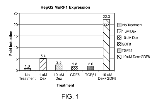
FIG. 1 is a bar graph showing that endogenous MuRF1 expression is
synergistically
upregulated by Dex and GDF8 treatment in HepG2 cells.
FIG. 2 is a graph showing that MuRF1 mRNA in HepG2 cells is induced within 4
hours of treatment with Dex and GDF8.
DEFINITIONS
The terms "determining", "measuring", "evaluating", "assessing" and "assaying"
are
used interchangeably herein to refer to any form of measurement, and include
determining if
an element is present or not. These terms include both quantitative and/or
qualitative
determinations. Assessing may be relative or absolute. "Determining the
presence of'
includes determining the amount of something present, as well as determining
whether it is
present or absent.
The term "contacting" means to bring or put together. As such, a first item is
contacted with a second item when the two items are brought or put together,
e.g., by
touching them to each other or combining them in the same solution. Unless
otherwise
indicated, a cell that is contacted with an agent may be a cell in vivo, i.e.,
within a
multicellular organism, or a cell in vitro, i.e., a cultured cell.
The term "optically detectable protein" refers to a protein whose expression
can be
detected by the presence of an optical signal produced by the protein. An
optical signal is
produced by a protein, for example, when the protein is capable of being
excited by a
particular wavelength of light and emits another wavelength of light which is
detectable. An
optical signal is produced by a protein, for example, when the protein
catalyzes a reaction
which results in a light signal. Fluorescent proteins, luminescent proteins,
etc., are examples
of optically detectable proteins.
The term "gene" refers to a nucleic acid sequence comprised of a promoter
region, a
coding sequence, and a 3'UTR.
The terms "protein" and "polypeptide" are used interchangeably herein.
The term "nucleic acid" encompasses DNA, RNA, single stranded or double
stranded
and chemical modifications thereof. The terms "nucleic acid" and
"polynucleotide" are used
interchangeably herein.
A "non-human" animal refers to any mammal of a species that is not human.
3
CA 02786086 2012-06-29
WO 2011/087946 PCT/US2011/020266
The terms "rodent" and `rodents' refer to all members of the phylogenetic
order
Rodentia including any and all progeny of all future generations derived
therefrom.
The term "murine" refers to any and all members of the family Muridae,
including
rats and mice.
The term "operably-linked" refers to the association of nucleic acid sequences
on a
single nucleic acid fragment so that the function of one is affected by the
other. For example,
a promoter is operably-linked with a coding sequence when it is capable of
affecting the
expression of that coding sequence (i.e., the coding sequence is under the
transcriptional
control of the promoter). Similarly, when an IRES is operably-linked to a
coding sequence,
the IRES provides for translation of the mRNA transcribed from that coding
sequence.
"Unlinked" means that the associated genetic elements are not closely
associated with one
another and the function of one does not affect the other.
The term "luciferase" refers to an enzyme that emits light during the
oxidation of its
substrate luciferin. The terms luciferin and luciferase do not refer to a
particular luciferin or
luciferase. They are generic terms for a substrate and its associated enzyme
(or protein) that
catalyzes a light-producing reaction.
The term "induced" with respect to a promoter, is intended to encompass both
the
initiation of transcription of a downstream nucleic acid, as well as an
increase in the rate of
transcription of a downstream nucleic acid that is already being transcribed,
compared to an
uninduced state.
The term "endogenous" with reference to a gene, indicates that the gene is
native to a
cell, i.e., the gene is present at a particular locus in the genome of a non-
modified cell. An
endogenous gene may be a wild type gene present at that locus in a wild type
cell (as found
in nature). An endogenous gene may be a modified endogenous gene if it is
present at the
same locus in the genome as a wild type gene. An example of such a modified
endogenous
gene is a gene into which a foreign nucleic acid is inserted. An endogenous
gene may be
present in the nuclear genome, mitochondrial genome etc.
The term "construct" refers to a recombinant nucleic acid, generally
recombinant
DNA, that has been generated for the purpose of the expression of a specific
nucleotide
sequence(s), or is to be used in the construction of other recombinant
nucleotide sequences.
A construct might be present in a vector or in a genome.
The term "recombinant" refers to a polynucleotide or polypeptide that does not
naturally occur in a host cell. A recombinant molecule may contain two or more
naturally-
4
CA 02786086 2012-06-29
WO 2011/087946 PCT/US2011/020266
occurring sequences that are linked together in a way that does not occur
naturally. A
recombinant cell contains a recombinant polynucleotide or polypeptide.
The term "expression", as used herein, refers to the process by which a
polypeptide is
produced based on the nucleic acid sequence of a gene. The process includes
both
transcription and translation.
The term "introduced" in the context of inserting a nucleic acid sequence into
a cell,
means "transfection", or `transformation" or "transduction" and includes
reference to the
incorporation of a nucleic acid sequence into a eukaryotic or prokaryotic cell
wherein the
nucleic acid sequence may be incorporated into the genome of the cell (e.g.,
chromosome,
plasmid, plastid, or mitochondrial DNA), converted into an autonomous
replicon, or
transiently expressed (e.g., transfected mRNA).
"Treating" or "treatment" of a condition or disease includes providing a
clinical
benefit to a subject, and includes: (1) preventing at least one symptom of the
conditions, i.e.,
causing a clinical symptom to not significantly develop in a mammal that may
be exposed to
or predisposed to the disease but does not yet experience or display symptoms
of the disease,
(2) inhibiting the disease, i.e., arresting or reducing the development of the
disease or its
symptoms, or (3) relieving the disease, i.e., causing regression of the
disease or its clinical
symptoms.
The term "candidate agents" means oligonucleotides, polynucleotides, siRNA
(which
may be administered as a shRNA), gene products, polypeptides, small molecules,
e.g., up to
2500 Daltons (Da) in size, and pharmacological compounds that are combined
with the cells
or the animals described herein to screen for their effect on muscle atrophy.
In certain cases,
a candidate agent may be delivered as a nucleic acid that is transcribed
and/or translated to
provide the candidate agent, for example, a RNAi molecule or a polypeptide.
The term "coding sequence" refers to a nucleic acid sequence that once
transcribed
and translated produces a protein, for example, in vivo, when placed under the
control of
appropriate regulatory elements. A coding sequence as used herein may have a
continuous
ORF or might have an ORF interrupted by the presence of introns or non-coding
sequences.
In this embodiment, the non-coding sequences are spliced out from the pre-mRNA
to
produce a mature mRNA.
The phrase "muscle cell", as used herein, refers to muscles cells of all
kinds, such as
including skeletal, smooth and cardiac, precursors of these muscle cells, any
intermediate
cell existing during the differentiation of a muscle precursor cell, muscle
fibers, muscle cell
lines, etc. Examples of muscle cells include myoblasts, myotubes, myocytes,
cardiac muscle
5
CA 02786086 2012-06-29
WO 2011/087946 PCT/US2011/020266
cells, skeletal muscle cells, myofibers etc. A muscle cell may be present in
vivo (in an
animal) or in vitro (in a cell culture).
The phrase "atrogen gene" refers to a gene whose expression is induced in
muscle
cells in response to an atrophy-inducing stimulus (e.g., fasting, etc.) prior
to a detectable
muscle atrophy phenotype, i.e., a detectable loss of muscle mass, shriveling
of cells, etc., is
observable. MuRF1 and MAFbx, which encode ubiquitin-protein ligases, are
examples of
atrogen genes, although others exist. The molecular mechanisms that regulate
muscle
atrophy have been extensively reviewed in, e.g., Siu et al, Front. Biosci.
2009 14:432-52;
Murton et al, Biochim. Biophys. Acta 2008 1782:730-43; Tisdale, Curr. Opin.
Support
Palliat. Care 2007 1:287-92; Zhang et al, Med. Hypotheses. 2007 69:310-21; Cao
et al, Int. J.
Biochem. Cell Biol. 2005 37:2088-97; Nader, Int. J. Biochem. Cell Biol. 2005
37:1985-96;
Glass, Int. J. Biochem. Cell Biol. 2005 37:1974-84; Du et al, Int. J. Biochem.
Cell Biol. 2005
37:2147-55; Franch et al, Curr. Opin. Clin. Nutr. Metab. Care. 2005 8:271-5;
Glass, Trends
Mol. Med. 2003 9:344-50; and Glass, Nat. Cell Biol. 2003 5:87-90.
The phrase "atrogen promoter" refers a promoter that is induced in muscle
cells
exposed to an atrophy-inducing stimulus (e.g., fasting, etc) prior to a
detectable muscle
atrophy phenotype i.e., a detectable loss of muscle mass, shriveling of cells,
etc., is
observable. An atrogen promoter may be the promoter of a wild type atrogen
gene, or an
active variant thereof that is, for example, at least 95% identical to a wild
type atrogen
promoter.
The term "atrophy response" refers to any quantitatively or qualitatively
observable
muscle atrophy-related response of a cell. An atrophy response may be
observable at the
molecular level and includes an altered gene expression, e.g., of an
endogenous muscle
related gene or of a reporter protein driven by a muscle-related promoter such
as an atrogen
promoter. An atrophy response may also be observable at the cellular level,
e.g., by
observing an altered cell phenotype such as cell shriveling, cell death or
altered cell staining,
or at the tissue level, e.g., by observing mass of a muscle, fiber size, cross-
sectional area, etc.
A decrease in the mass of the muscle is usually accompanied with a weakening
of the
muscles. An atrophy response may be observed in vitro (in a cultured cell ) or
in vivo (in an
multicellular animal), for example. In an animal, muscle atrophy may be caused
by fasting,
cachexia, diabetes, dexamethasone treatment, myostatin treatment, being on a
ventilator after
surgery, muscular dystrophy, sarcopenic frailty of the elderly and
amylotrophiic lateral
sclerosis, as well as a variety of other muscle-wasting diseases, conditions
and treatments.
6
CA 02786086 2012-06-29
WO 2011/087946 PCT/US2011/020266
The term "glucocorticoid receptor", also known as GR, GCR and NR3C1 (nuclear
receptor subfamily 3, group C, member 1) is the receptor that cortisol and
other
glucocorticoids bind to and activates. The glucocorticoid receptor is
expressed in almost
every cell in the body and regulates genes controlling development,
metabolism, and
immune response. When the GR binds to a glucorticoid, its primary mechanism of
action is
the regulation of gene transcription (Lu et al, Pharmacol. Rev. 2006 58: 782-
97; Rhen et al,
N. Engl. J. Med. 2005 353: 1711-23). The unbound receptor resides in the
cytosol of the
cell. After the receptor is bound to glucocorticoid, the receptor-glucorticoid
complex can
take either of two paths. The activated GR complex up-regulates the expression
of anti-
inflammatory proteins in the nucleus or represses the expression of pro-
inflammatory
proteins in the cytosol (by preventing the translocation of other
transcription factors from the
cytosol into the nucleus). The human GR protein and encoding mRNA are provided
by
Genbank accession nos NP_000167 and NM_000176, respectively. The mouse GR
protein
and encoding mRNA are provided by Genbank accession nos. NP_03219 and
NM_008173,
respectively. In the human genome, the GR gene is located on chromosome 5 at
142.64 -
142.8 M. The glucocorticoid receptor is described in Kumar (Steroids 1999 64:
310-9) and
Kumar (J. Steroid Biochem. Mol. Biol.) 2005 94: 383-94, for example. A ligand
for the
glucocorticoid receptor activates the glucocorticoid receptor. Dexamethasone
is an example
of a ligand for the glucocorticoid receptor although, as will be discussed
below, there are
many others.
"GDF8", also known as myostatin (MSTN) or growth differentiation factor 8, is
a
secreted TGF(3 protein family member that inhibits muscle differentiation and
growth.
Myostatin is produced primarily in skeletal muscle cells, circulates in the
blood and acts on
muscle tissue, by binding a cell-bound receptor called the Activin type II
receptor. The
sequence of GDF8 has been determined for a variety of organisms. The human
GDF8
protein and encoding mRNA are provided by Genbank accession numbers NP_005250
and
NM_005259, respectively. The mouse GDF8 protein and encoding mRNA are provided
by
Genbank accession numbers NP_034964 and NM_010834, respectively. In the human
genome, the GDF8 gene is located on chromosome 2 at 190.63 - 190.64 Mb. In the
mouse
genome, the GDF8 gene is located on chromosome 1 at 53.12 - 53.12 Mb. GDF8 is
described in McPherson (Nature 1997 387: 83-90) and Rodgers (Am. J. Physiol.
Endocrinol.
Metab. 2007 292: E371-2), for example.
The term "myostatin receptor" refers to the receptor through which GDF8 acts.
The
myostatin receptor is thought to be an activin type II receptor, including the
activin IIA
7
CA 02786086 2012-06-29
WO 2011/087946 PCT/US2011/020266
receptor (ActRIIA) and activin IIB receptor (ActRIIB). The myostatin receptor
is reviewed
in Tsuchida et al (Endocr J. 2008 55:11-21), Joulia-Ekaza et al (Curr. Opin.
Pharmacol. 2007
7:310-5), Walsh et al (Biochem. Soc. Trans. 2005 33:1513-7) and Tsuchida et al
(Immune
Endocr. Metabol. Disord. 2004 4:157-66). A ligand for the myostatin receptor
activates that
receptor. GDF8 is an example of a ligand for the myostatin receptor although,
as will be
discussed below, there are many others.
DESCRIPTION OF EXEMPLARY EMBODIMENTS
Before the present subject invention is described further, it is to be
understood that
this invention is not limited to particular embodiments described, as such
may, of course,
vary. It is also to be understood that the terminology used herein is for the
purpose of
describing particular embodiments only, and is not intended to be limiting,
since the scope of
the present invention will be limited only by the appended claims.
Where a range of values is provided, it is understood that each intervening
value, to
the tenth of the unit of the lower limit unless the context clearly dictates
otherwise, between
the upper and lower limit of that range and any other stated or intervening
value in that
stated range is encompassed within the invention.
Unless defined otherwise, all technical and scientific terms used herein have
the same
meaning as commonly understood by one of ordinary skill in the art to which
this invention
belongs. Although any methods and materials similar or equivalent to those
described
herein can be used in the practice or testing of the present invention, the
preferred methods
and materials are now described. All publications mentioned herein are
incorporated herein
by reference to disclose and describe the methods and/or materials in
connection with which
the publications are cited.
It must be noted that as used herein and in the appended claims, the singular
forms
"a", "and", and "the" include plural referents unless the context clearly
dictates otherwise.
Thus, for example, reference to "a cell" includes a plurality of cells and
reference to "a
candidate agent" includes reference to one or more candidate agents and
equivalents thereof
known to those skilled in the art, and so forth. It is further noted that the
claims may be
drafted to exclude any optional element. As such, this statement is intended
to serve as
antecedent basis for use of such exclusive terminology as "solely", "only" and
the like in
connection with the recitation of claim elements, or use of a "negative"
limitation.
The publications discussed herein are provided solely for their disclosure
prior to the
filing date of the present application. Nothing herein is to be construed as
an admission that
8
CA 02786086 2012-06-29
WO 2011/087946 PCT/US2011/020266
the present invention is not entitled to antedate such publication by virtue
of prior invention.
Further, the dates of publication provided may be different from the actual
publication dates
which may need to be independently confirmed.
All publications and patents cited in this specification are herein
incorporated by
reference as if each individual publication or patent were specifically and
individually
indicated to be incorporated by reference and are incorporated herein by
reference to
disclose and describe the methods and/or materials in connection with which
the
publications are cited. The citation of any publication is for its disclosure
prior to the filing
date and should not be construed as an admission that the present invention is
not entitled to
antedate such publication by virtue of prior invention. Further, the dates of
publication
provided may be different from the actual publication dates which may need to
be
independently confirmed.
As will be apparent to those of skill in the art upon reading this disclosure,
each of
the individual embodiments described and illustrated herein has discrete
components and
features which may be readily separated from or combined with the features of
any of the
other several embodiments without departing from the scope or spirit of the
present
invention. Any recited method can be carried out in the order of events
recited or in any
other order which is logically possible.
As noted above, a method that generally comprises contacting a mammalian cell
with
a glucocorticoid receptor ligand and a myostatin receptor ligand. A screening
assay
involving the same, is also provided. In the following description the
glucocorticoid and
myostatin receptor ligands are described first, followed by a description of a
method in
which those ligands may be employed.
Glucocorticoid receptor ligands
The glucocorticoid receptor ligand employed in the subject method may be any
compound that binds to and activates the glucocorticoid receptor. The
mechanism by which
activation of the glucocorticoid receptor initiates downstream response is
known. In general
terms, upon binding of the ligand to the receptor, the receptor-ligand complex
translocates
into the cell nucleus, where it binds to glucocorticoid response elements
(GRE) in the
promoter region of the target genes resulting in the regulation of gene
expression. This
process is reviewed in, for example, Newton (Thorax 2000 55: 603-13).
Activation of the
glucocorticoid receptor inhibits the ability of NF-K B and AP-1 to stimulate
transcription
9
CA 02786086 2012-06-29
WO 2011/087946 PCT/US2011/020266
(see, e.g., Jonat, Cell 1990 62, 1189; Yang-Yen, Cell 1990 62, 1205; Diamond,
Science 1990
249, 1266; and Caldenhoven, Mol. Endocrinol. 1995 9, 401).
A glucocorticoid receptor ligand may be steroidal or non-steroidal. Various
exemplary classes of glucocorticoid receptor ligands are described in the
following
published U.S. patent applications: US20090227548, US20090170898,
US20090137655,
US20090105292, US20090075995, US20090074675, US20080090792, US20070281959,
US20080076795, US20070281928 and US20070149577, which publications are
incorporated by reference for disclosure of the glucocorticoid receptor
ligands described
therein.
In some embodiments, the glucocorticoid receptor ligand may be a
glucocorticoid
such as dexamethasone, betamethasone, cortisone, hydrocortisone,
methylprednisolone,
prednisolone, triamcinolone, fludrocortisone acetate, triamcinolone,
fluocortolone,
clobetasol, diflorasone, mometasone, desoximetasone, including salts, solvates
and hydrates
thereof. In particular embodiments, the glucocorticoid receptor ligand may
have a potency of
at least 10 times the potency of hydrocortisone, as reviewed by Begg (Med J.
Aust. 1987
146:37-41).
Glucocorticoids and their mechanism of action are reviewed in the following
books:
Glucocorticoid Hormone: Mechanisms of Action by Y. Sakamoto (Editor)
Publisher:
Springer-Verlag (June 1986); Glucocorticoid Action: Basic and Clinical
Implications
(Hardcover) by Tomoshige Kino (Editor), Publisher: New York Academy of
Sciences;
second edition (Aug 30 2004); Glucocorticoids by Goulding (Author) Publisher:
Springer/Sci-Tech/Trade; 1 edition (May 11 2001); and Recent Advances in
Glucocorticoid
Receptor Action by A. Cato (Editor), Publisher: Springer; 1 edition (Nov 11
2002), which
are incorporated by reference in their entireties.
Dosages and routes of administration for glucocorticoid receptor ligands are
known.
Myostatin receptor ligands
The myostatin receptor ligand employed in the subject method may be any
compound
that binds to and activates the myostatin receptor. Such compounds include
peptide and non-
peptidic compounds, including GDF8 peptides defined by the following NCBI
accession
numbers: GI:9506907 (Rattus norvegicus), GI:6754752 (Mus musculus),
GI.=4885259 (Homo
sapiens), GI.=48314966 (Bos Taurus), GI.=260809331 (Branchiostoma floridae),
GI.=51783959
(Sus scrofa), GI.=47825371 (Gallus gallus), GI:18858751 (Danio rerio),
GI:121583758
(Macaca mulatta), GI.=50950173 (Canis lupusfamiliaris), GI:120952608 (Pan
troglodytes),
CA 02786086 2012-06-29
WO 2011/087946 PCT/US2011/020266
GI:198417205 (Ciona intestinalis) and GI:57164247 (Ovis aries), including
active variants
and peptidomimetic variants thereof. The structure of human GDF8 is set forth
as MMDB
ID: 75808 in NCBI's structure database. In certain cases, a myostatin receptor
ligand used
herein may have an amino acid sequence that is at least 50% identical to,
e.g., at least 60%
identical, at least 70% identical, at least 80% identical, at least 85%
identical, at least 90%
identical, at least 95% identical, or at least 98% identical, to a wild type
myostatin receptor
ligand.
Assays for identifying myostatin receptor ligands are known and include those
described in published U.S. patent applications US20090220491, US20090098114,
US20070149458, US20060216279, US20050272028 and US20040248121, which
publications are incorporated by reference for disclosure of those assays.
The role of GDF8 in muscle degeneration by activating the myostatin receptor
has
been reviewed in a variety of publications, including Tsuchida (Expert Opin.
Biol. Ther.
2006 6:147-54), Wagner (Curr. Opin. Rheumatol. 2005 17:720-4), Tsuchida (Curr.
Drug
Targets Immune Endocr. Metabol. Disord. 2004 4:157-66), and Bellinge (Anim.
Genet. 2005
36:1-6), which publications are incorporated by reference herein.
Dosages and routes of administration for myostatin receptor ligands are known
Methods
The above-described glucocorticoid receptor ligand and myostatin receptor
ligand
may be contacted with a mammalian cell in vivo (i.e., by administering the
compounds to an
animal) or in vitro (i.e., by contacting the compounds with cells grown in
culture). The
compounds may be contacted with the cell simultaneously (e.g., the compounds
may be
mixed together prior to contacting the compounds with the cell, or the
compounds may be
separately combined with the cell at the same time) or at different times.
Exemplary in vitro
and in vivo methods are described below.
In in vitro methods, the glucocorticoid receptor ligand and myostatin receptor
ligand
may each be independently contacted with a cultured mammalian cell at a
concentration that
is consistent with the use of the same compounds individually at a
concentration sufficient to
effect a response from an isolated cell, as is known in the art. Exemplary
effective
concentrations and are described in the references cited above as well as many
others, and
are generally in the range of about 0.1 to 1000 g/mL, although concentrations
outside of
this range may be employed in certain circumstances. In general terms, the
contacting is
done by mixing the compounds with culture medium.
11
CA 02786086 2012-06-29
WO 2011/087946 PCT/US2011/020266
The cultured cell employed in the assay may be any cell that expresses a
glucocorticoid receptor and myostatin receptor. If a cell does not express
both receptors
endogenously, then the receptors may be expressed using recombinant means.
Cultured cells
from any animal, e.g., cultured mammalian cells, may be employed, including
but not
limited to: monkey kidney cells (COS cells), monkey kidney CVI cells
transformed by SV40
(COS-7, ATCC CRL 165 1); human embryonic kidney cells (HEK-293, Graham et al.
J.
Gen Virol. 36:59 (1977)); baby hamster kidney cells (BHK, ATCC CCL 10);
chinese
hamster ovary-cells (CHO, Urlaub and Chasin, Proc. Natl. Acad. Sci. (USA)
77:4216,
(1980); mouse sertoli cells (TM4, Mather, Biol. Reprod. 23:243-251 (1980));
monkey
kidney cells (CVI ATCC CCL 70); african green monkey kidney cells (VERO-76,
ATCC
CRL-1587); human cervical carcinoma cells (HELA, ATCC CCL 2); canine kidney
cells
(MDCK, ATCC CCL 34); buffalo rat liver cells (BRL 3A, ATCC CRL 1442); human
lung
cells (W138, ATCC CCL 75); human liver cells (hep G2, HB 8065); mouse mammary
tumor
(MMT 060562, ATCC CCL 51); TRI cells (Mather et al., Annals N. Y. Acad. Sci
383:44-68
(1982)); NIH/3T3 cells (ATCC CRL-1658); and mouse L cells (ATCC CCL-1).
Additional
cell lines will become apparent to those of ordinary skill in the art. A wide
variety of cell
lines are available from the American Type Culture Collection, 10801
University Boulevard,
Manassas, Va. 20110-2209. In particular embodiments, the cultured cell may be
a cultured
myocyte, e.g., a cultured cell of skeletal muscle, smooth muscle, or cardiac
muscle origin. In
exemplary embodiments, the cultured cell may be an HL-1 cell (Claycomb PNAS
1998 95:
2979-2984, a BWEM or CLEM cell (Enelmann et al Molecular and Cellular
Biochemistry
1996 157), an L6 myoblasts, or a C2C12, SM3, Aza2, BC3H-1, BD1, BD2, BD10,
TD33,
TD38, TD45, TG1, C2, or AT-1 cell, for example. Methods for culturing such
cells are
known.
Contacting a cultured cell with the glucocorticoid receptor ligand and
myostatin
receptor ligand activates the glucocorticoid receptor and the myostatin
receptor of the cell,
thereby altering a phenotype of the cell. In certain embodiments, the method
comprises
maintaining the cell in the presence of the compounds for a time sufficient
for the cell to
exhibit a phenotype that is not produced in the absence of the compounds. In
certain cases,
the phenotype may be a cell proliferation phenotype, a cell death (apoptosis)
phenotype, a
change to the cells shape or size, an inflammatory response (observed as an
altered
production of an inflammatory mediator, for example), an altered staining
pattern, or altered
gene expression. In particular embodiments, the phenotype may be an atrophy
response, as
defined above.
12
CA 02786086 2012-06-29
WO 2011/087946 PCT/US2011/020266
In certain cases, the cells may contain a reporter system for evaluating gene
expression in the cell. For example, the cell may contain a coding sequence
for a reporter
protein (e.g., luciferase or GFP), operably linked to a promoter (e.g., a
promoter that is
induced or repressed during muscle cell development or muscle wasting), where
contacting
the cell with the compounds induces or represses expression of the reporter
protein. In
certain embodiments, the promoter may be an atrogen promoter, and contacting
the cell with
a glucocorticoid receptor ligand and myostatin receptor ligand induces
production of the
reporter protein. In particular embodiments, the genome of the cell may be may
be altered,
e.g., by inserting a coding sequence for a reporter protein into an endogenous
gene (e.g., an
atrogen gene) such that the expression of the reporter is operably linked to
the endogenous
promoter, or by inserting a recombinant nucleic acid containing both a
promoter and
reporter-encoding sequence into the genome of the cell.
In in vivo methods, the glucocorticoid receptor ligand and myostatin receptor
ligand
may be contacted with a mammalian cell by administering the compounds at
independent
concentrations that are consistent with the use of the same compounds
individually at a
concentration sufficient to effect a response from the animal, as is known in
the art.
Exemplary effective concentrations and are described in the references cited
above as well as
many others, and are generally independently in the range of about 0.01 to 500
milligrams of
the compounds per kilogram of animal per dose, e.g., from at least about 0.1
to 100
milligrams agent/kilogram, although concentrations outside of this range may
be employed
in certain circumstances. In general terms, the contacting is done by
administering the
compounds to the animal, e.g., orally or by injection (which may be
intravenous or
intramuscular), locally or systemically. The animal employed in the assay may
be any
animal, particularly a mammal such as a rodent (e.g., a mouse or rat).
Administering the glucocorticoid receptor ligand and myostatin receptor ligand
to the
animal activates the glucocorticoid receptor and the myostatin receptor in
cells of the animal,
thereby altering a phenotype of the animal. In certain embodiments, after the
compounds
have been administered, the method may comprises maintaining the animal for a
time
sufficient for the animal to exhibit a phenotype that is not produced in the
absence of the
compounds. In certain cases, the phenotype may be a cancer-related phenotype
(a cell
proliferation, cell death, or metastasis-related phenotype) or an inflammatory
response-
mediated phenotype (e.g., a change in the response of the immune system to a
challenge). In
particular embodiments, the phenotype may be an atrophy response, as defined
above, where
in certain embodiments may be observed as an change in muscle mass, a change
in muscle
13
CA 02786086 2012-06-29
WO 2011/087946 PCT/US2011/020266
cross-section, or a down regulation of myosin synthesis, an activation of a
myosin
breakdown pathway (e.g., via activation of the ATP-dependent,
ubiquitin/proteasome
pathway or induction of an E3 ubiquitin ligase).
In certain cases, the cells may contain a recombinant reporter system for
evaluating
gene expression in the animal. For example, the animal may contain a coding
sequence for a
reporter protein (e.g., luciferase or GFP), operably linked to a promoter
(e.g., a promoter that
is induced or repressed during muscle cell development or muscle wasting),
where
contacting the cell with the compounds induces or represses expression of the
reporter
protein. In certain embodiments, the promoter may be an atrogen promoter, and
administering a glucocorticoid receptor ligand and myostatin receptor ligand
to the animal
induces production of the reporter protein. In particular embodiments, the
genome of the
animal may be may be altered, e.g., by inserting a coding sequence for a
reporter protein into
an endogenous gene (e.g., an atrogen gene) such that the expression of the
reporter is
operably linked to the endogenous promoter, or by inserting a recombinant
nucleic acid
containing both a promoter and reporter-encoding sequence into the genome of
the cell.
Screening assays
The above-described method may be employed in a screening assay to identify an
agent that modulates the phenotype induced by the glucocorticoid receptor
ligand and
myostatin receptor ligand. In particular embodiments, the method may be
employed to
identify an agent that modulates the initiation of muscle cell atrophy. In
exemplary
embodiments, the method involves contacting a subject cell (i.e., a cell
contacted with the
glucocorticoid receptor ligand and myostatin receptor ligand, which cell can
be present in
vitro or in vivo) with a candidate agent, and determining the effect, if any,
of the candidate
agent on the phenotype induced by the the glucocorticoid receptor ligand and
myostatin
receptor ligand. In a particular embodiment, the phenotype may be assessed by
evaluating
the production of a reporter protein. In some embodiments, the method involves
contacting a
cell (in vivo or in vitro) with a candidate agent in the presence of a
glucocorticoid receptor
ligand an a myostatin receptor ligand; and determining if the candidate agent
alters the
phenotype of the cell, where the phenotype is produced in response to the
glucocorticoid
receptor ligand and myostatin receptor ligand.
The term "agent" as used herein describes any molecule, e.g. protein or non-
protein
organic or inorganic pharmaceutical. Agents of particular interest are those
that inhibit
initiation of muscle cell atrophy. A plurality of assays is run in parallel
with different agent
14
CA 02786086 2012-06-29
WO 2011/087946 PCT/US2011/020266
concentrations to obtain a differential response to the various
concentrations. One of these
concentrations may serve as a negative control, i.e. at zero concentration or
below the level
of detection.
The terms "candidate agent", "test agent", "agent", "substance" and "compound"
are
used interchangeably herein. Candidate agents encompass numerous chemical
classes,
typically synthetic, semi-synthetic, or naturally-occurring inorganic or
organic molecules.
Candidate agents include those found in large libraries of synthetic or
natural compounds.
For example, synthetic compound libraries are commercially available from
Maybridge
Chemical Co. (Trevillet, Cornwall, UK), ComGenex (South San Francisco, CA),
and
MicroSource (New Milford, CT). Alternatively, libraries of natural compounds
in the form
of bacterial, fungal, plant and animal extracts are available from Pan Labs
(Bothell, WA) or
are readily producible.
Candidate agents may be small organic or inorganic compounds having a
molecular
weight of more than 50 and less than about 2,500 Da. Candidate agents may
comprise
functional groups necessary for structural interaction with proteins,
particularly hydrogen
bonding, and may include at least an amine, carbonyl, hydroxyl or carboxyl
group, and may
contain at least two of the functional chemical groups. The candidate agents
may comprise
cyclical carbon or heterocyclic structures and/or aromatic or polyaromatic
structures
substituted with one or more of the above functional groups. Candidate agents
are also
found among biomolecules including peptides, saccharides, fatty acids,
steroids, purines,
pyrimidines, derivatives, structural analogs or combinations thereof.
Candidate agents are obtained from a wide variety of sources including
libraries of
synthetic or natural compounds. For example, numerous means are available for
random
and directed synthesis of a wide variety of organic compounds and
biomolecules, including
expression of randomized oligopeptides. Alternatively, libraries of natural
compounds in the
form of bacterial, fungal, plant and animal extracts are available or readily
produced.
Additionally, natural or synthetically produced libraries and compounds are
readily modified
through conventional chemical, physical and biochemical means, and may be used
to
produce combinatorial libraries. Known pharmacological agents may be subjected
to
directed or random chemical modifications, such as acylation, alkylation,
esterification,
amidification, etc. to produce structural analogs. New potential therapeutic
agents may also
be created using methods such as rational drug design or computer modeling.
CA 02786086 2012-06-29
WO 2011/087946 PCT/US2011/020266
Screening may be directed to known pharmacologically active compounds and
chemical analogs thereof, or to new agents with unknown properties such as
those created
through rational drug design.
Agents that modulate a phenotype may decrease the phenotype by at least 10%,
at
least 20%, at least 30%, at least 40%, at least 50%, at least 60%, at least
70%, at least 80%,
or at least 90%, or more, relative to a control that has not been exposed to
the agent.
Agents that modulate the phenotype may be subjected to directed or random
and/or
directed chemical modifications, such as acylation, alkylation,
esterification, amidification,
etc. to produce structural analogs. Such structural analogs include those that
increase
bioavailability, and/or reduced cytotoxicity. Those skilled in the art can
readily envision and
generate a wide variety of structural analogs, and test them for desired
properties such as
increased bioavailability and/or reduced cytotoxicity, etc.
In a particular embodiment, an in vitro method for identifying agents that
modulate
initiation of muscle cell atrophy is provided. This method generally involves
contacting a
cultured cell that produces a reporter protein upon initiation of muscle cell
atrophy with a
candidate agent in the presence of a glucocorticoid receptor ligand an a
myostatin receptor
ligand; and determining if the candidate agent decreaes the production of the
reporter protein
by the cell as compared to a control cell not treated with the candidate
agent.
The cell may be contacted with the glucocorticoid receptor ligand an a
myostatin
receptor ligand prior to, after or simultaneous with contacting the cell with
a candidate agent.
The production of reporter protein(s) may be monitored at different points
before and
after subjecting the cells to conditions that induce the phenotype. Similarly,
the effect of a
candidate agent may be determined by measuring the phenotype at several time
points. For
example, the production of reporter protein(s) may be measured 5 mins, 30
mins, 1 hr, 2 hrs,
4 hrs, 8 hrs, 12 hrs, 24 hrs, 36 hrs, 48 hrs, 72 hrs, 120 hrs, 1 week, 2 week,
and up to 1 month,
after contacting the cell with a candidate agent.
An in vivo screening assay for identifying agents that modulate initiation of
muscle
cell atrophy is provided. This method generally involves administering to an
animal that
produces a reporter protein upon initiation of muscle cell atrophy: a
candidate agent, a
glucocorticoid receptor ligand and a myostatin receptor ligand; and
determining if the
candidate agent decreaes the production of the reporter protein by the animal
as compared to
a control animal to which the candidate agent has not been administered.
Any phenotype produced in the in vivo system be monitored at different points
before
and after administering the candidate agent to the animal. For example, the
effect of a
16
CA 02786086 2012-06-29
WO 2011/087946 PCT/US2011/020266
candidate agent may be determined by measuring the reporter proteins at
several time points.
For example, the production of reporter protein(s) may be measured at time
Ohrs, 12hrs, 24
hrs, 36 hrs, 48 hrs, 72 hrs, 120 hrs, 1 week, 2 week, 1 month, 2 months, 3
months, 5 months,
etc., after contacting the cell with a candidate agent. In certain
embodiments, the
measurement of the reporter proteins may be complimented by measuring the
expression of
the atrogen gene(s), as well as measuring cell/fiber size, morphology, muscle
strength, etc.
Any agent identified by above-described method may be further tested in an
animal
model. For example, in one embodiment, an animal may be subjected to an
atrophy inducing
stimuli and contacted with a candidate agent. A number of conditions known to
induce
atrophy may be used as an atrophy. In an animal, muscle cell atrophy may be
initiated by a
number of stimuli including but not limited to fasting, ageing, diabetes,
advanced cancer,
renal failure, sepsis, cachexia, arthritis, osteoporosis, diabetes,
denervation, immobilization,
muscle unloading, spinal cord injury, glucocorticoid treatment, and the like.
In vitro, muscle
cell atrophy may be initiated by starving, exposure of cells to for example,
glucocorticoids,
or to viruses.
Also provided is a composition comprising a mammalian cell, a glucocorticoid
receptor ligand and a myostatin receptor ligand. The cell and ligands that may
be present in
the composition are discussed in greater detail above.
Utility
The in vivo and in vitro assays presented herein provide for methods to
identify and
test agents that modulate a variety of phenotypes, including those that
decrease muscle cell
atrophy. These agents may be used in formulations that may be used to treat
subjects with
muscle cell atrophy. In addition, these agents may be given prophylactically
to subjects at
risk for developing muscle cell atrophy, e.g., prior to a surgery in which the
patient will be
put on a ventilator. A subject that may benefit from an agent identified by
the methods
provided herein may have or be at risk for developing muscle cell atrophy
caused by a
variety of stimuli. These stimuli include but are not limited to fasting,
ageing, advanced
cancer, renal failure, sepsis, cachexia, arthritis, osteoporosis, and
diabetes. Atrophy of
muscles may also be a result of their disuse or denervation, e.g.,
immobilization, muscle
unloading, spinal cord injury, etc. In certain embodiments, the subject may
have a health
problem that is exacerbated by muscle cell atrophy, such as, HIV, chronic
heart failure,
chronic kidney disease, liver cirrhosis, burn injuries, osteoporosis,
arthritis, etc. The methods
of using cells and animal models to screen for candidate compounds that
inhibit muscle cell
17
CA 02786086 2012-06-29
WO 2011/087946 PCT/US2011/020266
atrophy may be used identify agents that improve protein content, fiber
diameter, force
production, and fatigue resistance of muscles in subjects with muscle cell
atrophy.
EXAMPLES
The following examples are provided in order to demonstrate and further
illustrate
certain embodiments and aspects of the present invention and are not to be
construed as
limiting the scope thereof.
Example 1
Cultured HepG2 cells were exposed to 1 M dexamethasone, 10 M dexamethasone,
100 ng/ml GDF8, 100 ng/ml TGF(31, and 10 M dexamethasone + 100 ng/ml GDF8,
and
expression of endogenous MuRF1 was evaluated by RT-PCR using Taqman RT-PCR
assay.
Results are shown in Fig. 1. Endogenous MuRF1 was induced 22x over the no
treatment
control. 10 M dexamethasone induced expression of endogenous MuRF1 2.5x over
the no
treatment control, and 100 ng/ml GDF8 induced expression of endogenous MuRF1
1.8x
over the no treatment control. Since treatment with a combination of both
dexamethasone
and GDF8 caused an induction of MuRF1 that is well above the induction of
MuRF1 by
dexamethasone and GDF8 individually, endogenous MuRF1 expression is
synergistically
upregulated by dexamethasone and GDF8.
Fig. 2 shows a time course of MuRF1 induction after a HepG2 cell is treated
with
both 10 M dexamethasone and 100 ng/ml GDF8. MuRF1 induction is observable at
four
hours of treatment, as compared to controls.
18