Note: Descriptions are shown in the official language in which they were submitted.
CA 02816489 2013-05-15
WO 2004/085630 PCT/AU2004/000416
PERIVASCULAR MESENCHYMAL PRECURSOR CELLS
FIELD OF THE INVENTION
This invention relates to mesenchymal precursor cells, and the isolation of a
subpopulation
BACKGROUND OF THE INVENTION
Numerous attempts at isolating and enriching mesenchymal precursor cells have
been
attempted because of the potential that these cells have for medicinal use.
Pittinger et al.,
mesenchymal precursor cells from a wide range of tissues.
SUMMARY OF THE INVENTION
The present invention arises from the finding that a population of
multipotential mesenchmal
SUBSTITUTE SHEET (RULE 26) RO/AU
CA 02816489 2013-05-15
WO 2004/085630 PCT/AU2004/00041C
2
In a first form of a first aspect the invention might be said to reside in a
method of enriching
for mesenchymal precursor cells (MPCs), the method including the step of
preparing a single
cell suspension from a vascularised source tissue and the step of enriching
based on the
presence of an early perivascular cell marker.
In a second form of the first aspect the invention might be said to reside in
a method of
enriching for mesenchymal precursor cells, the method including the step of
preparing a
single cell suspension from a, non-bone marrow, vascularised source tissue and
separating
the tissue into separate cells and the step of enriching based one of the
presence or level of
one or more early developmental markers and the absence of one or more surface
markers
indicative of commitment.
In a third form of the first aspect the invention might be said to reside in a
method of
enriching for mesenchymal precursor cells (MPCs), the method including the
step of
preparing a single cell suspension from a vascularised source tissue and the
step of enriching
based on the presence of markers expressed in the vascularized tissue by pen-
vascular cells.
In a second aspect the invention might be said to reside in an enriched
population of cells
enriched for mesenchymal precursor cells (MPCs) said MPCs having a phenotype
of 3G5,
MUC18, VCAM-1, STRO-1 and a smooth muscle actin.
In a first form of a third aspect the invention might be said to reside in an
isolated
mesenchymal precursor cells (MPCs) said MPCs having a phenotype of 305, MUC18,
VCA1VI-1, STRO-1' and a smooth muscle actin.
In a second form of the third aspect the invention might be said to reside in
an isolated
mammalian cell that is multipotent and that is positive for the surface marker
305.
SUBSTITUTE SHEET (RULE 26) ROIAU
CA 02816489 2013-05-15
WO 2004/085630 PCT/AU2004/000416
3
In a third form of the third aspect the invention might be said to reside in a
mesenchymal
precursor cell (MPC), capable of forming a clonogenic colony and
differentiating to three or
more mesenchymal tissue types, isolated from a tissue of the group comprising,
but not
limited to, adipose tissue, teeth, dental pulp, skin, liver, kidney, heart,
retina, brain, hair
follicles, intestine, lung, spleen, lymph node, thymus, pancreas, bone,
ligament, bone
marrow, tendon, and skeletal muscle, and which is positive for the surface
marker STRO-1.
In a fourth form of the third aspect the invention might be said to reside in
an unexpanded
population of cells enriched for mesenchymal precursor cells (MPCs), capable
of forming a
clonogenic colony and differentiating to three or more mesenchymal tissue
types, said MIICs
co-expressing the surface markers MUC18/CD146 and alpha-smooth muscle actin.
In a fourth aspect the invention might be said to reside in a differentated
progeny cell arising
from the third aspect of the invention preferably wherein the progeny cell is
at least an
osteoblast, odontoblast, dentin-producing, chondrocyte, tendon, ligament,
cartilage,
adipocyte, fibroblast, marrow stroma, osteoclast- and hematopoietic-supportive
stroma,
cardiac muscle, smooth muscle, skeletal muscle, pericyte, vascular,
epithelial, glial,
neuronal, astrocyte or oligodendrocyte cell.
BRIEF DESCRIPTION OF THE DRAWINGS
Figure 1. Properties of STRO-1+ MACS-isolated cells co-labeled with anti-
CD146
(CC9). (A) Sort region, R1, represents the double positive STRO-1'/CD146+
population. (B) The incidence of clonogenic cell colonies (>50 cells) based
on STRO-18"/CD146+ expression was determined by limiting dilution
analysis of 24 replicates per cell concentration using Poisson distribution
analysis from 5 independent experiments. Forward (size) and perpendicular
(granularity) light scatter characteristics of BMMNCs (C), ST1.O-1'/CD146-
cells (D) and STRO-1BRT/CD146+ cells (E). (F) RT-PCR analysis of STRO-
18RT/CD146+ sorted marrow cells for CBFAI (lane 2), osteocalcin (lane 4) and
SUBSTITUTE SHEET (RULE 26) RO/AU
CA 02816489 2013-05-15
WO 2004/085630 PCT/AU2004/00041k
4
GAPDH (lane 6) transcripts. Control cells (BMSSC cultures grown in the
presence of dexamethasone) expressing CBFA1 (lane 1), osteocalcin (lane3),
and GAPDH (lane 5) is also shown. Reaction mixes were subjected to
electrophoresis on a 1.5% agarose gel and visualised by ethidium bromide
staining. (G) In situ expression of CD146 on blood vessel (by) walls (arrow)
in human bone marrow (bm) sections near the bone (b) surface 20X. Sections
were counter stained with Hematoxylin. (H) Dual Immunofluorescence
staining demonstrating reactivity of the STRO-1 antibody labeled with Texas
red and the CC9 antibody labeled with fluorescein isothiocyanate, reacting to
blood vessel walls in frozen sections of human bone marrow.
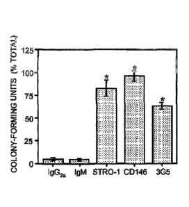
Figure 2. Immunophenotypic analysis of DPSCs in vivo. The bar graph
depicts the
number of clonogenic colonies retrieved from single cell suspensions of dental
pulp following imnumomagnetic bead selection based on reactivity to
antibodies that recognize STRO-1, CD146, and 3G5 and isotype-matched
negative control antibodies. The data are expressed as the number of colony-
forming units obtained in the bead positive cell fractions as a percentage of
the total number of colonies in unfractionated pulp cells averaged from three
separate experiments. Statistical significance (*) was determined using the
student t-test (p 0.01) comparing the percent total number of colonies for
each
antibody with the corresponding isotype-matched control.
Figure 3. Reactivity of perivascular makers in dental pulp. (A)
Immunolocalization of
the STRO-1 antigen on blood vessels (small arrows) in human dental pulp (p)
and around perineurium (large arrow) surrounding a nerve bundle (nb) 20X.
(B) Dual Immunofluorescence staining demonstrating reactivity of the STRO-
1 antibody labeled with Texas Red to dental pulp perineurium (arrow) in
combination with an anti-neurofilament antibody labeled with fluorescein
isothiosyanate staining the inner nerve bundle (nb), 40X. (C)
Immunolocalization of the CD146 antigen to blood vessel walls in human
SUBSTITUTE SHEET (RULE 26) ROIAU
CA 02816489 2013-05-15
WO 2004/085630
PCT/A1.J2004/000416
dental pulp tissue 20X. (D) Dual Immunofluorescence staining demonstrating
reactivity of the STRO-1 antibody labeled with Texas red to a blood vessel
and the CC9 antibody labeled with fluorescein isothiosyanate. (E)
Immunohistochemical staining of pulp tissue with a rabbit polyclonal anti-
5 DSP antibody (arrow) to the odontoblast outer layer (od). 20X. (F)
3G5
reactivity to a single pericyte (arrow) in a blood vessel (by) wall 40X.
Tissue
sections were counter stained with Hematoxylin.
Figure 4. 3G5 reactivity to BMSSCs. (A) The representative histogram
depicts a typical
dual-color FACS analysis profile of whole bone marrow mononuclear cells
(BMMNCs) expressing CD146 (PE) and 3G5 (FITC). (B) Colony efficiency
assays were performed for all the different expression patterns observed
(regions "R" 1-6). The data are expressed as the mean incidence of colony-
forming units for each cell fraction averaged from three separate experiments.
Figure 5. Developmental potential of purified BMSSCs and DPSCs in vivo.
Cytospin
preparations of MACS/FACS isolated STRO-1'/CD146+ marrow cells
(arrow) stained with an antibody specific to a -smooth muscle actin (A) and
von Willebrand Factor (B). CD146 pulp cells (large arrow) isolated by
immunomagnetic bead selection (magnetic beads depicted by small arrows),
stained with an. antibody specific to a -smooth muscle actin (C) and von
Willebrand Factor. (D). (E) Ectopic bone formation (b) and
haematopoietic/adipogenic marrow (bm) by ex vivo expanded cells derived
from STRO-1BRT/CD1464- BMSSCs transplanted with HA/TCP into
immunocompromised mice for three months (E). (F) Ectopic formation of
dentin (d) and fibrous pulp tissue (p) by ex vivo expanded cells derived from
CD146+ DPSCs transplanted with HA/TCP into inununocompromised mice
for three months. Sections were stained with Hematoxylin & Eosin.
SUBSTITUTE SHEET (RULE 26) ROIAU
CA 02816489 2013-05-15
WO 2004/085630 PCT/AU2004/00041
6
Figure 6 Expression of CD34, CD45 and Glycophorin-A on STRO-1 positive
bone
marrow mononuclear cells. Representative histograms depicting typical dual-
colour flow cytometric analysis profiles of STRO-1 positive bone marrow
mononuclear cells isolated initially by magnetic activated sorting and co-
stained with antibodies directed against CD34 (A), CD45 (B) or Glycophorin-
A (C). The STRO-1 antibody was identified using a goat anti-murine IgM-
fluorescein isothiocyanate while CD34, CD45 and Glycophorin-A were
identified using a goat anti-murine IgG- phycoerythrin. The high expressing
STRO-1 fraction which contained the clonogenic MPC population was
isolated by fluorescence activated cell sorting based on regions R1 and R2.
Figure 7 Bone marrow MPC are STRO-1 bright, CD34 negative, CD45
negative and
Glycophorin-A negative. The graph depicts the results of in vitro adherent
colony formation assays performed for each of the different sorted STRO-1
bright populations selected by their co-expression or lack of either the CD34,
.CD45 or Gycophorin-A antigens, based on regions R1 and R2 as indicated in
Figure 6. These data are expressed as the mean incidence of colony-forming
units for each cell fraction averaged from two separate experiments.
Figure 8 Reactivity of perivascular makers in different human tissues. Dud-
colour
immunofluorescence staining demonstrating reactivity of (A) STRO-1 and
CD146, (B) STRO-1 and alpha-smooth muscle actin, and (C) 3G5 and
CD146, on blood vessels and connective tissue present on spleen, pancreas
(Panel 1), brain, kidney (Panel 2), liver, heart (Panel 3) and skin (Panel 4)
20X. The STRO-1 and 3G5 antibodies were identified using a goat anti-
murine IgM-Texas Red while CD146 and alpha-smooth muscle actin were
identified using a goat anti-murine or IgG-fluorescein isothiocyanate. Co-
localization is indicated by overlaping areas of yellow and orange
fluorescence (white arrows).
SUBSTITUTE SHEET (RULE 26) RO/AU
CA 02816489 2013-05-15
WO 2004/085630 PCT/A1J2004/000416
7
Figure 9 Isolation of adipose-derived MPC by FACS. Representative flow
cytometirc
histograms depicting the expression of STRO-1, CD146 and 305 in fresh
preparations of peripheral adipose-derived single-cell suspensions generated
following collagenase/dispase digestion as previously described (Shi and
Gronthos 2003). The antibodies were identified using either a goat anti-
murine IgM or IgG-phycoerythrin. Cell populations were then selected by
FACS, based on their positivity (region R3) or negativity (region R2) to each
marker and then plated into regular growth medium to assess the incidence of
adherent colony-forming cells in each cell fraction.
Figure 10 Clonogenic adipose-derived MPC are positive for S'TRO-
1/305/CD146. The
bar graph depicts the number of clonogenic colonies retrieved from single cell
suspensions of enzymatically digested human peripheral adipose tissue,
following fluorescence activated cell sorting, based on their reactivity to
antibodies that recognize STRO-1, CD146, and 305 (Figure 9), then cultured
in standard growth medium as previously described for bone marrow and
dental pulp tissue (Shi and Gronthos 2003). The data are expressed as the
number of colony-forming units obtained per 105 cells plated in the positive
and negative cell fractions averaged from two separate experiments.
Figure 11 Immunophenotypic analysis of adipose-derived MPC.
Representative flow
cytometric histograms depicting the co-expression of STRO-1 and CD146 (A)
and 305 and CD146 in fresh preparations of peripheral adipose-derived
single-cell suspensions generated following collagenase/dispase digestion.
The STRO-1 and 305 antibodies were identified using a goat anti-murine
IgM-phycoerythrin while CD146 was identified using a goat anti-murine IgG-
fluorescein isothiocyanate. Approximately 60% and 50% of the CD146
positive cells co-express STRO-1 and 305, respectively. These data suggest
that 10% or more of the CD164 positive cells co-express S1'RO-1 and 305.
SUBSTITUTE SHEET (RULE 26) RO/AU
CA 02816489 2013-05-15
WO 2004/085630 PCT/AU2004/00041c
8
Figure 12 Developmental potential of purified Adipocyte-derived MPC in
vitro.
Preparations of primary MPC cultures derived from STRO-1/CD146 adipose
cells were re-cultured either in standard culture conditions (A), osteogenic
inductive medium (B), Adipogenic inductive medium (C) or condrogenic
conditions (D) as previously described Gronthos et al. 2003. Following two
weeks of multi-differentiation induction, the adipocyte-derived MPC
demonstrated the capacity to form bone (B; Alizarin positive mineral
deposits), fat (C, Oil Red 0 positive lipid) and cartilage (I): collagen type
II
matrix).
=
Figure 13 Isolation of skin-derived MPC by FACS. Representative flow
cytometirc
histograms depicting the expression of STRO-1, CD146 and 305 in fresh
preparations of full thickness skin-derived single-cell suspensions generated
following collagenase/dispase digestion. The antibodies were identified using
either a goat anti-murine IgM or IgG-phycoerythrin. Cell populations were
then selected by FACS, based on their positivity (region R3) or negativity
(region R2) to each marker and then plated into regular growth medium to
assess the incidence of adherent colony-forming cells in each cell fraction.
Figure 14 Clonogenic skin-derived MPC are positive for STRO-1/3G5/CD146.
The bar
graph depicts the number of adherent colonies recovered from single cell
suspensions of enzymatically digested human skin, following fluorescence
activated cell sorting, based on their reactivity to antibodies that recognize
STRO-1, CD146, and 305 (Figure 6), then cultured in standard growth
medium as previously described for bone marrow and dental pulp tissue (Shi
and (Ironthos 2003). The data are expressed as the number of colony-forming
units obtained per 105 cells plated in the positive and negative cell
fractions
averaged from two separate experiments.
SUBSTITUTE SHEET (RULE 26) ROIAU
CA 02816489 2013-05-15
WO 2004/085630 PCT/A1J2004/000416
9
Figure 15 A. Immunophenotypic expression pattern of ex vivo expanded bone
marrow
MPC. Single cell suspensions of ex vivo expanded bone marrow MPC were
prepared by trypsin/EDTA treatment then incubated with antibodies
identifying cell lineage-associated markers. For those antibodies identifying
intracellular antigens, cell preparations were fixed with cold 70% ethanol to
permeanbiliz,e the cellular membrane prior to staining for intracellular
markers. Isotype matched control antibodies were treated under identical
conditions. Flow cytometric analysis was performed using a COULTER
EPICS instrument. The dot plots represent 5,000 listmode events indicating
the level of fluorescence intensity for each lineage cell marker (bold line)
with
reference to the isotype matched negative control antibodies (thin line).
B. Gene expression profile of cultured MPC. Single cell
suspensions of
ex vivo expanded bone marrow MPC were prepared by trypsin/EDTA
treatment and total cellular RNA was prepared. Using RNAzolB extraction
method total RNA was isolated and used as a template for cDNA synthesis,
prepared using standard procedure. The expression of various transcripts was
assessed by PCR amplification, using a. standard protocol as described
previously (Gronthos et al. 2003). Primers sets used in this study are shown
in
Table 2. Following amplification, each reaction mixture was analysed by
1.5% agarose gel electrophoresis, and visualised by ethidium bromide
staining. Relative gene expression for each cell marker was assessed with
reference to the expression of the house-keeping gene, GAPDH, using
ImageQuant software.
Figure 16. Ex vivo expanded STRO-1b4 MPC can develop into arterioles in
vitro. Single
cell suspensions of ex vivo expanded bone marrow STRO-lbfi MPC were
prepared by trypsin/EDTA treatment then plated into 48-well plates
containing 200p1 of matrigel. The STRO-lbfi MPC were plated at 20,000 cells
per well in serum-free medium (Gronthos et al. 2003) supplemented with the
SUBSTITUTE SHEET (RULE 26) RO/AU
CA 02816489 2013-05-15
WO 2004/085630 PCT/AU2004/00041.
growth factors PDGF, EGF, VEGF at lOng/ml. Following 24 hours of culture
at 37 C in 5% CO2, the wells were washed then fixed with 4%
parafonnaldehyde. Immunohistochemical studies were subsequently
performed demonstrated that the cord-like structures expressed alpha-smooth
5 muscle actin identified with a goat-anti-murine 1ga horse radish
peroxidase
antibody.
DETAILED DESCRIPTION OF THE ILLUSTRATED AND EXEMPLIED
EMBODIMENTS OF THE INVENTION
10 The present invention relates to mesenchmal precursor cells, in
particular those that may be
present in the perivascular compartment of vascularised tissue. Such
mesenchymal cells may
be identified by the presence of the 3G5 surface marker, and perhaps
additionally or
separately by other early developmental markers such as CD146 (MUC18), VCAM-1
and
STRO-1.
Precursor cells are early cells that are substantially at a pre-expansion
stage of development.
These are cells that have yet to differentiate to fully committed cells,
however they need not
be stem cells in a strict sense, in that they are necessarily able to
differentiate into all types of
cells. Partially differentiated precursor cells have a benefit in that they
have a greater
proliferative potential than stem cells.
The present precursor cells are somewhat differentiated in that they are
committed to
mesenchymal tissue, as opposed, for example, to haemopoietic tissues. It is
evident from the
data produced that the MPCs that have been isolated lack markers associated
with
haemopoietic cells such as CD34, and additionally their differentiation
potential does not
extend to haemopoietic lines. Additionally they need not necessarily have the
potential to
differentiate into all mesenchymal cell type, rather, they may be able to
differentiate into one,
two three or more cell types.
SUBSTITUTE SHEET (RULE 26) ROIAU
CA 02816489 2013-05-15
WO 2004/085630 PCT/AU2004/000416
11
It is anticipated that these precursor cell harvested from the tissues
concerned may be useful
for regenerating tissue for cells types from which they have been sourced.
Thus precursor
cells isolated from heart may be reintroduced to regenerate heart tissue,
however their
potential need not be so limited, precursor cells isolated from one tissue
type might be useful
for regenerating tissue in another tissue type. The microenvironrnent in which
an
undifferentiated cell finds itself is known to exert an influence on the route
of differentiation
and therefore the reintroduction need not necessarily be tissue specific.
The data presented show that MPCs have been harvested and then re-introduced
to produce
bone and bone marrow and dentin and pulp respectively, in addition aterioles,
cord like
structures, have been produced after ex vivo expansion of isolated MPCs.
It is anticipated that a wide range of cells might be produced based on gene
expression of
markers characteristic for certain cell types. It is thus anticipated that
under appropriate
culture conditions the range of cell types that can be generated from the
perivascular MPCs
of the present invention include but are not limited to the following,
osteoblast, odontoblast,
dentin-producing, chondrocyte, tendon, ligament, cartilage, adipocyte,
fibroblast, marrow
stroma, osteoclast- and hematopoietic-supportive stroma, cardiac muscle,
smooth muscle,
skeletal muscle, pericyte, vascular, epithelial, glial, neuronal, astrocyte or
oligodendrocyte
cell.
One of the benefits of the finding that MPCs can be isolated from perivascular
cells is that
this greatly expands the range of source tissues from which MPCs can be
isolated or enriched
and there is no longer an effective restriction on the source of MPCs to bone
marrow. The
tissues from which these MPCs have been isolated in the exemplifications of
this invention
are human bone marrow, dental pulp cells, adipose tissue and skin. In addition
in situ
staining and histological studies have identified that MPC are present in the
perivascular
compartment of spleen, pancreas, brain, kidney, liver and heart. Given this
wide and diverse
range of tissue types where perivascular MPCs are present, it is proposed that
lVfPC will also
be present from an even wider range of tissue which may include, adipose
tissue, teeth,
SUBSTITUTE SHEET (RULE 26) RO/AU
CA 02816489 2013-05-15
WO 2004/085630 PCT/AU2004/00041C -
12
dental pulp, skin, liver, kidney, heart, retina, brain, hair follicles,
intestine, lung, spleen,
lymph node, thymus, pancreas, bone, ligament, bone marrow, tendon, and
skeletal muscle.
These precursor cells of the present invention are distinguished from other
blown MPCs in
that they are positive for 3G5 or perhaps that they carry another perivascular
markers. They
can be isolated by enriching for an early developmental surface marker present
on
perivascular cells, in particular the presence of one or more of CD146(MUC18),
VCAM-1
and alternatively or additionally high level expression of the marker
recognised by the
monoclonal antibody STRO-1. Alternatively or additionally enrichment may be
carried out
using 3G5.
Markers associated with perivascular cells may also be present on the MPCs,
for example
alpha smooth muscle actin (aSMA).
Other early developmental markers associated with MPCs may also be present.
These may
include but are not necessarily limited to the group consisting of THY-1, VCAM-
1, ICAM-1,
PECAM-1, CD49a/CD49b/CD29, CD49c/CD29, CD49d/CD29, CD29, CD61, integrin beta
5,6-19, thrombomodulin, CD10, CD13, SCF, STRO-lbri, PDGF-R, EGF-R, IGF1-R, NGF-
R, FGF-R, Leptin-R (STRO-2). Positive expression of one or more of these
markers may be
used in methods of enriching for MPCs from source tissue.
The MPCs of the present invention may also be characterised by the absence of
markers
present in differentiated tissue, and enrichment may be based on the absence
of such
markers.
Similarly it is preferred that the enriched cell populations are not of
haemopoietic origin and
thus it is preferred that these cells are not present. Markers
characteristically identified as
not present include but are not limited to CD34, CD45 and glycophorin A.
Additional other
markers for this purpose might include CD20 and CD19 (B lymphocyte markers),
CD117 (c-
SUBSTITUTE SHEET (RULE 26) ROIAU
CA 02816489 2013-05-15
- WO 2004/085630 PCT/AU2004/000416
13
kit oncoprotein) present on hemopoietic stem cells and angioblasts, CD14
(macrophage),
CD3 and CD4 (T cells).
It may be desirable to use the relatively quiescent, directly enriched or
isolated perivascular
MCPs. Alternatively it has been discovered that expansion of the enriched
population can be
carried out and have the beneficial effect of resulting in much greater
numbers of cells. The
effect of expansion of the directly enriched pool of cells is, however, that
some
differentiation of the initial MCPs will occur. Expansion over a 5 week period
might result in
an increase of 103 fold. Other periods might be chosen to expand the
population to between
102 to 103 fold. This potential might be directed by culturing them is media
containing
cytolcines and other factors directing the differentiation to a particular
tissue type for example
PDGF and VEGF forming smooth muscle alpha cords. These could then be introduce
into a
tissue with, for example, an insult to assist with repair. Alternatively it
may be desired after
expansion to re select cells on the basis of an early developmental marker,
that might be
STRO-1" to increase the proportion of MPCs in the population.
It is found that an essentially pure population of MCPs is not necessary to
provide for
formation of differentiated cells to form desired tissue structures. The
enriched population
may have levels of MCPs of greater than about 0.001, 0.01, 0.02, 0.05, 0.1,
0.2, 0.5 or 1% or
higher as a proportion of total cell numbers in the enriched population. This
order of
enrichment can be achieved by the use of a single marker for selection of the
enriched MCP
population. This is particularly so where the source tissue has an inherently
high level of
peiivascular MCPs. It is found that considerably more 305 pos MCPs are present
in certain
tissue, for example dental pulp, than in bone marrow. Thus in bone marrow 305
positive
MPCs constitute about 15% of MPC based on STR1" colony forming cells, whereas
in
dental pulp that are found to constitue 65% and greater than 90% in fat and
skin tissues.
Expansion of the population and then re-enrichment using a single marker coung
result in
higher leves of MPCs, perhaps levels greaer than about 0.1,0.5, 1,2, 5 or 10%
SUBSTITUTE SHEET (RULE 26) ROIAU
CA 02816489 2013-05-15
WO 2004/085630 PCT/AU2004/00041x
14
Whilst it is considered desirable that a substantial proportion and preferably
a majority of
precursor cells are perivascular MPCs, it is not considered essential for
certain forms of the
invention for perivascular Miles to be the sole precursor cell form. Other
forms of
precursors may also be present without unduly interfering with the capacity of
the
perivascular MPCs to undergo the desired differentiation. Such other forms may
include
haemopoietic precursors or non-perivascular MPCs, perhaps being negative for
305.
Certain forms of the present invention provide perivascular MPCs substantially
free of
endothelial cells. In that context substantially free might be considered to
be less than about
5,2, 1, or 0.1% endothelial cells. Alternatively the context might be an
assessment that the
enriched population is von Willebrand Factor negative.
It will be understood that recognition of cells carrying the cell surface
markers that form the
basis of the separation can be effected by a number of different methods,
however, all of
these methods rely upon binding a binding agent to the marker concerned
followed by a
separation of those that exhibit binding, being either high level binding, or
low level binding
or no binding. The most convenient binding agents are antibodies or antibody
based
molecules, preferably being monoclonal antibodies or based on monoclonal
antibodies
because of the specificity of these latter agents. Antibodies can be used for
both steps,
however other agents might also be used, thus ligands for these markers may
also be
employed to enrich for cells carrying them, or lacking them.
The antibodies may be attached to a solid support to allow for a crude
separation. The
separation techniques should maximise the retention of viability of the
fraction to be
collected. Various techniques of different efficacy may be employed to obtain
relatively
crude separations. The particular technique employed will depend upon
efficiency of
separation, associated cytotoxicity, ease and speed of performance, and
necessity for
sophisticated equipment and/or technical skill. Procedures for separation may
include, but
are not limited to, magnetic separation, using antibody-coated magnetic beads,
affinity
chromatography and "panning" with antibody attached to a solid matrix.
Techniques
providing accurate separation include but are not limited to PACS.
SUBSTITUTE SHEET (RULE 26) R0fAU
CA 02816489 2013-05-15
= WO 2004/085630
PCT/AU2004/000416
= 15
It is in the context of these methods that a cell be either negative or
positive. The positive
cells may either be low(lo) or a hi (bright) expresser depending on the degree
to which the
marker is present on the cell surface, the terms relate to intensity of
fluoresence or other
color used in the color sorting process of the cells. The distinction of lo
and bri will be
understood in the context of the marker used on a particular cell population
being sorted.
The method of enriching for perivascular MPCs might include the step of making
a first
partially enriched pool of cells by enriching for the expression of a first of
the markers, and
then the step of enriching for expression of the second of the markers from
the partially
enriched pool of cells.
It is preferred that the method comprises a first step being a solid phase
sorting step, based
on recognition of one or more of the markers. The solid phase sorting step of
the illustrated
embodiment utilises MACS recognising high level expression of STRO-1. This
then gives
an enriched pool with greater numbers of cells than if a high accuracy sort
was used as a first
step. If for example FACS is used first, many of the precursor cells are
rejected because of
their association with other cells. A second sorting step can then follow
using an accurate
separation method. This second sorting step might involve the use of two or
more markers.
Thus in the illustrated embodiment two colour FACS is used to recognise high
level
expression of the antigen recognised by STRO-1 as wells as the expression of
CD146. The
windows used for sorting in the second step can be more advantageously
adjusted because
the starting population is already partially enriched.
The method of enriching for perivascular MPCs might also include the
harvesting of a source
of the stem cells before the first enrichment step using known techniques.
Thus the tissue
will be surgically removed. Cells comprising the source tissue will then be
seprated into a so
called single cells suspension. This separation may be achieved by physical
and or enzymic
means.
The preferred source of such perivascular MPCs is human, however, it is
expected that the
invention is also applicable to animals, and these might include agricultural
animals such as
cows, sheep, pigs and the like, domestic animals such as dogs, laboratory
animals such as
mice, rats, hamsters, and rabbits or animals that might be used for sport such
as horses.
SUBSTITUTE SHEET (RULE 26) ROIAU
CA 02816489 2013-05-15
WO 2004/085630 PCT/AU2004/0004k
16
In a further form the invention might be said to reside a method of generation
tissue in a
mammal comprising the step of enriching a population of precursor cells as in
the first aspect
of the invention, and introducing the enriched population into the mammal, and
allowing the
enriched population to generate the tissue in the mammal.
Another potential use for enriched cells of the present invention is as a
means of gene
therapy, by the introduction of exogenous nucleic acids for expression of
therapeutic
substances in the tissue types concerned.
In the context of the present invention the term isolated cell may mean that
perivascular
MPCs comprise at least 30, 40,50, 60, 70, 80, or 95% of total cells of the
population in
which they are present.
EXAMPLE 1 Isolation and expansion of precursor cells
Stem cell niches identified in a number of different adult tissues including
skin, hair follicles,
bone marrow, intestine, brain, pancreas and more recently dental pulp, are
often highly
vascularized sites.' ) The maintenance and regulation of normally quiescent
stem cell
populations is tightly controlled by the local microenvironment according to
the
requirements of the host tissue!z3) Both the supportive connective tissues of
bone marrow
and dental pulp contain stromal stem cell populations with high proliferative
potentials
capable of regenerating their respective microenvironments with remarkable
fidelity,
including the surrounding mineralized structures of bone and dentin!" In the
postnatal
organism, bone marrow stoma exists as a loosely woven, highly vascularized
tissue that
supports and regulates hematopoiesis!") At a time when many tissues have lost
or
decreased their ability to regenerate, adult bone marrow retains a capacity
for continuous
renewal of haematopoietic parenchymal tissue and is responsible for remodeling
the
adjoining bone surfaces!9'10) In contrast, the inner pulp chamber of teeth is
comprised of a
non-hematopoietic, compact fibrous tissue, infiltrated by a microvascular
network, that is
SUBSTITUTE SHEET (RULE 26) RO1AU
CA 02816489 2013-05-15
WO 2004/085630 PCT/AU2004/000416
17
entombed by mineralized dentin.(1143) Following tooth maturation, dental pulp
becomes
relatively static, acting only in a reparative capacity in response to a
compromised dentin
matrix caused by insults such as caries or mechanical trauma.
Precursors of functional osteoblasts (BMSSCs: bone marrow stromal stem cells)
and
odontoblasts (DPSCs: dental pulp stem cells), both forms of MPCs identified by
their source
tissue, were initially identified by their capacity to form clonogenic cell
clusters in vitro, a
common feature amongst different stem cell populations.(434-") The progeny of
ex vivo
expanded BMSSCs and DPSCs share a similar gene expression profile for a
variety of
transcriptional regulators, extracellular matrix proteins, growth
factors/receptors, cell
adhesion molecules, and some, but not all lineage markers characteristic of
fibroblasts,
- endothelial cells, smooth muscle cells and osteoblasts.(" However, previous
studies have
documented that individual BMSSC colonies demonstrate marked differences in
their
proliferation rates in vitro and developmental potentials in vivo.(5=14"
Similar to these
findings, we have recently observed comparable levels of heterogeneity in the
growth and
developmental capacity of different DPSC colonies." Together, these studies
infer a
hierarchical arrangement of stromal precursor cells residing in bone marrow
and dental pulp,
headed by a minor population of highly proliferative pluri-potential stem
cells that give rise
to committed bi- and uni-potential progenitor cell populations.(n)
Despite our extensive knowledge about the properties of cultured BMSSCs and
DPSCs, we
still do not know if their in vitro characteristics are an accurate portrait
of their true gene
expression patterns and developmental potentials in situ. In addition, it is
not formally
known if all of the colony-forming cells within each tissue are derived from
one pluri-potent
stem cell pool or whether they arise from committed progenitors belonging to
distinct
lineages. There is also a lack of information regarding the precise anatomical
location of
BMSSCs and DPSCs in their respective tissues. This is mainly attributed to the
rarity of stem
cells and the absence of specific markers that identify different
developmental stages during
osteo genesis and odontogenesis, particularly for primitive subpopulations. It
has previously
SUBSTITUTE SHEET (RULE 26) RO/AU
CA 02816489 2013-05-15
WO 2004/085630 PCT/AU2004/00041t.
18
been hypothesized that one possible niche for precursors of osteoblasts and
odontoblasts may
be the microvasculature networks of bone marrow and dental pulp,
respectively.(23.24)
MATERIALS AND METHODS
Tissue Samples
Iliac crest-derived bone marrow mononuclear cells (BMMNCs), from normal human
adult
volunteers were obtained under guidelines set by the Royal Adealaide Hospital
Human
Ethics Committee. Noma' human impacted third molars were collected from young
adults
the University of Adelaide Dental Clinic Research under approved guidelines
set by the
University of Adelaide Human Ethics Committee, respectively. Discarded full
thickness skin
and peripheral adipose tissue were obtained from routine plastic surgery
procedures from the
Skin Cell Engineering Laboratory, under the guidelines set by the Royal
Adelaide Hospital
Human Ethics Committee. The pulp tissue was separated from the crown and root
as
previously described. Single cell suspensions of dental pulp, skin and
adipose tissue were
prepared by enzymatic digestion in a solution of 3 mg/ml collagenase type I
(Worthington
Biochem, Freehold, NJ) and 4 mg/m1 dispase (Boehringer Mal:minim, GMBH,
Germany) for
one to three hours at 37 C. Single cell suspensions were obtained by passing
the cells
through a 70 pm strainer (Falcon, BD Labware, Franklin Lakes, NJ). Cell (0.01
to 1 x
105/well) preparations of bone marrow, dental pulp, skin and adipose were then
used for
either, immunolselection, RNA extraction, or direct culture in 6-well plates
(Costar,
Cambridge, MA) as described below.
Other human tissue specimens (Brain, liver, heart , kidney, lung, spleen,
thymus, lymph node,
pancreas, skin) were obtained from autopsies carried out at the Royal Adelaide
Hospital
during routine pathological examinations under approved guidelines set by the
Royal
Adelaide Hospital Human Ethics Committee. Small specimens approximately 0.5
cm2 of each
tissue type were placed into Tissue-Tek IITZTyomoulds 25 mm x 20 mm x 5 mm
(Miles
Laboratories; Naperville, IL) and embedded with O.C.T. compound medium (Miles
Laboratories) by immersion into a 150m1 to 200m1 pyrex glass beaker of iso-
pentane (BDH
SUBSTITUTE SHEET (RULE 26) ROIAU
CA 02816489 2013-05-15
WO 2004/085630 PCT/AU2004/000416
19
Chemicals, Poole, England) pre-cooled by suspending a glass beaker into a bath
of liquid
nitrogen. The isopentane has cooled when the bottom of the glass is white. The
frozen
sections were immediately stored at -800C. Frozen sections of nerve and muscle
tissue were
obtained from the Histopathology Department of the I.M.V.S., South Australia
and sections of
foreskin were obtained from the Immunology Department of the I.M.V.S., South
Australia.
Sections of formalin fixed, paraffin embedded human foetal limb (52 days) were
kindly
provided by Dr. T.J. Khong from the Department of Histopathology, Women's and
Children's
Hospital, Adelaide, South Australia.
Colony Efficiency Assay and Culture
Single cell suspensions were plated at low plating densities (between 1,000
and 10,000 cells
per well, as triplicates in six well plates) to assess colony-forming
efficiency of different
immunoselected cell fractions. The cells were cultured in alpha-Modification
of Eagle's
Medium supplemented with 20% foetal calf serum, 2mM L-Glutamine, 100 i.tM L-
ascorbate-
2-phosphate, 100 U/ml penicillin and 100 g/m1 streptomycin at 37 C in 5% CO2.
Day 14
cultures were fixed with 4% formalin, and then stained with 0.1% toluidine
blue. Aggregates
of equal to or greater than fifty cells were scored as clonogenic colonies
equivalent to colony
forming units-fibroblastic (CFU-F).
Magnetic-Activated Cell Sorting (MACS)TM
This procedure is a modification of that described elsewhere.21 Briefly,
approximately 1 x
108 BMMN-Cs were incubated with STRO-1bri supernatant (murine anti-human
BMSSCs,
IgM)(") (1/2) for 1 hour on ice. The cells were then washed with PBS/5% FBS
and
resuspended in a 1/50 dilution of biotinylated goat anti-mouse IgM (p.-chain
specific; Caltag
Laboratories, Burlingame, CA) for 45 minutes on ice. After washing, the cells
were
incubated with streptavidin microbeads (Miltenyi Biotec, Bergisch Gladbach,
F.R.G.) for 15
TM
minutes on ice, then separated on a Mini MACS magnetic column (Miltenyi
Biotec)
according to the manufacturers recommendations.
SUBSTITUTE SHEET (RULE 26) RO/AU
CA 02816489 2013-05-15
WO 2004/085630 PCT/A1J2004/00041,
Fluorescence activated Cell Sorting (FAGS)
STRO-lbri MACS isolated cells were incubated with a streptavidin-FM conjugate
(1/50;
CALTAG Laboratories) for 20 minutes on ice then washed with PBS/5% FBS. Single-
color
5 fluorescence activated cell sorting (FACS) was performed using a
FAcstarP's Tm flow
cytometer (Becton Dickinson, Surmyvale,CA). Dual color-FACS analysis was
achieved by
incubating MACS-isolated STRO-lbd BMMNCs with saturating (1:1) levels of CC9
antibody supernatant (mouse anti-human CD146/MUC-18/Mel-CAM, IgG2., Dr. Stan
Gronthos) for one hour on ice. After washing with PBS/5% FBS, the cells were
incubated
10 with a second label goat anti-mouse IgG2a (7-chain specific)
phycoeryduin (PE) conjugate
antibody (1/50, CALTAG Laboratories) for 20 minutes on ice. The cells were
then sorted
using the automated cell deposition unit (ACDU) of a FAcstarP"s Tm flow
cytometer. Limiting
dilution assay: seeded 1,2, 3 4, 5, & 10 cells per well, 24 replicates,
cultured in serum-
deprived medium for 10 days as previously described (26). Similarly, freshly
prepared
15 unfractionated BMIVINCs were incubated with CC9 (Ig028) and 305 (IgM)
antibodies or
isotype-matched negative control antibodies for one hour on ice. After washing
with
PBS/5% FBS, the cells were incubated with a second label goat anti-mouse 102.
(7-chain
specific) phycoetythrin (PE) and IgM (1/50; CALTAG Laboratories) conjugated
antibodies
'for 30 minutes on ice. Cells were washed in PBS/%5 FBS prior to being
analysed using a
20 FAC StarP"s TM flow cytometer. Positive reactivity for each antibody was
defined as the level
of fluorescence greater than 99% of the isotype matched control antibodies.
Flow Cytometric Analysis
Single cell suspensions of ex vivo expanded bone marrow M2C were prepared by
trypsin/EDTA treatment then incubated with neat STRO-1 supernatant or
antibodies
identifying different cell lineage-associated markers (10 mind) for one hour
on ice. The cells
were then washed in PBS/5% FBS then incubated either with a goat anti-murine
IgM-
phycoerythrin (1/50, SouthernBiotechnologies), goat anti-murine or anti-rabbit
IgG-
phycoerythrin (Caltag Laboratories). For those antibodies identifying
intracellular antigens,
SUBSTITUTE SHEET (RULE 26) RO/AU
CA 02816489 2013-05-15
- WO 2004/085630 PCT/A1J2004/000416
21
cell preparations were permeanbilize the cellular membrane prior to staining
for intracellular
markers. Isotype matched control antibodies were treated under identical
conditions. Flow
cytometric analysis was performed using a COULTER EPICS instrument. The dot
plots
represent 5,000 listmode events indicating the level of fluorescence intensity
for each lineage
cell marker with reference to the isotype matched negative control antibodies.
Immunhistochemistry
Human tissue sections (Ian) were de-waxed in xylene and rehydrated through
graded ethanol
into PBS. Frozen tissue sections (gm) and cytospin preparations were fixed
with cold
acetone at -20 C for 15 minutes then washed in PBS. The samples were
subsequently treated
with PBS containing 1.5% of hydrogen peroxide for 30 minutes, washed then
blocked with
5% non-immune goat serum for 1 hour at room temperature. Samples were
incubated with
primary antibodies for 1 hour at room temperature. Antibodies used: Mouse
(IgGi & IgG2)
controls (Caltag, Burlingame, CA); Rabbit (Ig) control, 1A4 (anti-a smooth
muscle actin,
Ig01), 2F11 (anti-neurofilament, IgGi), F8/86 (murine anti-von Willebrand
Factor, IgGi)
(Dako, Carpinteria, CA); STRO-1; CC9 (anti-CD146); LF-151 (rabbit anti-human
dentinsialoprotein; Dr. L. Fisher, NIDCR/NIH, MD). Working dilutions: rabbit
serum
(1/500), monoclonal supernatants (1/2) and purified antibodies (10 jig/m1).
Single staining
was performed by incubating the samples with the appropriate secondary
antibody,
biotinylated goat anti-mouse IgM, IgGi, IgG26 or biotinylated goat anti-rabbit
for one hour at
room temperature (Caltag Laboratories). Avidin-Peroxidase-complex and
substrate were
then added according to the manufacturer instructions (Vectastain ABC Kit
standard, Vector
Laboratories). Samples were counterstained with hematoxylin and mounted in
aqueous
media. Dual-fluorescence labeling was achieved by adding the secondary
antibodies, goat
anti-mouse IgM-Texas Red
and IgG-FITC (CALTAG Laboratories), for 45 minutes at room temperature. After
washing
the samples were mounted in VECTASHIELD fluorescence mountant.
=
SUBSTITUTE SHEET (RULE 26) RO/AU
CA 02816489 2013-05-15
WO 2004/085630 PCT/AU2004/00041k.
22
Immunomagnetic bead selection
Single cell suspensions of dental pulp tissue were incubated with antibodies
reactive to
STRO-1 (1/2), CD146 (1/2), or 305 (1/2) for 1 hour on ice. The cells were
washed twice
with PBS/1%BSA then incubated with either sheep anti-mouse IgG-conjugated or
rat anti-
mouse IgM-congugated magnetic Dynabead;m(4 beads per cell: Dynal, Oslo,
Norway) for 40
minutes on a rotary mixer at 4 C. Cells binding to beads were removed using
the MPC-1
magnetic particle concentrator (Dynal) following the manufactures recommended
protocol.
Matrigel-Arteriole Assay
Single cell suspensions of ex vivo expanded bone marrow STRO-Pright MPC were
prepared
by trypsin/EDTA treatment then plated into 48-well plates containing 200 1 of
matrigel. The
STRO-Plight MPC were plated at 20,000 cells per well in serum-free medium
(Gronthos et
al. 2003) supplemented with the growth factors PDGF, EGF, VEGF at lOng/ml.
Following
24 hours of culture at 37 C in 5% CO2, the wells were washed then fixed with
4%
paraformaldehyde. Immunohistochemical studies were subsequently performed for
alpha-
smooth muscle actin identified with a goat-anti-murine IgG horse radish
peroxidase
antibody/Vectastaining Kit as described above.
Osteogenic, Adipogenic and Chondrogenic Differentiation of MPC in vitro
Single cell suspensions of ex vivo expanded adipose-derived MPC were cultured
in aMEM
supplemented with 10% FCS, 100 M L-ascorbate-2-phosphate, dexamethasone 104M
and 3
mM inorganic phosphate previously shown to induce bone marrow MPC to form a
mineralized bone matrix in vitro (Gronthos et al., 2003). Mineral deposits
were identified by
positive von Kossa staining. Adipogenesis was induced in the presence of 0.5
mM
methylisobutylmethylxanthine, 0.5W hydrocortisone, and 601.1.M indomethacin as
previously described (Gronthos et al. 2003). Oil Red 0 staining was used to
identify lipid-
laden fat cells. Chondrogenic differentiation was assessed in aggregate
cultures treated with
10 ng/ml TGF-433 as described (Pittenger et al., 1999)
SUBSTITUTE SHEET (RULE 26) RO/AU
CA 02816489 2013-05-15
WO 2004/085630 PCT/AU2004/000416
23
In vivo transplantation studies
Approximately 5.0x10⁶ of ex vivo expanded cells derived from either STRO-
11ri/CD146+
BMSSCs or CD146+ DPSCs were mixed with 40 mg of hydroxyapatite/tricalcium
phosphate
(HA/TCP) ceramic powder (Zimmer Inc, Warsaw, IN) and then transplanted
subcutaneously
into the dorsal surface of 10-week-old immunocompromised beige mice (N1H-bg-nu-
xid,
Harlan Sprague Dawley, Indianapolis, IN) as previously described.(4) These
procedures were
performed in accordance to specifications of an approved animal protocol
(NIDCR #00-113).
Reverse transcription-polymerase chain reaction.
Total RNA was prepared from STRO-1m/CD146+ sorted BMMNCs, and control cells
(primary BMSSC cultures grown in the presence of 104 M dexamethasone for three
weeks)
using RNA STAT-60 (TEL-TEST Inc. Friendswood TX). First-strand cDNA synthesis
was
performed with a first-strand cDNA synthesis kit (GIBCO BRL, Life
Technologies) using an
oligo-dT primer. First strand cDNA (2 I) was added to 46 I of a 1X PCR
master reaction
mix (Roche Diagnostics, Gmbh Mannheim Germany) and 10 pMol of each human
specific
primer sets: CBFA1 (632bp, and three smaller alternative splice variants)27
sense 5'-
CTATGGAGAGGACGCCACGCCTGG-3' [SEQ ID NO. 1], antisense,
CATAGCCATCGTAGCCTTGTCCT-3' [SEQ ID NO. 2]; osteocalcin (310bp)(4) sense, 5%
CATGAGAGCCCTCACA-3' [SEQ ID NO. 3], antisence, 5'-AGAGCGACACCCTAGAC-
3' [SEQ ID NO. 4]; GAPDH (800bp) (4) sense, 5'-AGCCGCATCTTC1TITGCGTC-3'
[SEQ ID NO. 5]; antisense 5'-TCATATTTGGCAGGTTTTTCT-3' [SEQ ID NO. 6]. The
TM
reactions were incubated in a PCR Express Hybaid thermal cycler (Hybaid,
Franklin, MA) at
95 C for 2 minutes for 1 cycle then 94 C/(30 sec), 60 C/(30 sec), 72 C/(45
sec) for 35 cycles,
with a final 7 minute extension at 72 C. Following amplification, each
reaction was
analyzed by 1.5% agarose gel electrophoresis, and visualized by ethidium
bromide staining.
SUBSTITUTE SHEET (RULE 26) RO/AU
CA 02816489 2013-05-15
WO 2004/085630 PCT/AU2004/000410
24
RESULTS
BMSSCs and DPSCs express vascular associated antigens STRO-1 and CD146 in
vivo.
We have previously demonstrated the efficacy of magnetic activated cell
sorting (MACS), to
isolate and enrich for all detectable clonogenic colonies from aspirates of
human marrow,
based on their high expression of STRO-1 antigen.(25.26) To further
characterize BMSSCs we
incubated the STRO-lbri MACS isolated cells with another monoclonal antibody,
CC9,(28)
that recognizes the cell surface antigen CD146, also known as MUC-18, Mel-CAM
and
Sendo-1, that is present on endothelial and smooth muscle cells. These studies
determined
that CC9, selectively bound the STRO-1 bright expressing fraction (STRO-ln
from the
total STRO-1+ population by dual-color FACS analysis (Figure 1A). Cloning
efficiency
assays using Poisson distribution statistics, yielded a marked increase in the
incidence of
BMSSCs (1 colony per 5 STRO-1BRT/CD146+ cells plated), and achieved a 2 x 10³
fold
enrichment of the clonogenic colony population when compared to unfractionated
marrow
(Figure 1B). No colony formation could be detected in STRO-1'/CD146- cell
fraction
(data not shown).
The light scatter properties of STRO-1BRT/CD146+ marrow cells were typically
larger and
more granular than the nucleated erythroid cells and B-lymphocytes comprising
the bulk of
the STRO-1+ popu1ation(29) (Figure 1C-E). Cytospitimpreparations of STRO-
1'/CD146+
sorted cells were found to be negative for the erythroid (g,lycophorin-A) and
leukocyte
(CD45) associated markers (data not shown). Confnmation that BMSSCs
represented an
early osteogenic precursor population was obtained by RT-PCR analysis of
highly purified
MACS/FACS-isolated STRO-1'/CD146+ cells, which failed to detect the early and
late
osteogenic, markers CBFA1 and osteocalcin, respectively (Figure 1F). However,
the
progeny of STRO-1mT/CD146+ sorted BMSSCs were found to express both CBFA1 and
osteocalcin, following ex vivo expansion. Immunolocalization studies
demonstrated that the
CD146 antigen was predominantly expressed on blood vessel walls in sections of
human
bone marrow (Figure 1G). Localization of both STRO-1 and CD146 was confined to
large
blood vessels in frozen sections of human bone marrow trephine (Figure 1H).
SUBSTITUTE SHEET (RULE 26) RO/AU
CA 02816489 2013-05-15
WO 2004/085630 PCT/A1J2004/000416
Immunoselection protocols were subsequently used to determine if human DPSCs
also
expressed STRO-1 and CD146 in situ. The use of either MACS or FACS analysis to
isolate
DPSCs was restrictive due to the rarity of these cells (1 colony-forming cell
per 2 x 10³ cells
plated) compounded by the limited number of pulp cells (approximately 105
cells per pulp
5 sample) obtained following processing. To circumvent this, we pooled
several pulp tissues
obtained from 3 to 4 different third molars per experiment and employed
immunomagnetic
bead selection on single cell suspensions of pulp tissue, based on their
expression of either
the STRO-1 or CD146 antigens. The STRO-1+ fraction represented approximately
6% of the
total pulp cell population. Comparative studies demonstrated that growth rates
of individual
10 colonies were unperturbed in the presence of magnetic beads (data not
shown). Colony
efficiency assays indicated that the majority of dental pulp derived colony-
forming cells
(82%) were represented in the minor, STRO-1+ cell fraction analogous to BMSSCs
(Figure
2). The mean incidence of DPSCs in the STRO-1 positive fraction (329 colony-
forming
cells per 105 cells plated 56 SE, n=3) was six-fold greater than
unfractionated pulp cells
15 (55 colony-forming cells per 105 cells plated 14 SE, n=3). Using a
similar strategy,
different fractions of human dental pulp cells were selected based on their
reactivity with the
antibody, CC9. Colony efficiency assays showed that a high proportion (96%) of
dental
pulp-derived clonogenic colonies were also present in the CD146 population,
using
immunomagnetic Dynal bead selection (Figure 2). The mean incidence of
clonogenic
20 colonies in the CD1464- fraction (296 colony-forming cells per 105 cells
plated 37 SE, n=3)
was seven-fold greater than unfractionated pulp cells (42 colony-forming cells
per 105 cells
plated 9 SE, n=3).
Immunolocalization studies showed that STRO-1 expression was restricted to
blood vessel
25 walls and perineurium surrounding the nerve bundles, but was not present
in the mature
odontoblast layer or fibrous tissue, in frozen sections of human dental pulp
tissue (Figure
3A-B). Furthermore, co-localization of CD146 with STRO-1 was detected on the
outer
blood vessel cell walls, with no reactivity to the surrounding fibrous tissue,
odontoblast
layer, and the perineurium of the nerve (Figure 3C-D). Importantly, expression
of human
odontoblast-specific differentiation marker, dentinsialoprotein (DSP), was
restricted to the
SUBSTITUTE SHEET (RULE 26) RO/AU
CA 02816489 2013-05-15
WO 2004/085630 PCT/AU2004/0004k
26
outer pulpal layer containing mature odontoblasts (Figure 3E) and was absent
in fibrous
tissue, nerve bundles and blood vessels.
Differential expression of the perivascular marker 3G5 by BMSSCs and DPSCs.
In the present study, flow cytometric analysis revealed that the cell surface
antigen, 305, was
highly expressed by a large proportion (54%) of hematopoietic marrow cells
(Figure 4A).
This observation eliminated 305 as a candidate marker for isolating purified
populations of
BMSSCs directly from aspirates of human marrow. In addition, dual-FACS
analysis based
on 305 and STRO-1 expression was not possible since both antibodies shared the
same
isotype. Nevertheless, in vitro colony efficiency assays for different
3G5/CD146 FACS
sorted subfractions demonstrated that only a minor proportion (14%) of bone
marrow
clonogenic colonies expressed the 305 antigen at low levels (Figure 4B).
Conversely, a
larger proportion (63%) of clonogenic DPSCs (192 colony-forming cells per 105
cells plated
18.4 SE n=3) were present in the 3G5+ cell fraction following immunomagnetic
bead
selection (Figure 2). 305 demonstrated specific reactivity to pericytes in
frozen sections of
human dental pulp tissue (Figure 3F).
We next analyzed the expression of more specific markers of endothelial cells
(von
Willebrand Factor) and smooth muscle cells/pericytes (a -smooth muscle actin)
on cytospin
preparations using freshly isolated STRO-0"/CD146+ BMSSCs and CD146+
expressing
DPSCs. A large proportion of purified BMSSCs (67%), were found to be positive
for a -
smooth muscle actin (Figure 5A), but lacked expression of von Willebrand
Factor (Figure
5B). Similarly, the majority of isolated DPSCs (85%) were also found to
express a -smooth
muscle actin, but not von Willebrand Factor (Figure 5C, 5D). Purified
populations of STRO-
18"/CD146+ BMSSCs and CD146+ DPSCs were subsequently expanded in vitro then
transplanted into immunocompromised mice to assess their developmental
potentials in vivo.
The progeny of cultured BMSSCs and DPSCs displayed distinct capacities,
capable of
regenerating the bone marrow and dental/pulp raicroenvironments, respectively
(Figure 5E,
SUBSTITUTE SHEET (RULE 26) RO/AU
CA 02816489 2013-05-15
WO 2004/085630 PCT/AU2004/000416
27
F), and appeared identical to the developmental potential of non-selected
multi-colony
derived BMSSCs and DPSCs (4).
DISCUSSION
The present study provides direct evidence that two mesenchymal stem cell
populations,
distinct in their ontogeny and developmental potentials, are both associated
with the
microvasculature of their respective tissues.
We employed different immunoselection protocols to demonstrate that BMSSCs and
DPSCs
could be efficiently retrieved from bone marrow aspirates and enzyme digested
pulp tissue
respectively, based primarily on their high expression of the STRO-1 antigen.
This cell
surface antigen is present on precursors of various stromal cell types
including, marrow
fibroblasts, osteoblasts, chondrocytes, adipocytes, and smooth muscle cells
isolated from
human adult and fetal bone marrow.c"." Previous studies have implicated STRO-1
as a
marker of pre-osteogenic populations, where its expression is progressively
lost following
cell proliferation and differentiation into mature osteoblasts in
vitro.c27.35'36) The STRO-1
antigen was also found to be present on the outer cell walls of human bone
marrow and
dental pulp blood vessels, in accord with previous studies that localized STRO-
1 on large
blood vessels, but not capillaries, in different adult tissues such as brain,
gut, heart, kidney,
liver, lung, lymphnode, muscle, thymus. (6) Therefore, STRO-1 appears to be an
early marker
of different mesenchymal stem cell populations and infers a possible
perivascular niche for
these stem cell populations in situ.
To determine if BMSSCs and DPSCs were associated directly with blood vessels
we utilized
another antibody (CC9),(28) which recognizes the immunoglobrilin super family
member,
CD146 (MUC-18/Mel-CAM), known to be present on smooth muscle, endothelium,
myofibroblasts and Schwann cells in situ, as well as being a marker for some
human
neoplasms.m Notably, CD146 is not expressed by bone marrow hematopoietic stem
cells,
nor their progenitors. While the precise function of CD146 is not known, it
has been linked
SUBSTITUTE SHEET (RULE 26) RO/AU
CA 02816489 2013-05-15
WO 2004/085630 PCT/AU2004/00041,
28
to various cellular processes including cell adhesion, cytoskeletal
reorganization, cell shape,
migration and proliferation through transmembrane signaling.
In order to dissect the BMSSC population, STRO-1' expressing marrow cells were
further
distinguished from STRO-1+ hematopoietic cells (predominantly glycophorin-A'
nucleated
erythrocytes) based on their expression of CD146, using dual-FACS analysis.
Purified
STRO-1'/CD146+ human BMSSCs displayed light scatter properties characteristic
of large
granular cells. Our study supports the findings of Van Vlasselaer and
colleagues (1994)8)
.who isolated partially purified BMSSCs from murine bone marrow following 5-
fluoracil (5-
FU) treatment, and identified this population as having high perpendicular and
forward light
scatter characteristics. Interestingly, freshly isolated 5-FU resistant murine
BMSSCs were
also found to be positive for two perivascular markers Sab-1 and Sab-2.38
Conversely, more .
recent studies have shown that when BMSSCs are cultivated in vitro, the most
primitive
populations display low perpendicular and forward light scatter properties(")
and therefore
may not reflect the true morphology of BMSSC in situ. In the present study,
STRO-
18RT/CD146' sorted human BMSSCs lacked the expression of CBFA1 and osteocalcin
that
identify committed early and late osteogenic populations, respectively
,(40,4n indicating that
BMSSCs exhibit a pre-osteogenic phenotype in human bone marrow aspirates. We
found
that a high proportion of freshly isolated STRO-1'/CD146+BMSSCs expressed a -
smooth
muscle actin, but not the endothelial specific marker von Willebrand Factor,
providing direct
evidence that this primitive precursor population displays a characteristic
perivascular
phenotype.
The present study also demonstrated the efficacy of using magnetic bead
selection to isolate
and enrich for DPSCs directly from human dental pulp tissue based on their
expression of
either STRO-1 or CD146. Immunolocalization of CD146 appeared to be specific to
the
microvasculature within dental pup. Co-localization of both STRO-1 and CD146
on the
outer walls of large blood vessel in dental pulp tissue, implied that the
majority of DPSCs
arise from the microvasculature. However, since the STRO-1 antibody also
reacted with the
SUBSTITUTE SHEET (RULE 26) RO/AU
CA 02816489 2013-05-15
WO 2004/085630 PCT/A1J2004/000416
29
perineurium in dental pulp and peripheral nerve bundles (unpublished
observations), further
investigation is required to determine the role of this antigen in neural cell
development.
Analogous to BMSSCs, freshly isolated CD146+ DPSCs were found to express a -
smooth
muscle actin but not von Willebrand Factor. DPSCs were also shown to be an
immature pre-
odontogenic population both by their location distal from the dentin forming
surface and by
their lack of expression of the human odontoblast-specific dentin sialoprotein
(DSP), which
is restricted to the outer pulpal layer containing differentiated
odontoblasts. We have
previously described that ex vivo expanded human DPSCs do not express the
precursor
molecule, dentinsialophosphoprotein (DSPP), in vitro when cultured under non-
inductive
conditions (4) Similar studies have shown that DSPP inRNA was highly expressed
in freshly
isolated,odontoblast/pulp tissue, but was not detect in cultured dental
papilla cells derived
from rat incisors.(43'" It is only when DPSCs are induced, either in
vitro,(45) or by in vivo
transplantation to form an ordered dentin matrix that DSPP is expressed.(4)
In vitro studies of ex vivo expanded BMSSCs and DPSCs supported the notion
that their
progeny were morphologically similar to cultured perivascular cells having a
bi-polar
fibroblastic, stellar or flat morphology, rather than a polygonal endothelial-
like appearance.
In addition, we have previously shown that the progeny of BMSSC- and DPSC-
derived
colonies exhibit heterogeneous staining for both CD146 and a -smooth Muscle
actin, but
lack expression of the endothelial markers, CD34 and von Willebrand Factor, in
vitro .(4)
The observations that two different mesenchymal stem cell populations such as
BMSSCs and
DPSCs harbour in perivascular niches may have further implications for
identifying stem cell
populations in other adult tissues. Recent findings have identified human
"reserve" multi-
potent mesenchymal stem cells in connective tissues of skeletal muscle, and
dermis derived
from human fetal and adult samples. (5 However the exact location,
developmental potential
and ontogeny of these stem cells is still largely unknown. In the present
study, identification
of mesenchymal stem cell niches in bone marrow and dentin pulp may help
elucidate the
SUBSTITUTE SHEET (RULE 26) RO/AU
CA 02816489 2013-05-15
WO 2004/085630 PCT/AU2004/00041.
fundamental conditions necessary to selectively maintain and expand primitive
multi-
potential populations in vitro, in order to direct their developmental
potentials in vivo.
EXAMPLE 2
5 Adult human bone marrow MPG are distinct from stromal precursor cells,
haematopoietic
stem cells and angloblasts by their high expression of the STRO-1 antigen and
lack of CD34
expression
Postnatal bone marrow appears to be a hub of residential stem and precursor
cell types
responsible for blood cell formation (haematopoietic stem cells), endothelial
development
10 (angioblast), and connective tissue/stromal differentiation (stromal
precursor cells/bone
marrow stromal stem cells/mesenchymal stem cells). Recent work by our group
(Gronthos et
al. 2003; Shi and Gronthos 2003) has, for the first time, purified and
characterised human
multipotential bone marrow mesenchymal precursor cells (MPC) based on their
high
expression of the STRO-1 antigen and by their co-expression of the
immunoglobulin
15 superfamily members, VCAM-1 (CD106) and MUC-18 (CD146). Early studies by
Simmons
and Torok-Storb (1991a and b), have shown that bone marrow-derived STRO-14
stromal
precursor cells, with the capacity to form adherent colonies in vitro, also
expressed the
haematopoietic stem cell marker, CD34, albeit at low levels. These studies
used CD34
antibody-complement mediated cell lysis to eliminate a high proportion of
adherent colony-
20 forming cells in marrow aspirates (Simmons and Torok-Storb 1991b). It is
important to note
that while the STRO-1 antibody was generated following immunisation of mice
with human
CD34 + bone marrow cells, this may have arisen due to the fact that the STRO-1
antigen is
also expressed at moderate to low levels on CD3e/Glycophorin-A+ nucleated red
cells and
CD3e/CD20+ B-lymphocytes. We now offer direct evidence, using sophisticated
25 fluorescence activated cell sorting technology that multipotential adult
human bone marrow
MPC express high levels of STRO-1, but lack expression to the stromal
precursor cell,
haematopoietic stem cell and angioblast maker (CD34), the leukocyte antigen
(CD45), and
the nucleated red cell marker (Glycophorin-A) (Figure 6A-C). These data
demonstrate that
adult human bone marrow-derived MPC are a novel stem cell population, distinct
from more
30 mature stromal precursor cells, haematopoietic stem cells and angioblast
(Figure 7).
SUBSTITUTE SHEET (RULE 26) ROIAU
CA 02816489 2013-05-15
WO 2004/085630 PCT/AU2004/000416
31
Unless otherwise indicated the materials and methods of this example are the
same as those
for Example 1.
Figure 6. Expression of CD34, CD45 and Glycophorin-A on STRO-1 positive bone
marrow
mononuclear cells. Representative histograms depicting typical dual-colour
flow cytometric
analysis profiles of STRO-1 positive bone marrow mononuclear cells isolated
initially by
magnetic activated sorting and co-stained with antibodies directed against
CD34 (A), CD45
(B) or Glycophorin-A (C). The STRO-1 antibody was identified using a goat anti-
murine
IgM-fluorescein isothiocyanate while CD34, CD45 and Glycophorin-A were
identified
using a goat anti-murine IgG- phycoerythrin. The high expressing STRO-1
fraction which
contained the clonogenic MPC population was isolated by fluorescence activated
cell sorting
based on regions R1 and R2.
Figure 7. Bone marrow MPC are STRO-1 bright, CD34 negative, CD45 negative and
Glycophorin-A negative. The graph depicts the results of in vitro adherent
colony formation
assays performed for each of the 'different sorted STRO-1 bright populations
selected by
their co-expression or lack of either the CD34, CD45 or Gycophorin-A antigens,
based on
regions R1 and R2 as indicated in Figure 6. These data are expressed as the
mean incidence
of colony-forming units for each cell fraction averaged from two separate
experiments.
EXAMPLE 3. Identification of multipotential MPC in different human tissues
While the existence and precise location of MPC in different tissues is
largely unknown, we
have recently demonstrated that MPC appear to reside in a perivascular niche
in human bone
marrow and dental pulp tissues (Shi and Gronthos 2003). These observations
were based on
a combination of immunohistochemical and immunoselection methods to identify
and
isolate different MPC populations based on their expression of the mesenchymal
stem cell
marker, STRO-1, the smooth muscle and pericyte markers, CD146, alpha-smooth
muscle
actin and the pericyte specific marker, 3G5. We have now extended these
studies
demonstrating the co-localization of STRO-1/CD146, STRO-1/alpha-smooth muscle
actin,
SUBSTITUTE SHEET (RULE 26) ROIAU
CA 02816489 2013-05-15
WO 2004/085630 PCT/AU2004/00041.
32
and 3G5/CD146 antigens in a wider variety of tissues including heart, liver,
kidney, skin,
spleen, pancreas, lymph node (Figure 8).
To confirm our earlier findings that MPC can be derived from non-bone marrow
tissue such
as dental pulp, we used fluorescence activated cell sorting to isolate
different MPC
populations from adult human peripheral adipose. Single cell suspensions were
obtained
following digestion of the adipose tissue with collagenase and dispase as
previously
described (Shi and Gronthos 2003). The adipose-derived cells were then
incubated with
antibodies reactive against STRO-1, CD146 and 3G5. Cell populations were then
selected
by FACS, based on their positivity (region R3) or negativity (region R2) to
each marker and
then plated into regular growth medium (Shi and Gronthos 2003) to assess the
incidence of
adherent colony-forming cells in each cell fraction (Figure 9). Following 12
days of culture,
colonies (aggregates of 50 cells or more) were scored and displayed as the
number of
colonies per 105 cells plated for each cell fraction. Our data demonstrated
that MPC can be
derived from adipose tissues based on their expression of STRO-1/3G5/CD146
antigens
(Figure 10). Dual colour flow cytometric analysis confirmed that only a minor
proportion of
adipose-derived cells co-expressed STRO-1/CD146 and 3G5/CD146 (Figure 11).
These
findings are consistent with our previous observations that MPC can be
isolated from both
bone marrow and dental pulp tissue based on the same set of perivascular
markers (Shi and
Gronthos 2003). Furthermore, we provide evidence demonstrating that adipose
derived
MPC isolated by CD146 selection have the capacity to differentiate into
different tissues
such as bone, fat and cartilage (Figure 12), as previous described (Gronthos
et al. 2003).
Recent findings examining the existence of MPC in unrelated tissues such as
skin has also
been examined to further strengthen our hypothesis. Single cell suspensions
were obtained
following digestion of full thickness human skin with collagenase and dispase
as described
above for human adipose tissue. The skin-derived cells were then incubated
with antibodies
reactive against STRO-1, CD146 and 3G5 identified using either a goat anti-
murine IgM or
IgG- phycoerythrin. Cell populations were then selected by FACS, based on
their positivity
(region R3) or negativity (region R2) to each marker and then plated into
regular growth
SUBSTITUTE SHEET (RULE 26) RO/AU
CA 02816489 2013-05-15
WO 2004/085630 PCT/A1J2004/000416
33
medium (Shi and Gronthos 2003) to assess the incidence of adherent colony-
forming cells in
each cell fraction (Figure 13). Following 12 days of culture, colonies
(aggregates of 50 cells
or more) were scored and displayed as the number of colonies per 10 cells
plated for each
cell fraction. The data demonstrated that MPC can also be derived from skin
based on their
expression of STRO-1/3G5/CD146 antigens (Figure 10). Collectively these data
suggest that
multipotential MPC can be identified and isolated in virtually all
vascularised tissues
derived from postnatal human tissue based on a common phenotype.
Unless otherwise indicated the materials and methods of this example are the
same as those
for Example 1.
Figure 8. Reactivity of perivascular makers in different human tissues. Dual-
colour
immunofluorescence staining demonstrating reactivity of (A) STRO-1 and CD146,
(B)
STRO-1 and alpha-smooth muscle actin, and (C) 3G5 and CD146, on blood vessels
and
connective tissue present on spleen, pancreas (Panel I), brain, kidney (Panel
II), liver, heart
(Panel III) and skin (Panel IV) 20X. The STRO-1 and 3G5 antibodies were
identified using
a goat anti-murine IgM-Texas Red while CD146 and alpha-smooth muscle actin
were
identified using a goat anti-murine or IgG-fluorescein isothiocyanate. Co-
localization is
indicated by overlaping areas of yellow and orange fluorescence (white
arrows).
Figure 9. Isolation of adipose-derived MPC by FACS. Representative flow
cytometric
histograms depicting the expression of STRO-1, CD146 and 3G5 in fresh
preparations of
peripheral adipose-derived single-cell suspensions generated following
collagenase/dispase
digestion as previously described (Shi and Gronthos 2003). The antibodies were
identified
using either a goat anti-murine IgM or IgG-phycoerythrin. Cell populations
were then
selected by FACS, based on their positivity (region R3) or negativity (region
R2) to each
marker and then plated into regular growth medium to assess the incidence of
adherent
colony-forming cells in each cell fraction.
SUBSTITUTE SHEET (RULE 26) ROIAU
CA 02816489 2013-05-15
WO 2004/085630 PCT/AU2004/00041" -
34
Figure 10. Clonogenic adipose-derived MPC are positive for STRO-1/3G5/CD146.
The bar
graph depicts the number of clonogenic colonies retrieved from single cell
suspensions of
enzymatically digested human peripheral adipose tissue, following fluorescence
activated
cell sorting, based on their reactivity to antibodies that recognize STRO-1,
CD146, and 3G5
(Figure 9), then cultured in standard growth medium as previously described
for bone
marrow and dental pulp tissue (Shi and Gronthos 2003). The data are expressed
as the
number of colony-forming units obtained per 105 cells plated in the positive
and negative
cell fractions averaged from two separate experiments.
Figure 11. Imm.unophenotypic analysis of adipose-derived MPC. Representative
flow
cytometric histograms depicting the co-expression of STRO-1 and CD146 (A) and
305 and
CD146 in fresh preparations of peripheral adipose-derived single-cell
suspensions generated
following collagenase/dispase digestion. The STRO-1 and 305 antibodies were
identified
using a goat anti-murine IgM-phycoerythrin while CD146 was identified using a
goat anti-
murine IgG-fluorescein isothiocyanate. Approximately 60% and 50% of the CD146
positive
cells co-express STRO-1 and 305, respectively. These data suggest that 10% or
more of the
CD164 positive cells co-express STRO-1 and 305.
Figure 12, Developmental potential of purified Adipocyte-derived MPC in vitro.
Preparations of primary MPC cultures derived from STRO-1/CD146+ adipose cells
were re-
cultured either in standard culture conditions (A), osteogenic inductive
medium (B),
Adipogenic inductive medium (C) or condrogenic conditions (D) as previously
described
Gronthos et al. 2003. Following two weeks of multi-differentiation induction,
the adipocyte-
derived MPC demonstrated the capacity to form bone (B; Alizarin positive
mineral
deposits), fat (C; Oil Red 0 positive lipid) and cartilage (D: collagen type
II matrix).
Figure 13. Isolation of skin-derived MPC by FACS. Representative flow
cytometirc
histograms depicting the expression of STRO-1, CD146 and 305 in fresh
preparations of
full thickness skin-derived single-cell suspensions generated following
collagenase/dispase
digestion. The antibodies were identified using either a goat anti-murine IgM
or IgG-
SUBSTITUTE SHEET (RULE 26) RO/AU
CA 02816489 2013-05-15
WO 2004/085630 PCT/AU2004/000416
phycoerythrin. Cell populations were then selected by PACS, based on their
positivity
(region R3) or negativity (region R2) to each marker and then plated into
regular growth
medium to assess the incidence of adherent colony-forming cells in each cell
fraction.
5 Figure 14. Clonogenic skin-derived MPC are positive for STRO-
lbri/3G5/CD146. The bar
graph depicts the number of adherent colonies recovered from single cell
suspensions of
enzymatically digested human skin, following fluorescence activated cell
sorting, based on
their reactivity to antibodies that recognize STRO-1, CD146, and 3G5, then
cultured in
standard growth medium as previously described for bone marrow and dental pulp
tissue
10 (Shi and Gronthos 2003). The data are expressed as the number of colony-
forming units
obtained per 105 cells plated in the positive and negative cell fractions
averaged from two
separate experiments.
EXAMPLE 4. Immunophenolypic analysis of ex vivo expanded human bone marrow
15 mesenchymal precursor cells
We have previously reported that multipotential mesenchymal precursor cells
(MPC) can be
purified from adult human bone marrow mononuclear cells based on the phenotype
STRO-
1brighWCA1VI-1 (CD106)+ or STRO-Pight/MUC-18 (CD146)+ (Gronthos et al. 2003;
Shi and
Gronthos 2003). The MPC population can be readily propagated in vitro under
defined
20 culture conditions (Gronthos et al. 2003). We now present data
characterising the ex vivo
expanded MPC progeny based on markers associated with different cell lineages,
at both the
mRNA and protein level, using reverse transcriptase-polymerase chain reaction
(RT-PCR)
and flow cytomeiiic analysis, respectively.
25 In the first series of experiments, semi-quantitative RT-PCR analysis
was employed to
examine the gene expression profile of various lineage-associated genes
present in the
cultured MPC populations (Figure 15). Relative gene expression for each cell
marker was
assessed with reference to the expression of the house-keeping gene, GAPDH,
using
ImageQuant software (Figure 15 B). In addition, single-colour flow cytometric
analysis was
30 used to examine the protein expression profile of ex vivo expanded MPC
based on their
SUBSTITUTE SHEET (RULE 26) RO/AU
CA 02816489 2013-05-15
WO 2004/085630 PCT/AU2004/00041;
36
expression of cell lineage-associated markers (Figure 15 A). A summary of the
general
phenotype based on the gene and protein expression of the cultured MPC is
presented in
Table 1. Direct comparison of the gene expression profile of MPC described in
the present
patent demonstrated clear differences between this cell population and
mesenchymal stem
cells (MSC) previously described by Pittenger et al. 1999, (Table 1).
Unless otherwise indicated the materials and methods of this example are the
same as those
for Example 1.
Figure 15 A. Immunophenotypic expression pattern of ex vivo expanded bone
marrow
MPC. Single cell suspensions of ex vivo expanded bone marrow MPC were prepared
by
trypsin/EDTA treatment then incubated with antibodies identifying cell lineage-
associated
markers. For those antibodies identifying intracellular antigens, cell
preparations were fixed
with cold 70% ethanol to permeanbilize the cellular membrane prior to staining
for
intracellular markers. Isotype matched control antibodies were treated under
identical
conditions. Flow cytometric analysis was performed using a COULTER EPICS
instrument.
The dot plots represent 5,000 listmode events indicating the level of
fluorescence intensity
for each lineage cell marker (bold line) with reference to the isotype matched
negative
control antibodies (thin line).
Figure 15 B. Gene expression profile of cultured MPC. Single cell suspensions
of ex vivo
expanded bone marrow MPC were prepared by trypsin/EDTA treatment and total
cellular
RNA was prepared. Using RNAzolB extraction method total RNA was isolated and
used as
a template for cDNA synthesis, prepared using standard procedure. The
expression of
various transcripts was assessed by PCR amplification, using a standard
protocol as
described previously (Gronthos et al. 2003). Primers sets used in this study
are shown in
Table 2. Following amplification, each reaction mixture was analysed by 1.5%
agarose gel
electrophoresis, and visualised by ethidium bromide staining. Relative gene
expression for
each cell marker was assessed with reference to the expression of the house-
keeping gene,
GAPDH, using ImageQuant software.
SUBSTITUTE SHEET (RULE 26) ROIAU
CA 02816489 2013-05-15
WO 2004/085630 PCT/A1.J2004/000416
37
Figure 16. Ex vivo expanded STRO-1" MPC can develop into arterioles in vitro.
Single cell
suspensions of ex vivo expanded bone marrow STRO-1" and STRO-1" MPC were
prepared
by trypsinJEDTA treatment then plated into 48-well plates containing 200111 of
matrigel. The
STRO-1" (A) and STRO-1" (B) MPC were plated at 20,000 cells per well in serum-
free
medium (Gronthos et al. 2003) supplemented with the growth factors PDGF, EGF,
VEGF at
lOng/ml. Following 24 hours of culture at 37 C in 5% CO2, the wells were
washed then fixed
with 4% paraformaldehyde. hnmunohistochemical studies were subsequently
performed
demonstrated that the cord-like structures expressed alpha-smooth muscle actin
identified
with a goat-anti-murine IgG horse radish pemddase antibody.
Table 1. Comparison between cultured human Mesenchymal Precursor Cells (MCP'
s) and
cultured human Mesenchymal Stem Cells (MSC's) following ex vivo expansion.
Antigens
found to be present on cell surface, intracellular or in the extra cellular
matrix. MPCs express
markers of tissues with different developmental origin, ie. ECT-ectoderm, MES-
mesoderm
and END ¨ endoderm.
ANTIGEN MSC MPC Differentiated Cell
Type.
STRO-1 -ve +ve
Collagen II -ye +ve Chondrocyte (MES)
Collagen IV -ve +ve Fibroblast (MES)
Laminin -ye +ve Fibroblast (MES) _
Bone Sialoprotein (BSP) -ve +ve Osteoblast (MES)
Osteocaicin (OCN) -ye +ve Osteoblast (MES)
Nestin ND +ve Neural (ECT)
Glial Fibriliary Acidic ND +ve Neural (ECT)
Protein (GFAP)
CBFA1 -ve +ve Osteoblast (MES)
Osterix (OSX) ND +ve Osteoblast (MES)
=
Osteocalcin (OCN) -ve +ve Osteoblast (MES)
Sox9 ND +ve Chondrocyte (MES)
Collagen X (COL X) +ve +ve Chondrocyte (MES)
Leptin ND +ve Adipose (MES)
GATA-4 ND +ve Cardiomyocyte
(MES)
Transferrin (TFN) ND +ve Hepatocyte (END)
Flavin Containing ND +ve Hepatocyte (END)
Honoony mese (FCM)
SUBSTITUTE SHEET (RULE 26) RO/AU
CA 02816489 2013-05-15
WO 2004/085630 PCT/AU2004/00041L
38
Table 2. RT-PCR primers and conditions for the specific amplification of human
mRNA
Product
Target Sense/ Antisense (5 -3 ) Primer Sequences
Gene
GAPDH CACTGACACGTTGGCAGTGG/ [SEQ ID NO. 7] 417
CATGGAGMGGCTGGGGCTC [SEQ ID NO. 8]
Leptin ATGCATTGGGMCCCTGTGC/ [SEQ ID NO. 9] 492
GCACCCAGGGCTGAGGTCCA [SEQ ID NO. 10]
CBFA-1 GTGGACGAGGCAAGAGTTTCA/ [SEQ ID NO. 11] 632
TGGCAGGTAGGTGTGGTAGTG [SEQ ID NO. 121
OCN ATGAGAGCCCTCACACTCCTC/ [SEQ ID NO. 13] 289
CGTAGAAGCGCCGATAGGC [SEQ ID NO. 14]
GFAP CTGTTGCCAGAGATGGAGGTT/ [SEQ ID NO. 15] 370
TCATCGCTCAGGAGGTCCTT [SEQ ID NO. 16]
Nestin GGCAGCGTTGGAACAGAGG.TTGGA/ [SEQ ID NO. 17] 460
CTCTAAACTGGAGTGGTCAGGGCT [SEQ ID NO. 18]
GATA-4 GACTTCTCAGAAGGCAGAG/ [SEQ ID NO. 19] 800
CTATCCTCCAAGTCCCAGAG [SEQ ID NO. 20]
PDGFI3- AATGTCTCCAGCACCTTCGT/ [SEQ ID NO. 21] 650
AGCGGATGTGGTAAGGCATA [SEQ ID NO. 22]
Osterix GGCACAAAGAAGCCGTACTC/ [SEQ ID NO. 23] 247
CACTGGGCAGACAGTCAGAA [SEQ ID NO. 24]
COL X AGCCAGGGTTGCCAGGACCN [SEQ ID NO. 25] 387
TTTTCCCACTCCAGGAGGGC [SEQ ID NO. 26]
SOX9 CTC TGC GIG TTT GGA CU TGT/ [SEQ ID NO. 27] 598
CCT TTG CT!' GCC TTT TAG CTC [SEQ ID NO. 28]
Ang-1 CCAGTCAGAGGCAGTACATGCTA AGAATTGAGTTA/ 300
[SEQ ID NO. 29]
G'TTTTCCATGGTTTTGTCCCGCAGTA [SEQ ID NO. 30]
=
SUBSTITUTE SHEET (RULE 26) ROIAU
CA 02816489 2013-05-15
WO 2004/085630 PCT/AU2004/000416
39
REFERENCES
1. Spradling et al., (2001). Nature 414(6859):98-104.
2. Bianco and Robey (2001) Nature 414(6859):118-121.
3. Fuchs and Segre (2000) Cell100(1):143-55.
4. Gronthos etal., (2000) Proc Natl Acad Sci USA 97(25):13625-30.
5. Kuznetsov et ad., (1997). J Bone Miner Res 12(9):1335-47.
6. Bianco etal., (2001) Stem Cells 19(3):180-92.
7. Lichtman. (1981) Exp Hematod 9(4):391-410.
8. Weiss (1976) Anatomical Record 186:161-84.
9. Weiss and Sakai H (1984)Am J Anat 170(3):447-63.
10. Dexter and Shadduck (1980) J Cell Physio1102(3):279-86.
11. Orchardson amd Cadden (2001) Dent Update 28(4):200-6, 208-9.
12. Peters and Balling (1999) Trends Genet 15(2):59-65.
13. Thesleff and Aberg (1999) Bone 25(1):123-5.
14. Friedenstein etal., (1974) Transplantation 17(4):331-40.
15. Castro-Malaspina etal., (1980) Blood 56(2):289-301.
16. Weissman (2000) Cel1100(1):157-68.
17. Uchida et al., (2000) Proc Natl Acad Sci USA 97(26):14720-5.
18. Kuznetsov et al., (2001) J Cell Bio1153(5):1133-40.
19. Shi et ad. (2001) Bone 29(6):532-39.
20. Pittenger etal., (1999) Science 284(5411):143-7.
21. Gronthos et al., (2002)J Dent Res 81(8):531-5.
22. Owen and Friedenstein (1988) Ciba Found Symp 136(29):42-60.
23. Doherty et al., (1998)J Bone Miner Res 13(5):828-38.
24. Bianco and Cossu (1999). Exp Cell Res 251(2):257-63.
25. Gronthos etal., (1998) Isolation, purification and in vitro
manipulation of human
bone marrow stromal precursor cells. In: Beresford JN and Owen ME (ed) Marrow
stromal cell culture. Cambridge University Press, Cambridge, UK, pp 26-42.
26. Gronthos and Simmons (1995) Blood 85(4):929-40.
27. Gronthos etal., (1999) J Bone Miner Res 14(1):47-56.
SUBSTITUTE SHEET (RULE 26) RO/AU
CA 02816489 2013-05-15
WO 2004/085630 PCT/AU2004/00041k.
28. Filshie et aL, (1998) Leukemia 12(3):414-21.
29. Simmons and Torok-Storb (1991). Blood 78(1):55-62.
30. Canfield and Schor (1990 Osteogenic potential of vascular pericytes.
Beresford
IN and Owen ME (ed) Marrow stromal cell culture. Cambridge University Press,
5 Cambridge, UK, pp 128-148.
31. Riminueci and Bianco (1998) The bone marrow stroma in vivo: ontogeny,
structure,
cellular composition and changes in disease. In: Beresford IN and Owen ME (ed)
Marrow stromal cell culture. Cambridge University Press, UK, Cambridge, UK, pp
10-25.
10 32. Gronthos et al., (1994) Blood 84(12):4164-73.
33. Oyajobi etal., (1999) J Bone Miner Res 14(3):351-61.
34. Dennis etal., (2002). Cells Tissues Organs 170(2-3):73-82.
35. Stewart etal., (1999) J Bone Miner Res 14(8):1345-56.
36. Ahdjoudj etal., (2001) J Cell Biochem 81(1):23-38.
15 37. Shih (1999) J Pathol 189(1):4-11.
38. Van. Vlasselaer et al., (1994) Blood 84(3):753-63.
39. Prockop et ai.,(2001). Cytotherapy 3(5):393-6.
40. Duey et al., (1997) Cell 89(5):747-54.
41. Komori et al., (1997) Cell 89(5):755-64.
20 42. Woodbury et al., (2000) J Neurosci Res 61(4):364-70.
43. Dey etal., (2001) Arch Oral Biol 46(3):249-60.
44. Ueno et al., (2001) Matrix Biol 20(5-6):347-55.
45. Couble et al., (2000) Calcif Tissue Int 66(2):129-38.
46. Nehls and Drenckhahn (1993) Histochemistry 99(1):1-12.
25 47. Schor etal., (1995) Clin Orthop 313:81-91.
48. Pugach et al., (1999) Arkh Patol 61(4):18-21.
49. Nehls et cll., (1992) Cell Tissue Res 270(3):469-74.
50. Brighton et al., (1992) Clin Orthop 275:287-99.
51. Nayak etal., (1988) J Exp Med 167(3):1003-15.
30 52. Andreeva etal., (1998) Tissue Cell 30(1):127-35.
SUBSTITUTE SHEET (RULE 26) RO/AU
CA 02816489 2013-05-15
WO 2004/085630
PCT/AU2004/000416
41
53. Cattoretti et al., (1993) Blood 81(7):1726-38.
54. Charbord et al., (2000) J Hematother Stem Cell Res 9(6):935-43.
55. Dennis and Charbord (2002) Stem Cells 20(3):205-14.
56. Young et al., (2001) Anat Rec 263(4):350-60.
Gronthos et al., (2003). Journal of Cell Science 116: 1827-1835.
Pittenger et aL, (1999). Science 234, 143-7.
Simmons and Torok-Storb (1991a). Blood 73(1):55-62.
Simmons and Torok-Storb (1991b). Blood 78:2848.
Shi and Gronthos. (2003). Journal of Bone and Mineral Research, 18(4): 696-
704.
SUBSTITUTE SHEET (RULE 26) RO/AU