Note: Descriptions are shown in the official language in which they were submitted.
CA 02826646 2013-08-06
WO 2012/112951
PCT/US2012/025736
COMPOSITIONS COMPRISING PEROXY a -KETOCARBOXYLIC
ACID AND METHODS FOR PRODUCING AND USING THE SAME
CROSS-REFERENCE TO RELATED APPLICATIONS
[0001] This application claims the priority benefit of U.S. Provisional
Application
Nos. 61/444,111, filed February 17, 2012, and 61/565,986, filed December 2,
2011, all of
which are incorporated herein by reference in their entirety.
FIELD OF THE INVENTION
[0002] The present invention relates to compositions comprising peroxy a-
ketocarboxylic acid and methods for using and producing the same. In some
particular
embodiments, compositions of the invention also include a-ketoesters.
BACKGROUND OF THE INVENTION
[0003] The skin is the body's largest organ and serves as the primary
protective
barrier to the outside world. Any physical disruption (i.e., wound) to this
organ must
therefore be quickly and efficiently repaired in order to restore tissue
integrity and function.
Quite often proper wound healing is impaired with devastating consequences
such as severe
morbidity, amputations, or death. In humans and animals, protection from
mechanical injury,
chemical hazards, and bacterial invasion is provided by the skin because the
epidermis is
relatively thick and covered with keratin. Secretions from sebaceous glands
and sweat glands
also benefit this protective barrier. In the event of an injury that damages
the skin's protective
barrier, the body triggers a response called wound healing.
[0004] The classical model of wound healing is divided generally into
four
sequential, yet overlapping, phases: (1) hemostasis, (2) inflammatory, (3)
proliferative and
(4) remodeling. The hemostasis phase involves platelets (thomboctytes) to form
a fibrin clot
to control active bleeding. The inflammatory phase involves migration of
phagocytes to the
wound to kill microorganisms and release of subsequent signaling factors to
involve the
migration and division of cells involved in the proliferative phase. The
proliferative phase
involves vascular cell production for angiogenesis, fibroblast cells to
excrete collagen and
fibronectin to form an extracellular matrix, and epithelial cells to reform
the external
epidermis. In addition, the wound is made smaller by myofibroblasts. Finally,
collagen is
- 1 -
CA 02826646 2013-08-06
WO 2012/112951 PCT/US2012/025736
remodeled and cells that are no longer needed are removed by programmed cell
death (i.e.,
apoptosis).
[0005] The process of wound healing can be divided into two major phases:
early
phase and cellular phase. The early phase includes hemostasis that involves
vasoconstriction,
temporary blockage of a break by a platelet plug, and blood coagulation, or
formation of a
clot that seals the hole until tissues are repaired. The early phase also
includes the generation
of stimuli to attract the cellular responses needed to instigate inflammation.
In the
inflammation phase, white blood cells, or leukocytes, are attracted to the
wound site by
platelet-derived growth factor (PDGF), and these cells of the immune system
are involved in
defending the body against both infectious disease and foreign materials.
[0006] Currently, there are 18 other known proteins involved in the
inflammatory
phase which interact to regulate this response. For example, IL-4, IL-10, and
IL-13 are potent
activators of B lymphocytes. However, IL-4, IL-10, and IL-13 are also potent
anti-
inflammatory agents. The phagocytic cells engulf and then digest cellular
debris and
pathogens and stimulate lymphocytes and other immune cells to respond to the
wound area.
Once the invading microorganisms have been brought under control, the skin
proceeds
through the proliferative and remodeling stage by a complex cascade of
biochemical events
orchestrated to repair the damage. This involves the formation of a scab
within several hours.
The scab temporarily restores the integrity of the epidermis and restricts the
entry of
microorganisms. After the scab is formed, cells of the stratum basale begin to
divide by
mitosis and migrate to the edges of the scab. A week after the injury, the
edges of the wound
are pulled together by contraction. Contraction is an important part of the
healing process
when damage has been extensive, and involves shrinking in size of underlying
contractile
connective tissue, which brings the wound margins toward one another. In a
major injury, if
epithelial cell migration and tissue contraction cannot cover the wound,
suturing the edges of
the injured skin together, or even replacement of lost skin with skin grafts,
may be required to
restore the skin. Interruption of this healing process by a breakdown in any
of these wound
healing processes will lead to a chronic wound. Depending on the severity of
the wound, the
proliferative phase and final maturation of the wound to complete scar tissue
can take from
days up to years.
[0007] The molecular events in the wound healing process of acute,
chronic and burn
wounds continue to be studied. It has been found that wound healing exhibits
an extremely
- 2 -
CA 02826646 2013-08-06
WO 2012/112951 PCT/US2012/025736
complex array of biochemical events involving a regulated cascade of inter and
intra cellular
events. One of the rapidly growing fields in wound healing research is based
on cellular
growth factors and the use of these factors for the treatment of wounds. The
biochemical
response at the cellular level is a process involving intricate interactions
among different cell
functions that include energy production, structural proteins, growth factors,
and proteinases.
The treatment of wounds with known cellular growth factors has a potential to
help heal
wounds by stimulating the cellular processes involved in angiogenesis,
cellular proliferation,
regulating the production and degradation of the extracellular matrix, and
attracting the
inflammatory cells and fibroblasts to the wound. While many biochemical
reactions
involving wound healing has been discovered, the entire process of wound
healing is not
fully understood at this point.
[0008] Currently, many wound treatment protocols involve the use of
molecular
stimulators such as nucleotides, polysaccharides, and/or proteins (generally
referred to as
growth factors), and antioxidants. These cellular molecules function to incite
cellular, matrix,
angiogenesis and other response(s) within the wound to enhance the healing
process. Since
there are numerous metabolic events that occur during wound healing processes,
it is
generally believed that none of the conventional wound healing methods are all
en-
compassing solution to efficient and safe wound healing. Some of the
limitations for many
of conventional wound healing treatments are inability to efficiently deliver
some of these
compounds to deep wound cells involved in wound healing, inability to address
the problem
of infection control with sanitizers and/or antibiotics, and/or cost
justification for affordable
treatment plans and competition with anti-inflammatory medications.
[0009] Other skin wounds involve burns. Major burns are relatively common
injuries
that require multidisciplinary treatment for patient survival and recovery. It
is estimated that
more than 30,000 people die each year worldwide because of fire-related burn
injuries. Many
more are seriously injured, disabled, or disfigured because of burn injuries.
There have been
significant advances in medical care for burns over the last 15 years due to
fluid resuscitation,
wound cleaning, skin replacement, infection control, and nutritional support.
These changes
have primarily resulted from the use of early burn wound excursion, early
adequate nutrition,
and the use of surgical techniques that minimize blood and heat loss. Since
modern treatment
of burns has greatly advanced, sepsis has become the leading cause of death
after a burn
injury. Multiple antibiotic resistant bacteria now account for the bulk of
deaths due to sepsis
in burn victims, the etiology of which is believed to be due to antibiotic
resistant bacteria and
- 3 -
CA 02826646 2013-08-06
WO 2012/112951 PCT/US2012/025736
biofilm formation in the wound and extraneous nosocomial infections. It has
been estimated
that there is a 75% mortality rate in older burn patients due to sepsis
resulting from
Aspergillus niger infection. The common antiseptic treatment for burns, silver
sulfanide, will
not kill these spores in the burn wound, and therefore currently there is no
effective treatment
for this problem.
[0010] Impediments to wound healing include hypoxia, infection, presence
of debris
and necrotic tissue, use of inflammatory medications, a diet deficient in
vitamins or minerals
or general nutrition, tumors, environmental factors, and metabolic disorders
such as diabetes
mellitus. It is believed that the primary impediments to healing an acute
wound are hypoxia,
infection, wound debris, and/or anti-inflammatory medications. Typical
standard of care for
wounds generally involves wound debridement, dressing and administration of
antibiotics, if
infection occurs.
[0011] Despite many advances in wound treatment, there is a continuing
need for new
composition for treating wounds. And with rising cases of drug resistant
sepsis infection,
there is an urgent need for a composition that can effectively treat drug
resistant sepsis
infection.
SUMMARY OF THE INVENTION
[0012] Some aspects of the invention provide compositions comprising a
mixture of
an a-ketoester and a peroxy a-ketocarboxylic acid (PKCA). In some embodiments,
compositions of the invention also include an a-ketoacid. Within these
embodiments, in
some instances said a-ketoacid is a decarboxylated a-ketoacid of said PKCA. In
other
embodiments, said a-ketoester comprises an alkyl a-ketoester. Within these
embodiments,
in some instances said alkyl a-ketoester is an alkyl pyruvate ester. Yet in
other
embodiments, the molar ratio of said a-ketoester to said PKCA is from about
0.02:1 to about
10:1. Still in other embodiments, said PKCA comprises peroxy a-ketopyruvic
acid, peroxy
a-ketobutyric acid, peroxy a-ketovaleric acid, or a mixture thereof. Yet in
other
embodiments, said composition is formulate as a gel, a liquid, lotion, skin
patch, irrigation
gel, a spray, or a combination thereof
[0013] Other aspects of the invention provide methods for reducing the
amount of
microbe on a surface. Such methods typically comprise contacting the surface
with a
composition comprising an effective amount of a mixture of an a-ketoester and
a peroxy a-
- 4 -
CA 02826646 2013-08-06
WO 2012/112951 PCT/US2012/025736
ketocarboxylic acid. In some embodiments, the microbe comprises vegetative
bacteria. In
other embodiments, the microbe comprises bacterial spores, mycobacteria, gram-
negative
bacteria, vegetative gram-positive bacteria, or a combination thereof.
[0014] Yet other aspects of the invention provide methods for reducing
the number of
infectious vegetative bacteria on a substrate. Such methods generally include
contacting the
substrate with a composition comprising an effective amount of a mixture of an
a-ketoester
and a peroxy a-ketocarboxylic acid.
[0015] Still other aspects of the invention provide methods for
preventing and/or
reducing bacteria-related diseases in a mammal that result from the mammal's
contact with a
bacteria-infected substrate. Such methods comprise contacting the substrate
with a
composition comprising an effective amount of a mixture of an a-ketoester and
a peroxy a-
ketocarboxylic acid.
[0016] In other aspects of the invention provide methods for treating
wound in a
subject. Such methods comprise topically administering a composition
comprising a peroxy
a-ketocarboxylic acid to the wound area of the subject. In some embodiments,
the
composition further comprises an a-ketoester.
[0017] Still yet other aspects of the invention provide methods for
preventing sepsis
from a wound in a subject. Such methods comprise topically administering a
composition
comprising an effective amount of a peroxy a-ketocarboxylic acid to the wound
of the
subject. In some embodiments, the composition further comprises an a-
ketoester.
BRIEF DESCRIPTION OF THE DRAWINGS
[0018] Figure 1 is a graph showing efficacy of PKCA compounds against
Clostridium
difficile spores.
[0019] Figure 2 is a picture showing the results of various
concentrations of PPA
treatment on biofilm formation.
[0020] Figure 3 is a picture showing the results of various
concentrations of PPA-EP
treatment on biofilm.
[0021] Figure 4is a picture showing the results of various concentrations
of PPA and
PPA-EP on eliminating formed biofilm.
- 5 -
CA 02826646 2013-08-06
WO 2012/112951 PCT/US2012/025736
[0022] Figure5 is a graph showing the results PPA efficacy at different
concentrations
against MRSA in FBS.
[0023] Figure 6 is a graph showing the results of PPA efficacy at
different
concentrations against MRSA in PBS.
[0024] Figure 7 is a graph showing the results of PPA efficacy at
different
concentrations against A baumannii in PBS.
[0025] Figure 8 is a graph showing the results of PPA efficacy at
different
concentrations of Pseudomonas in egg yolk.
[0026] Figure 9 shows a composition of the invention that is formulated
in a variety
of different sized dissolvable film.
[0027] Figure 10 shows the blood agar plate that was treated with
different
concentrations of PPA dissolvable films.
DETAILED DESCRIPTION OF THE INVENTION
The Wound Healing Antagonist: Infection
Traumatic Wound Infections
[0028] Open wounds that are healing naturally are often contaminated by
skin flora
such as coagulase-negative staphylococci. The distribution and density of the
flora is
dependent on a variety of factors including, but not limited to, age and
environmental factors
such as temperature and humidity, which typically changes with the
geographical area.
Wounds resulting from trauma is often contaminated with the skin micro flora
and the
environmental micro flora present on the surfaces where the trauma occurs.
Although not
universal, a microbial load of? 105 bacteria per gram of tissue is considered
an infection.
Below these microbial levels, the term "colonization" is used to describe the
presence of non-
replicating bacteria on a wound surface that does not initiate a significant
host immune
response.
[0029] Wound infection is generally defined as the invasion and
multiplication of
microorganisms in a wound resulting in tissue injury and illiciting a host
immune reaction.
Without being bound by any theory, it is generally believed that when the
microbial
population in the wound exceeds 105, the presence of microorganism stimulates
a significant
host immune response in the form of a strong inflammatory response phase in
the wound
healing process. If a gross infection is not treated early and there are multi-
drug resistant
- 6 -
CA 02826646 2013-08-06
WO 2012/112951 PCT/US2012/025736
organisms (MDRO) within the wound, complications of the inflammatory immune
response
can occur, such as sepsis, subsequent morbidity, a chronic wound with
subsequent
amputation, or even mortality.
[0030] Again without being bound by any theory, it is believed that some
of the
factors leading to a complication in wound healing due to infection include,
but are not
limited to, prior or present history of antibiotic use, an infected in-
dwelling intravenous
catheter, previous history of an antibiotic resistant bacterial infection, an
impaired or
compromised immune system, and a continual open wound. Currently, the most
prevalent
pathogens involved in skin and soft tissue infections are believed to be
Staphylococcus
aureus (e.g., methicillin-resistant Staphylococcus aureus or MRSA),
Enterococcus sp,
coagulase-negative staphylocccus species, Escherichia coli, and Pseudomonas
aeruginosa.
Most of these bacteria are multidrug resistant organisms (MDRO). The fungal
spore
Aspergillus sp., which is resistant to the current therapeutic treatment, is
believed to be the
leading cause of sepsis and death after burn injuries. Staphylococcus aureus
and group A
streptococcus species are considered the pathogens most involved in infections
of the skin
outside of hospital settings. Wound infections often lead to long term care
with significant
costs to the patient, their family, and the medical treatment facilities.
Chronic Wound Infections
[0031] It has been estimated that 1-2% of the populations in Denmark and
the United
States have a non-healing wound. The predominant microorganisms involved in
chronic
wound infections include various faculative anaerobes such as Staphylococcus,
Corynebacterium, Pseudomonas, Serratia, Bacteroides and the anaerobes Pr
evotella,
Peptostreptococcus, and Porphyromonas. In some cases, microorganisms form a
biofilm,
i.e., an aggregate of microorganisms in which cells adhere to each other on a
surface. Two of
the primary biofilm forming infectious organisms are Staphylococcus aureus and
Pseudomonas aeruginosa. Bacteria living in biofilms are very well protected
against
antibiotics and other antimicrobial agents. Besides avoiding biocide
eradication, biofilm
forming bacteria, such as Pseudomonas aeruginosa can evade the body's defense
mechanism
by the up regulating synthesis of molecules that can eliminate host defense
cells such as
polymorphonuclear neutrophilic leukocytes (PMNs).
[0032] Bacteria living in biofilms are very well protected against
antibiotics and other
antimicrobial agents. Typically a wound is considered to be chronic if the
wound has not
- 7 -
CA 02826646 2013-08-06
WO 2012/112951 PCT/US2012/025736
shown 20%-40% reduction in area after 4 weeks. Some define a chronic wound as
those that
have not healed in 3 months. Microbial biofilm formation in wounds is now well
documented. There are many bacteria (as high as 95 species) within the wound
that progress
into producing a mature biofilm with a protective matrix and continued
maturity to enhance
survival against antimicrobial treatment methods including topical antibiotic
treatments. An
existing wound, when ineffectively treated, may progress into a chronic wound,
which may
continue to grow in size and severity.
[0033] In-hospital delay of elective surgery or long term hospital care
after surgery
has been associated with increase in infectious complications and mortality.
In recent years,
there has been a dramatic increase in instants of nosocomial bacterial
infections in hospitals.
It is estimated that nosocomial infections following surgical procedures or
incidental wounds
occur greater than 5,000 per hospital per year. A health care cost for such
nosocomial
infections is estimated to be nearly $100,000.00 per case and increasing. It
is believed that
these infections are primarily due to wound patient's exposure to other
contaminated patient,
contaminated surgical room surfaces, contaminated medical devices, and/or hand
carriage by
health care workers, patients and visitors.
[0034] As stated above, it is believed that the most problematic microbes
in the health
care facilities are the antibiotic resistant bacterium, such as Methicillin-
Resistant
Staphylococcus aureus (MRSA), Vancomycin Resistant Enterococci (VRE),
Acinetobacter
baumannii, and bacterial spores such as Clostridium difficile. It is believed
that Clostridium
difficile can persist for many months in Hospital environments and the
vegetative form can be
induced to the spore form with certain germicides such as detergents and
hypochlorites.
Hospital acquired infections from these particular microbes have increased
patient cost by
approximately 60% over 20 years and raised mortality rates from 5.7 per
million to 23.7 per
million. Wound patients, especially chronic wound patients, are clearly a high-
risk group for
the acquisition, carriage, and dissemination of antibiotic resistant
organisms. The cross
contamination risks (nosocomial infections) include patient to patient
exposure, handling of
contaminated inanimate objects, transmission or carrier by health care
personnel, long-term
use of antibiotics (resulting in bacterial resistance), and prolonged
residence time in hospitals
or nursing homes, which increases the probability of infection. In fact, some
studies has
shown that infections resulting from In-Hospital delays for elective surgery
increase by
6.68% after 1 day and 20.56% after 10 days.
- 8 -
CA 02826646 2013-08-06
WO 2012/112951 PCT/US2012/025736
Wound Disinfection Treatments
[0035] Several strategies have been employed to combat the significant
infectious
complication rates associated with wounds. However, to-date, these strategies
have been
mainly limited to improving surgical asepsis, surgical technique, and regimens
of
administration of pen-operative systemic antibiotics and local antibiotic
irrigation
procedures. New approaches are constantly being developed in hospitals
including vacuum-
sealed dressings, transparent film dressings, irrigation with antimicrobial
agents, use of the
port and cap, use of new agents such as deuteroporphyrin, gamma interferon
(IFN-y), silver
sulfadiazone water soluble gel, geomagnetic therapy, and natural remedies such
as
milliacynic oil and lysozyme. Unfortunately, only few of these innovations
have made a
major impact on infection and fatality rates. However, at least some of these
effective
approaches have also been shown to have cellular toxicity issues. Indeed, most
new
approaches involve delivery of antimicrobial compounds in some form of salve
or in
dressings, to which many wound pathogens are resistant. Also, these treatments
lend
themselves to continued production of antibiotic resistant bacteria that will
negatively affect
future therapies against resistive bacteria such as Methicillin-Resistant
Staphylococcus
aureus (MRSA), Vancomycin-resistant enterococci (VRE) and Acinetobacter
baumanni. It
is estimated that A. baumannii accounts for 6% of Gram-negative infections in
intensive care
facilities in the U.S. with mortality rates as high as 54% having been
reported. Isolation of
MDR Acinetobacter soared from 6.7% in 1993 to 29.9% by 2004, emphasizing the
need for
newer and better drugs. Out of 1,040 antibiotics tested only 20 (1.92%)
exhibited significant
antimicrobial activity and only five compounds exhibited activity against the
more resistant
BAA-1605 A baumanni. Today, it is believed that MRSA and C. difficile are the
leading
causes of nosocomial infection in most parts of the world. In 2003, S. aureus
was the leading
pathogen associated with skin and soft tissue infections. In the last 20
years, MRSA has moved
from an almost exclusively hospital-acquired pathogen (HA-MRSA) to a community-
acquired
pathogen, CA-MRSA.
[0036] Wound healing and "good" care of wounds has been synonymous with
topical
prevention and management of microbial contamination. Today's primary therapy
involves
the use of either topical application of antiseptics or systemic and topical
use of antibiotics.
The general perspective is that topical application of antibiotics to wounds
has no advantages
over the use of other antiseptic methods and may increase the risk of wound-
healing by
producing a sovereign bacteria that is resistant within the wound. The use of
silver-based
- 9 -
CA 02826646 2013-08-06
WO 2012/112951 PCT/US2012/025736
dressings for therapy against infections are widely used in chronic wound and
burn therapy.
There are several of these commercially available such as ActicoattTM,
Aquacels AgO,
Contreet0 Foam, PolyMem0 Silver, Urgotul0 SSD. Unfortunately, these silver
containing
dressings do not kill spores or biofilms and require long exposure times that
may result in
cytotoxicity to patient's own cells. The cytotoxic effect would explain, in
part, the clinical
observation of delayed wound healing or inhibition of wound epithelialization
after the use of
certain topical silver dressings.
[0037] The current FDA regulations state that to be rated as a
disinfectant/sterilant,
the compound has to be capable of destroying all microorganisms, including all
bacterial
spores. If used in an application with shorter exposure time, the disinfectant
must destroy all
viruses, vegetative bacteria, fungi, mycobacteria and some, but not all,
bacterial spores. In
addition, the disinfectant must be able to meet these microcidal requirements
within a
complex protein matrix such as that in a wound environment. If a compound does
not meet
these criteria then it can be registered as an antiseptic if it can kill 3
logs of a specified
bacteria species and labeled as such. As used herein, the term "kill" refers
to reducing the
amount or the level of microorganism. Typically, the term "kill" refers to
reducing at least 3
logs, typically at least 4 logs, often at least 5 logs, and more often at
least 6 logs of
microorganism within 15 minutes, typically within 10 minutes, often within 5
minutes, and
more often within 1 minute. As used herein, the term "x logs" refers to 10x.
For example, if
a composition is said to kill 6 logs of microorganism, it means that the
amount of
microorganism present after treatment is 1/106 or less of the original (i.e.,
pretreatment or
relative to the control) amount of microorganism.
[0038] There are a myriad of composition available that claim to kill
99.9% of MRSA
and other vegetative bacteria and some spores on surfaces and skin (e.g., hand
sanitizers).
However, contaminated surfaces can contain millions of bacteria, some of which
can be
contained within complex matrices such as blood drops, thus making them
difficult to kill.
Other types of bacteria, such as Bacillus subtilis, form biofilms on surfaces
of endoscopes
and other medical devices for insertion into the body, which significantly
reduces the
antibacterial activity of most disinfectants. These disinfectants are often
called sanitizers and
claim to kill 99.9% of the bacteria present. Typically, however, none of these
sanitizers will
kill all bacteria that are present, especially when bacteria are present in
high populations,
contained within a complex matrix, existing as a biofilm, or in vegetative or
spore form.
- 10 -
CA 02826646 2013-08-06
WO 2012/112951 PCT/US2012/025736
[0039] There are currently several topical antiseptics on the market that
are used to
treat or reduce bacterial infections in wounds. These include Betadine, which
is a mixture of
various compounds including Iodine, Polyhexanide (Prontosan ), chlorhexidine,
hydrogen
peroxide, as well as others. Most antiseptics are not suitable for continuous
treatment of open
wounds because they impede wound healing due to their cytotoxic effects on
keratinocytes
and fibroblasts. In general, current topical antiseptics have limited
bactericidal effect (e.g.,
only 3 log reduction of bacteria in 30 minute exposure) and nearly all have
some cytotoxicity
that varies with concentration and application time. Silver Nitrate solutions
are in the
antiseptic category and its cytotoxicity is well known
[0040] There are primarily five high level disinfectants/sterilants in
use today. These
include glutaraldehyde, orthopthalaldehyde, hypochlorite, hydrogen peroxide,
and peracetic
acid. The aldehydes are generally highly toxic and take a very long time to
affect a
>99.9999% (or 6 log kill) of spores. The most successful high level
disinfectants used today
appears to be oxidizers such as Hypochlorites, Hydrogen Peroxide and Peracetic
acid. It is
believed that the reactive advantage for disinfection by oxidation is the non-
specific free
radical damage to all components of the microbe, including proteins, lipids,
and DNA.
Therefore, microbial resistance to oxidation at high enough solution
concentration is virtually
non-existent. Safe and non-toxic concentrations of hydrogen peroxide are not
capable of
killing high populations of microbes. Hypochlorous acid, which is formed by
PMN by
myeloperoxidase-mediated peroxidation of chloride ions, is easily neutralized
at
physiological pH by nitrite, a major end-product of cellular nitric oxide (NO)
metabolism,
thereby reducing hypochlorous acid's bactericidal effects. Due at least in
part to this
neutralization in situ, it has been shown that hypochlorous acid is not as
effective as silver
sulfadiazine, a common topical wound sanitizer.
Microbial Infection in General
[0041] Systemic illness caused by microbial invasion of normally sterile
or physical
barrier parts of the body, such as the skin, is referred to as "sepsis." Any
opening of the
sterile or physical barrier body parts (i.e., a wound) must therefore be
quickly and effectively
repaired in order to restore tissue integrity and function. Quite often proper
healing is
impaired with devastating consequences such as sepsis that can lead to severe
morbidity and
possibly mortality. Some studies indicate an incidence of 3 cases of sepsis
per 1000
population per year or about 750,000 cases of sepsis a year in the United
States.
- 11 -
CA 02826646 2013-08-06
WO 2012/112951 PCT/US2012/025736
[0042] Very few pathogens, other than parasites such as malaria, multiply
preferentially in the bloodstream. Sepsis thus generally originates from a
breach of integrity
of the host barrier systems, either physical (such as damage or compromise to
the skin and
intact anatomical systems) or immunological (failure of the immune system to
effectively
recognize and eradicate an infective microorganism), and direct penetration of
the pathogen
into the bloodstream, creating the septic state.
[0043] Currently, there are no rapid and reliable techniques for
differentiating a
microbial from a non-microbial cause of systemic inflammation, and there are
no rapid
techniques for readily identifying the causative organism(s). Regardless of
availability of
rapidly identifying the cause of systemic inflammation due to wound, a broad
spectrum
disinfectant and wound healing would allow wound healing without the need for
a immediate
and definitive identification of the infectious organisms.
[0044] Accordingly, there is a need for a composition having a broad
spectrum
disinfectant and/or would healing activity to reduce the incidence of sepsis
resulting from a
wound in a subject.
Compositions of the Invention
[0045] Some aspects of the invention is based on a surprising and
unexpected
discovery by the present inventors that peroxy a-keto carboxylic acids (PKCAs)
can be used
to treat wound, promote wound healing, and have antimicrobial properties.
[0046] Representative examples of suitable PKCA for the invention are
disclosed in a
commonly owned U.S. Patent Application Nos. 12/618,605 filed November 13,
2009, and
12/760,940 filed April 15, 2010 as well as in a commonly owned U.S.
Provisional Patent
Application No. 61/444,111 filed February 17, 2011, all of which are
incorporated herein by
reference in their entirety.
[0047] In some embodiments, compositions of the invention comprise a
mixture of an
a-ketoester and PKCA. a-Ketoesters are ester compounds where the a-position
(i.e., the 2-
position or the position next to the ester functional group) of the molecule
is a carbonyl
group. In some instances, the a-ketoester is an alkyl a-ketoester. An alkyl a-
ketoester refers
to a-ketoester in which the ester functional group is an alkyl ester. In some
cases within
these instances, the alkyl a-ketoester is an ethyl a-ketoester or an alkyl
pyruvate. In one
particular instance, a-ketoester is ethyl pyruvate.
- 12 -
CA 02826646 2013-08-06
WO 2012/112951 PCT/US2012/025736
[0048] Surprisingly and unexpectedly, the present inventors have
discovered that a-
ketoesters have antimicrobial activity on their own. Furthermore, the presence
of a-
ketoesters in the mixture enhances tissue penetration of PKCA. In some
instances, a-
ketoesters also diminish cell's toxic anti-inflammatory response to pathogens.
More
surprisingly and unexpectedly, the present inventors have discovered that the
combination of
an a-ketoester and PKCA affords a synergistic antimicrobial activity as well
as synergistic
effect on wound treatment/healing and synergistic penetration of tissues.
[0049] It was discovered that compositions of the invention comprising a
PKCA or a
mixture of PKCA and a-ketoester simultaneously disinfect, stimulate immune
cellular
metabolism, decrease cellular hypoxia and promote early wound debridement
while
protecting against DNA damage. In some embodiments, compositions of the
invention also
include hydrogen peroxide and the carboxylate anion of the a-ketocarboxylic
acid of the
corresponding PKCA. These compounds are believed to exist in equilibrium with
PKCA and
thus are expected to be present and exert at least some activity within the
mixture to disinfect
and heal wounds according to each of their metabolic and cellular abilities.
Without being
bound to any theory, it is believed that the presence of an a-ketoester (such
as the a-ketoester
of the PKCA used) reduces inflammation of the cell that often results from the
by-products of
the dead bacteria.
[0050] The disinfecting capability of a pyruvate PKCA compound has been
tested
and shown to be a disinfectant/sterilant as defined by the Environmental
Protection Agency
(EPA). This test is the ASTM-E2197 method, which requires proof of killing 6
logs of
Clostridium difficile spores on a stainless steel surface within a very high
protein
environment in 10 minutes or less. Without being bound by any theory, it is
believed that the
antimicrobial property of PKCA is due to the peracid functional group, and
therefore PKCA
is expected to eliminate or minimize any possibility of developing resistance
by
microorganisms. The other chemical compound that may be present within the
compositions
of the invention (e.g., a-ketoester, hydrogen peroxide, and/or carboxylic
acid, etc.) may have
wound healing properties, although they themselves can also have antimicrobial
property.
[0051] In some embodiments, compositions of the invention include PKCA
and
optionally the corresponding a-ketoacid of the PKCA and/or the anion of such a-
ketoacid.
For example, if peroxy pyruvic acid (i.e., a compound of the formula
HOOC(=0)C(=0)CH3)
is the PKCA in the composition, the resulting composition can optionally also
include
- 13 -
CA 02826646 2013-08-06
WO 2012/112951
PCT/US2012/025736
pyruvic acid and/or the anion of pyruvic acid. This particular PKCA
composition is
hereinafter referred to as perpyruvic acid or PPA. Similarly, the composition
comprising
peroxy a-ketobutyric acid as the PKCA is sometimes referred to herein as
simply POKBA
and the composition comprising peroxy a-ketovaleric acid is sometimes referred
to herein as
simply POKVA.
[0052] In other aspects of the invention, compositions of the invention
consists of a
PKCA, an a-ketoester, and optionally one or more of the following: the parent
carboxylic
acid of PKCA and/or a salt thereof, decarboxylated derivative of PKCA, and
hydrogen
peroxide. The term "parent carboxylic acid of PKCA" refers to a carboxylic
acid having the
same number and carbon atom connections as that of PKCA except that the peroxy
(-00H)
moiety is replaced by a hydroxyl (¨OH) moiety. The term "decarboxylated
derivative of
PKCA" or "decarboxylated PKCA" or other similar terms are used interchangeably
herein
and refers to a compound in which the terminal peroxy carboxylic acid moiety
has been
removed, e.g., by hydrolysis. For example, a decarboxylated derivative of
peroxy pyruvic
acid (HOOC(=0)C(=0)CH3) refers to acetic acid (HOC(=0)CH3).
a-Ketoacid Anions
[0053] Studies have shown that cytotoxic oxidizers are released by cells
in the
inflammatory phase of a wound. These oxidizers are known as the reactive
oxygen species
(ROS) and include a singlet oxygen, superoxide anion, hydrogen peroxide,
hydroxyl radical,
and nitric oxide (NO). It is believed that one of the primary functions of the
ROS is to kill
microbial contamination. When a subject suffers a wound, polymorphonuclear
leukocytes
(PMN) gather at the wound site and release ROS. It was thought that the ROS
species are
only involved in killing bacteria within the wound. However, if a wound is
exposed to these
ROS for a prolonged period (e.g., because of inflammation due to infection),
then there is a
delay in wound healing due to ROS's toxicity to healthy cells.
[0054] Typically, the oxygen demand in wounds exceeds supply (hypoxia)
for a few
days following injury and a-keto pyruvate is well known for its protective
properties against
hypoxia. It has been shown that pyruvate improves the adaptive response and
resistance to
hypoxia in a multitude of metabolic ways. For example, pyruvate reduces
oxidative stress
(over production of oxidative molecules) caused by the release of
lipopolysaccharide (LPS)
from dead bacteria cell membranes (inflammation).
- 14 -
CA 02826646 2013-08-06
WO 2012/112951
PCT/US2012/025736
[0055] Pyruvate is believed to be one of the primary sources of energy
for hypoxic
cells. It is also believed that pyruvate reduces DNA damage during hypoxia
conditions.
Lactate, the end product of aerobic glycolysis and reduction of pyruvate, may
play a role in
cellular, regional, and whole body metabolism. Pyruvate in hypoxic cells then
becomes an
indirect metabolic contributor to other cellular functions through lactate
signaling for
collagen deposition and angiogenesis in wound healing. Furthermore, it has
been shown that
pyruvate and lactate together play a role in the up regulation of VEGF for an
angiogenic
response to hypoxia in wounds.
Hydrogen Peroxide
[0056] Of all the cytotoxic oxidizers produced by PMN cells in a wound
such as
singlet oxygen, superoxide anion, hydroxyl radical, nitric oxide and H202, it
has been shown
that only H202 has a long enough half-life to accumulate in the culture medium
of cells. It
has also been shown that H202 becomes a metabolic initiator for the
stimulation of
compounds essential for the wound healing process under certain conditions.
For example, it
has been demonstrated that H202 stimulates human macrophages to release high
levels of
vascular endothelial growth factor (VEGF). It has also been shown that
hydrogen peroxide
stimulates re-epithelialization of wounds, wound coagulation of neutrophils,
and monocyte
adhesion to the extracellular matrix and endothelial cells. In addition,
hydrogen peroxide
plays a role as a messenger in stimulating growth factors required for wound
healing such as
platelet derived growth factor (PDGF), tissue growth factor (TGF), epidermal
growth factor
(EGF), and vascular endothelial growth factor (VEGF).
[0057] The external addition of high levels of H202 to diminish microbial
infection is
known to be toxic to cells and therefore not recommended for continual use.
However, the
cells themselves produce very small extracellular H202 concentration
gradients. In some
embodiments of the invention, compositions of the invention comprise a
sufficient amount of
H202 needed to kill 6 logs (i.e., 106) of bacteria, e.g., in the micro molar
concentration which
is also a sufficient concentration to stimulate wound healing.
a-Keto esters
[0058] In some aspects, compositions of the invention comprise a-
ketoesters. a-
Ketoesters are ester compounds where the a-position (i.e., the 2-position or
the position next
to the ester functional group) of the molecule is a carbonyl group. In some
embodiments, the
a-ketoester is an alkyl a-ketoester. An alkyl a-ketoester refers to a-
ketoester in which the
- 15 -
CA 02826646 2013-08-06
WO 2012/112951 PCT/US2012/025736
ester functional group is an alkyl ester. In some instances within these
embodiments, the
alkyl a-ketoester is an ethyl a-ketoester. In one particular embodiment, a-
ketoester is ethyl
pyruvate. However, it should be appreciated that the scope of the invention is
not limited to
any particular a-ketoester. The present inventors have discovered that a-
ketoesters sublimate
the unpleasant odor of the PKCA. Without being bound by any theory, it is also
believed that
a-ketoesters such as ethyl pyruvate stabilize the PKCA solution by stabilizing
the hydrogen
peroxide that is present within the solution.
[0059] The amount of a-ketoester present relative to the PKCA in
compositions of
the invention typically ranges from about 0.1 mol% to about 20 mol%, often
from about 0.25
mol% to about 15 mol%, and more often from about 1 mol% to about 5 mol%.
Alternatively,
the amount of a-ketoester present in compositions of the invention ranges from
about 1 % by
weight to about 30 % by weight, typically from about 1.5 % by weight to about
15 % by
weight, and often from about 5 % by weight to about 12 % by weight of PKCA.
Utility
[0060] Conventionally widely used wound antiseptics do not always kill a
sufficient
amount of bacteria or spores required to promote wound healing and are often
cytotoxic at
longer term exposure. Some studies have been shown that irrigation of open
fracture wounds
with antibiotic solution offers no significant advantages over the use of a
nonsterile soap
solution and may actually increase cytotoxicity and inhibit wound healing. And
the use of
topical and systemic antibiotic treatment can sometimes lead to multi-drug
resistant
organisms.
[0061] Currently, there is no sufficiently suitable composition that is
available for
treating a wound with both an effective broad spectrum antimicrobial activity
and effective
enhanced healing property. There are treatments with wound healing stimulators
subsequent
to or in conjunction with cytotoxic antiseptics or antibiotics. While it may
be possible to
combine an antimicrobial compound such as antimicrobial nucleotides,
polysaccharides,
and/or proteins (generally referred to as growth factors), and an antioxidant
for use of as
molecular stimulators for wound healing, the cost of antimicrobial compound is
relatively
costly to produce and difficult to stabilize in the presence of an
antioxidant.
[0062] Compositions of the invention are of a relatively low cost and
stable broad
spectrum antimicrobial compositions that are substantially not cytotoxic, and
enhance wound
- 16-
CA 02826646 2013-08-06
WO 2012/112951
PCT/US2012/025736
healing. In addition, compositions of the invention are effective against
biofilms such as
those formed in chronic wounds. The present inventors have developed a family
of PKCAs
for use as a high level disinfectant/sterilant of vegetative bacteria, spores
and biofilms and are
described in the above incorporated by reference and commonly assigned patent
applications.
Table A below illustrates the ability of one particular PKCA compound to kill
(i.e., reduce
the amount or the level of) vegetative bacteria and spores at the
concentration or amount
acceptable to be called disinfectants and sterilants.
Table A. Antimicrobial Activity of PPA
Microorganism Logio kill Time
Concentration
Pseudonmas Aerginosa > 6.0 <1 min 500 ppm
MRSA > 6.0 <15 sec 100 ppm
Acinetobacter baumanii > 6.0 <15 sec 100 ppm
Candida albicans > 6.0 <15 sec 100 ppm
Clostridium difficile spores > 6.0 <5 min 1,500 ppm
Bacillus Subtillis Biofilm > 5.0 < 10 min 4,000 ppm
Influenza Virus > 5.0 <1 min 300 ppm*
Buckholder pseudomallei > 6.0 <1 min 50 ppm
Aspergillus spores > 6.0 <10 min 3,500 ppm
* lower concentration not tested
First three are antibiotic resistant bacteria.
A. baumanii is highly resistant to most antibiotics and prevalent in combat
wounds.
C. albicans is fungus that causes oral and genital infections in humans.
C. difficile spores is very difficult to kill bacterial spore that causes a
pathogenic infection
which can be fatal.
B. Subfilis is very difficult to kill bacteria spore that causes biofilms.
Influenza virus is commonly known as the flu virus.
B. pseudomallei is often considered as a potential bioterrorism organism that
literally "spits"
antibiotic out.
Aspergillus spore is fungal spore responsible for high mortality in burn
wounds and is not
significantly responsive to most conventional antiseptics.
[0063] It
has been discovered by the present inventors that unlike most other peroxy
carboxylic compounds, the PKCA compounds do not require an acid catalyst for
efficient
synthesis. Without the need for or the use of a toxic catalyst for synthesis,
compositions of
the invention have substantially no cytotoxic property when used in
therapeutically effective
amounts. In some embodiments, PKCA compound may be in equilibrium with the
corresponding a-keto acid, hydrogen peroxide, and the corresponding
decarboxylated
carboxylic acid, some of which are beneficial to healing of the wound. Many of
the parent
compounds of the PKCA's (e.g., pyruvic acid) are present within nearly all
living cells and
- 17 -
CA 02826646 2013-08-06
WO 2012/112951 PCT/US2012/025736
play significant roles in essential cellular metabolism. For example, the
parent compounds of
peroxypyruvic acid (i.e., pyruvic acid), peroxy Oxaloacetate (i.e., oxalic
acid), peroxy a-keto
glutarate (i.e., a-keto glutaric acid), are key compounds within the TCA
(i.e., Tricarboxylic
cycle also known as the Krebs cycle), the predominant energy producing
mechanism for
cellular metabolism. The parent compound of peroxy a-keto butyric acid (i.e.,
a-keto butyric
acid) is involved in the metabolic production of Succinyl-CoA, which is also
used in the TCA
cycle. a-Keto valeric acid, the parent compound of peroxy a-keto valeric acid,
is one of the
key intermediates in protein synthesis and the biosynthesis of the amino acids
such as leucine
and valine. a-Keto valeric acid is also involved in gluconeogenesis in cells.
Pyruvate is
involved in producing energy for hypoxic cells during wound healing through
glycolysis.
The potential harmful effects of the ROS can be mediated by the a-keto acid.
In addition,
pyruvate also has protective effect on DNA damage during hypoxia and is an
indirect
metabolic contributor to collagen deposition and angiogenesis in wound
healing.
Furthermore, pyruvic acid accelerates the debridement of the dead skin in both
wounds and
burns.
[0064] Topical antiseptics should have toxicity to bacteria but not to
underlying
tissues, and ideally, they should also preserve or enhance host defense
against infection.
Some aspects of the invention provide methods for treating a wound, e.g.,
surgical, traumatic,
chronic and burn wounds). In some embodiments, methods of the invention
include healing
and rapidly killing (i.e., reducing the level and/or the amount of or
eliminating completely of)
microorganisms such as, but not limited to, viruses, vegetative bacteria,
fungi, bacteria (e.g.,
mycobacteria) and spores. Unlike other conventional antiseptics available
today, in some
embodiments compositions of the invention eliminate substantially all
microorganisms and
enhance the wound healing process. It should be appreciated that each wound
type may be
unique in the optimum requirements for the PKCA and/or the a-ketoester used in
treating
wound. Some of the therapeutic uses of compositions of the invention include,
but are not
limited to, (i) use as an irrigation solution during early treatment of
traumatic or acute
wounds; (ii) use as an irrigation solution following completion of a surgical
procedure; (iii)
preventing nosocomial infections after surgery and treatment of acute wounds;
(iv) treating
wounds where biofilm colonization and/or antibiotic resistant infections have
or are expected
to resulted in a slow healing or chronic wound; (v) use in debridement,
antimicrobial therapy
and/or healing of burns, including chemical burns; (vi) treating infected
decubitus ulcers;
(vii) treating foot ulcers; (viii) treating venous ulcers; and (ix) treating
any type of wound
- 18 -
CA 02826646 2013-08-06
WO 2012/112951 PCT/US2012/025736
resulting from laser treatments, e.g., for the removal of scar and wrinkles.
In general, any
kind of skin or tissue damage can be treated with a composition of the
invention including,
but not limited to, sunburn, abrasions, surgical wounds, puncture wounds, etc.
[0065] A therapeutically effective amount of a composition of the
invention is
generally the amount that is sufficient to prevent and/or reduce further
injury to wounds
and/or increase the healing rate of the wounds. A therapeutic agent for wound
treatment
optionally can also include other wound treatment compounds such as the
metabolic growth
factors, antibiotics and/or antimycotics and stimulators. Compositions of the
invention can
be administered using a carrier solution. Exemplary suitable carrier solutions
include, but are
not limited to, physiological pH buffers, isotonic liquids and media.
Compositions of the
invention can also be formulated as a cream, gel, ointment, lotion, patch, and
the like.
Ointments and creams can, for example, be formulated with an aqueous or oily
base with the
addition of suitable thickening and/or gelling agents. Lotions can be
formulated with an
aqueous or oily base and will in general also contain one or more emulsifying
agents,
stabilizing agents, dispersing agents, suspending agents, thickening agents,
or coloring
agents.
[0066] Compositions of the invention can also be formulated for aerosol
administration, particularly as a spray on administration. The composition
will generally
have a small particle size for example of the order of five (5) microns or
less. Such a particle
size can be obtained by means known in the art, for example by micronization.
The
composition can be provided in a pressurized pack with a suitable propellant
such as a
chlorofluorocarbon (CFC), for example, dichlorodifluoromethane,
trichlorofluoromethane, or
dichlorotetrafluoroethane, or carbon dioxide or other suitable gas. The
aerosol can
conveniently also contain a surfactant such as lecithin. The dose of
composition can be
controlled by a metered valve or simply by the amount of spray time.
[0067] The amount of composition used in wound treatment can vary
depending on a
wide variety of factors including, but not limited to, the type and condition
of the wound
being treated, the size of the wound, age of the subject, amount of
contamination present in
the wound, weight of the subject, the form of administration and the
particular PKCA and a-
ketoester (if present) chosen, etc. In general, the physician can readily
determine the amount
of the composition of the invention that will be most suitable for a
particular wound treatment
for the patient.
- 19 -
CA 02826646 2013-08-06
WO 2012/112951 PCT/US2012/025736
[0068] As an example, a higher concentration of the composition of the
invention
may be more appropriate for treating a chronic wound than traumatic wound.
Therefore, the
amount of composition used would vary depending on the wound requirement. In
some
embodiments, the amount of PKCA in compositions of the invention ranges from
0.01 mM to
about 1 M, typically from about 1 mM to about 0.5 M, often from about 10 mM to
about 250
mM. Generally, for all types of wounds, the amount of PKCA in compositions of
the
invention used is from about 0.1 mM to about 200 mM. In one particular
embodiment for
treating all types of wounds, the amount of PKCA in the composition of the
invention ranges
from about 0.96 mM to about 192 mM.
[0069] For chronic wound treatment, the concentration of the PKCA in the
composition of the invention is typically from about 0.1 mM to 1 M, often from
about 1 mM
to about 0.5 M, and more often from about 10 mM to about 250 mM. In one
particular
embodiment for chronic wound treatment, the concentration of PKCA in the
composition of
the invention ranges from about 115 mM to about 154 mM. In another embodiment,
the
concentration of PKCA in the composition of the invention ranges from about 82
mM to
about 96 mM. Still in another embodiments, the concentration of PKCA in the
composition
of the invention ranges from about 38 mM to about 76 mM.
[0070] For non-chronic wound treatment, the concentration of the PKCA in
the
composition of the invention ranges typically from about 0.01 mM to about 1 M,
often from
about 0.1 mM to about 500 mM, and more often from about 0.1 mM to about 250
mM. In
one particular embodiment for non-chronic wound treatment, the concentration
of PKCA in
the composition of the invention ranges from about 38 mM to about 77 mM. In
another
embodiment, the concentration of PKCA in the composition of the invention
ranges from
about 19 mM to about 38 mM. Still in another embodiments, the concentration of
PKCA in
the composition of the invention ranges from about 4.2 mM to about 8.5 mM. Yet
in other
embodiments, the concentration of PKCA in the composition of the invention
rangesfrom
about 0.96 mM to about 2.1 mM.
[0071] As stated above, in some embodiments, the parent a-keto acid
and/or the
anion thereof of the PKCA may be present in compositions of the invention.
When and if
present, the amount of the parent a-keto acid and/or the anion thereof of the
PKCA is
typically in an equilibrium concentration amount. Alternatively, when and if
present, the
parent a-keto acid and/or the anion thereof of the PKCA present in
compositions of the
- 20 -
CA 02826646 2013-08-06
WO 2012/112951 PCT/US2012/025736
invention ranges from about 0.01 mM to about 10 M. In one particular
embodiment, the
amount of parent a-keto acid and/or the anion thereof of the PKCA present in
compositions
of the invention ranges from about 12.4 mM to about 7,352 mM. In another
embodiment, the
amount of parent a-keto acid and/or the anion thereof of the PKCA present in
compositions
of the invention ranges from about 2.5 mM to about 6.2 mM. Still in another
embodiment,
the amount of parent a-keto acid and/or the anion thereof of the PKCA present
in
compositions of the invention ranges from about 0.62 mM to about 1.2 mM. Yet
in another
embodiment, the amount of parent a-keto acid and/or the anion thereof of the
PKCA present
in compositions of the invention ranges from about 0.062 mM to about 0.31 mM.
[0072] As stated above, in some embodiment, hydrogen peroxide may also be
present
in compositions of the invention. When and if present, the amount of hydrogen
peroxide is
typically in an equilibrium concentration amount. Alternatively, when and if
present, the
amount of hydrogen peroxide in compositions of the invention ranges from about
0.01 mM to
about 10 M, typically from about 0.1 mM to about 5 M, and often from about 1
mM to about
M. In one particular embodiment, the amount of hydrogen peroxide present in
compositions of the invention ranges from 4.9 mM to about 2940 mM. In another
embodiment, the amount of hydrogen peroxide present in compositions of the
invention
ranges from about 586.4 mM to about 785.3 mM. Still in another embodiment, the
amount
of hydrogen peroxide present in compositions of the invention ranges from
about 418.2 mM
to about 489.6 mM. Yet in another embodiment, the amount of hydrogen peroxide
present
in compositions of the invention ranges from about 193.8 mM to about 387.6 mM.
[0073] For treating non-chronic wound, in one particular embodiment, the
amount of
hydrogen peroxide present in compositions of the invention ranges from about
193.8 mM to
about 392.7 mM. In another embodiment, the amount of hydrogen peroxide present
in
compositions of the invention ranges from about 43.3 mM to about 96.9 mM.
Still in another
embodiment, the amount of hydrogen peroxide present in compositions of the
invention
ranges from about 4.9 mM to about 21.4 mM.
[0074] Some aspects of the invention provide compositions that comprise
in addition
to PKCA an a-ketoester. In such compositions, the amount of a-ketoester ranges
from about
0.01 mM to about 1 M, typically from about 0.1 mM to about 0.5 M, often from
about 0.5
mM to about 250 mM. In one particular embodiment, the amount of a-ketoester
ranges from
about 0.72 mM to about 172 mM.
- 21 -
CA 02826646 2013-08-06
WO 2012/112951 PCT/US2012/025736
[0075] For chronic wound treatment, the amount of a-ketoester in
compositions of
the invention ranges from about 0.1 mM to about 500 mM, typically from about 1
mM to
about 250 mM, and often from about 10 mM to about 100 mM. In one particular
embodiment, the amount of a-ketoester in compositions of the invention ranges
from about
34 mM to about 46 mM. Yet in another embodiment, the amount of a-ketoester in
compositions of the invention ranges from about 23 mM to about 28.6 mM. Still
in another
embodiment, the amount of a-ketoester in compositions of the invention ranges
from about
11.5 mM to about 17.2 mM.
[0076] For non-chronic wound treatment, the amount of a-ketoester in
compositions
of the invention ranges from about 0.01 mM to about 500 mM, typically from
about 0.05 mM
to about 250 mM, often from about 0.1 mM to about 100 mM. In one particular
embodiment,
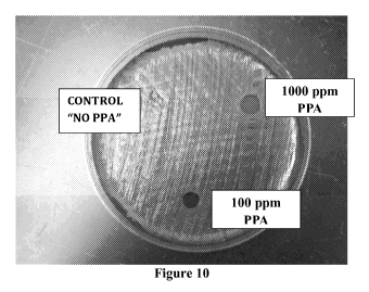
the amount of a-ketoester in compositions of the invention ranges from about
10 mM to
about 11.5 mM. In another embodiment, the amount of a-ketoester in
compositions of the
invention ranges from about 7.2 mM to about 8.6 mM. Yet in another embodiment,
the
amount of a-ketoester in compositions of the invention ranges from about 4.3
mM to about
6.4 mM. Still in another embodiment, the amount of a-ketoester in compositions
of the
invention ranges from about 0.29 mM to about 2.6 mM.
[0077] In some embodiments, compositions of the invention can kill at
least 105
amount of microorganisms within 10 minutes at a concentration of about 5,000
ppm or less
often at least 106 microorganisms within 10 minutes at a concentration of
about 5,000 ppm
including microorganism spores and microorganisms in biofilms. As used herein,
the term
"microorganism" includes bacteria, virus, fungi, algae, prion, and other
pathogenic organisms
known to one skilled in the art. Typically, the term microorganism refers to
bacteria, virus,
and fungi.
[0078] In other embodiments, compositions of the invention have
microorganism kill
activity of at least log 5 within 10 minutes, typically within 5 minutes and
often within 1
minute at a concentration of 4,000 ppm. Still in other embodiments,
compositions of the
invention have microorganism kill activity of at least log 6 within 10
minutes, typically
within 5 minutes and often within 1 minute at a concentration of 4,000 ppm.
Yet in other
embodiments, compositions of the invention have microorganism kill activity of
at least log 6
within 10 minutes at a concentration of about 4,000 ppm, typically at 3,000
ppm, often at
1,000 ppm, and more often at 500 ppm.
- 22 -
CA 02826646 2013-08-06
WO 2012/112951
PCT/US2012/025736
[0079] Surprisingly and unexpectedly, it has been found by the present
inventors that
compositions of the invention can also kill microorganism spores and biofilms
including, but
not limited to, those disclosed herein. See, for example, Table A above.
Conventional
antiseptics/detergents typically cannot kill microorganism spores and/or
biofilms in an
effective manner. In contrast, compositions of the invention have a broad
spectrum activity
and can effectively kill at least 70%, typically at least 80%, often at least
90%, more often at
least 95%, and still more often substantially all of bacteria in biofilm
within 10 minutes at a
concentration of about 5,000 ppm.
[0080] Additional objects, advantages, and novel features of this
invention will
become apparent to those skilled in the art upon examination of the following
examples
thereof, which are not intended to be limiting. In the Examples, procedures
that are
constructively reduced to practice are described in the present tense, and
procedures that have
been carried out in the laboratory are set forth in the past tense.
EXAMPLES
EXAMPLE 1
Disinfection of Spores on Medical Devices
[0081] This example demonstrates that the sporicidal efficacy of the PKCA
compounds in a dry, high protein environment, using the method described in
the ASTM E-
2197 procedure. Figure 1 illustrates sporicidal activity of peroxy a-keto
pyruvic acid (PPA),
peroxy a-keto valeric acid (POKVA), and peroxy a-keto butyric acid (POKBA).
Each of
these solutions was challenged to kill 6 logs of C. difficile spores in 10
minutes in a high
protein environment. The concentrations required were 1000 ppm (8.5 mM) for
PPA and
POKVA and 500 ppm (4.2 mM) for POKBA. In addition, 3 logs of C. difficile
spores were
killed with a PPA and POKVA at a concentration of 750 ppm (6.3 mM) and with
POKBA at
250 ppm (2.1 mM). These concentrations of PKCA are equivalent to a-keto acid
concentrations of 12.4 mM (1000 ppm), 9.3 mM (750 ppm), and 3.1 mM (250 ppm).
EXAMPLE 2
Disinfection of Biofilms on Medical Devices
[0082] The efficacy of PKCA against surface biofilm formation was tested
by the
AOAC 966.04 procedure. This procedure tests a candidate disinfectant against
Bacillus
subtillus spores dried onto ceramic penicylinders where they can form a
biofilm. Briefly, a
dilution of spore suspension in sterile distilled water is prepared at a final
concentration equal
- 23 -
CA 02826646 2013-08-06
WO 2012/112951 PCT/US2012/025736
to 1-4x10⁷CFU/mL. Using a sterilized hook, sterile penicylinders are placed in
the prepared
dilution and mixed well and then allowed to incubate for 10-15 minutes.
Afterwards, the
cylinders are removed, placed onto a sterilized screen in a sterile petri dish
and then placed in
a desiccator for at least 12-24 hours or until time of use. For disinfectant
testing, the
contaminated penicylinders and a non-contaminated control cylinder are placed
into vials
containing the PKCA mixture and allowed to set for 10 minutes. Afterwards, the
number of
spores are enumerated by placing a single cylinder into 10 mL of anaerobic
Brucella broth,
sonicated, and the appropriate dilution made on agar plates based upon the
expected count
(typically spiral plate 50 iut of a 1:1000 dilution). The cylinders should
contain 106
cfu/cylinder and subsequent loss in count from exposure to the PKCA mixture
reflects the log
kill of the spores. The results showed that each of the three PKCA solutions
containing PPA,
POKBA, and POKVA concentrations of 169 mM (2000 ppm ) were able to kill? 5.0
logs of
Bacillus subtilis on dried ceramic cylinders in 15 minutes.
EXAMPLE 3
Materials and Methods
Strains and growth condition
[0083] Pseudomonas aeruginosa PA01 (ATCC number: BAA-47), Enterococcus
faecalis V583 (ATCC number: 700802), and Staphylococcus aureus (ATCC number:
700699) were grown in Tryptic soy broth (TSB, Sigma Chemical Co., St. Louis,
MO, USA)
medium at 37 C with shaking for 16 hr.
Chemical treatment on biofilm formation
[0084] Bolton broth (Oxoid Ltd, Basingstock, Hampshire, England) and
Bovine
plasma (Biomeda, Foster City, CA, USA) were used for multi-species biofilm
formation.
Pseudomonas aeruginosa PA01, Enterococcus faecalis V583, and Staphylococcus
aureus
grown on TSB agar plates were inoculated into TSB broth and grown at 37 C in
a shaker for
16 hr. An aliquot was diluted in TSB broth to a series of dilutions for each
individual bacteria
type. The diluted bacteria were plated out to count colony forming units
(CFU). They were
further diluted tol x 106 cfu/mL. and mixed equally as inoculums. Bolton broth
with 50%
plasma was used for biofilm formation media. Glass 16 x 150 mm test tubes with
caps were
autoclaved, and 7 ml biofilm formation media aseptically dispensed in each
tube. The
normalized cultures of the three bacteria were mixed and 10 1 of the combined
and
normalized culture 1 x 10⁶ CFU/mL were inoculated into glass tubes. This was
done by
ejecting the pipette tips into the tubes. The pipette tip acts as a surface
for biofilm formation.
- 24 -
CA 02826646 2013-08-06
WO 2012/112951
PCT/US2012/025736
To the tubes, PPA/PPA-EP were added at 0 ppm, 400 ppm and 4000 ppm, and grown
at 37
C in a shaker for 24 hr at 150 rpm. Biofilm formation was subjectively
observed and the
biofilms were collected. The set of tubes with biofilm were placed in an oven
at 80 C for 48
h to obtain a dry weight. The biomass dry-weight was measured as the
difference of the total
weight minus the empty tube weight measured before use. Tests were performed
in triplicate
for each treatment group. A separate set of tubes in triplicate was used for
DNA extraction
and quantitative PCR analyses as described below.
PPA and PPA-EP incubation with formed biofilm
[0085] The formed biofilm were washed three times, and then treated with
8000 ppm
and 16000 ppm PPA and PPA with ethyl pyruvate (EP), incubated at 37 C with
shaking
(150 rpm) for 1 hr. The effects of PPA and PPA-EP incubation on formed biofilm
were also
evaluated using bacteria plate count. The biofilm were washed, sonicated for
10 min, and
vortexed. The process was repeated one more time. The supernatants were then
serially
diluted for bacteria plate count.
Designing specific primers for the three bacteria
[0086] Genome sequences of Pseudomonas aeruginosa PA01 (GenBank number:
AE004091), Enterococcus faecalis V583 (GenBank number: AE016830), and
Staphylococcus aureus (GenBank number: BA000017) were downloaded from NCBI
website. The individual genome sequence was used to BLAST against the whole
publicized
microbial genome sequences by using a Wnd-BLAST. The no-hit genes were used to
design
specific primers.
Real-time PCR analysis
[0087] Biofilm samples were homogenized by using a Qiagen TissueLyser
(QIAGEN, Santa Clara, CA, USA). A sterile 5 mm steel bead and 500 iut 0.1 mm
glass
beads were added to the tube with 500 iut TE buffer, and run at 30Hz for 5
min. Bacteria
DNA from the biofilm samples were then extracted by using a QIAamp DNA mini
kit
(QIAGEN). DNA samples were quantified using a Nanodrop spectrophotometer
(Nyxor
Biotech, Paris, France), and were diluted to 20 ng/ 1. DNA from three
individual bacteria
was also diluted to 20 ng/ 1 as positive control. Quantitative real-time PCR
was used to
assay specific gene levels for each bacterium representing the ratio of the
three bacteria in
biofilm samples. The levels of the genes were detected by using the Roche
LightCycler 480
(Roche, Mannheim, Germany). LightCycler 480 SYBR Green I Master Kit (Roche)
was
- 25 -
CA 02826646 2013-08-06
WO 2012/112951 PCT/US2012/025736
used for 20 pl real-time PCR reactions. Each sample was assayed three times.
The relative
gene level of each sample was calculated and analyzed. In brief, the threshold
cycle (Ct
value) of the target genes in different samples was obtained after
quantitative real-time PCR
reaction. The normalizer DNA Ct value was subtracted from the gene of interest
Ct (target
gene) to produce the dCt value of the sample. The dCt value of the calibrator
(the sample
with the highest dCt value) was subtracted from every other sample to produce
the ddCt
value. Two to the -ddCt power (2-ddct) was taken for every sample as the
relative gene levels.
The gene expression level of each bacterium represents relative ratios of each
bacterium
within a given DNA extracted sample.
Results
[0088] PPA and PPA-EP concentration of 400 ppm, 1000 ppm, 4000 ppm, 8000
ppm,
and 12000 ppm were initially used for testing the correct concentration for
further multi-
chemical treatment on biofilm formation (Figures 2 and 3). PPA and PPA-EP
demonstrated
an obvious and significant inhibitory effect on biofilm formation. So 400 ppm
and 4000 ppm
were used as the final concentration for the assays. Subjective observations
of the biofilm
formation were made and the biofilm biomass dry-weight was also measured to
provide a
more objective measurement. All of the treatments, exhibited lower biomass
formation,
based upon dry-weight, than the control biofilms, and the decrease of the
biomass correlated
with the visible reduction of the biofilm formation (Table 1). Adding 400 ppm
of PPA and
PPA-EP, the biofilm biomass dry-weight was decreased by 42.2% and 52.8%.
Adding 4000
ppm of PPA and PPA-EP, no biofilm growth was observed. The bacteria plate
count further
confirmed that 4000 ppm PPA and PPA-EP totally inhibit bacteria growth.
[0089] To further characterize the effects the chemicals on the
individual bacterial
populations within the multi-species biofilm, real-time PCR assay was
developed. This test
was used to compare untreated controls to the PPA and PPA-EP treated matrix,
The results
showed that PPA and PPA-EP did not completely inhibit growth of the 3 bacteria
at 400 ppm
but completely inhibited growth of the 3 bacteria at 4,000 ppm. See Table 1.
Table 1. Data summary of chemical treatment on biofilm prevention (N/A = no
counts)
biofilm dry
Sample/Treatment bacteria count weight (mg
(cfu/ml) SD) P. aeruginosa E..faecalis S.
aureus
CK (PPA) 1.0 Ell 112 16.1 58.7% 7.2% 16.9% 2.9% 24.5% 0.7%
400 ppm PPA 4.0 E9 64.7 2.5 62.3% 4.0% 19.7% 1.8% 20.5%
0.7%
4000 ppm PPA no growth No Biofilm NA NA NA
CK (PPA+EP) 1.0 Ell 115 12.8 55.1% 0.01% 12.3% 0.5% 32.1%
4.2%
- 26 -
CA 02826646 2013-08-06
WO 2012/112951 PCT/US2012/025736
400 ppm PPA+EP 3.0 E8 54.3 9.0 64.5% 5.7% 15.2% 4.1% 22.9%
1.6%
4000 ppm
PPA+EP no growth No Biofilm NA NA NA
[0090] In order to evaluate the effect of PPA/PPA-EP at eliminating
formed biofilm,
the 24 hr formed biofilm were treated with 8000 ppm and 16000ppm of PPA/PPA-EP
for 60
min. By subjectively observation, PPA/PPA-EP does show the effect to eliminate
formed
biofilm (Figure 4). All of the treatments, exhibited more biofilm degradation,
based upon
dry-weight, than the control biofilms, and the decrease of the biomass
correlated with the
visible reduction of the biofilm (Table 2). Adding 8000 ppm of PPA and PPA-EP,
the
biofilm biomass dry-weight was decreased by 62.4% and 60.8%. Adding 16000 ppm
of
PPA and PPA-EP, the biofilm biomass dry-weight was decreased by 49.6% and
64.2%. The
bacteria plate count further confirmed that 8000 ppm, 16000 ppm PPA and PPA-EP
totally
eliminate bacteria growth. To further characterize the effects the chemicals
on the individual
bacterial populations within the multi-species biofilm, real-time PCR assay
was developed.
This test was used to compare untreated controls to the PPA and PPA-EP treated
matrix, The
results showed that PPA and PPA-EP did not selectively eliminate these three
bacteria (Table
2).
Table 2. Data summary of PPA and PPA-EP treatment with formed biofilm
bacteria biofilm dry
count weight (mg
Sample/Treatment (cfu/ml) SD) P. aeruginosa E. faecalis S. aureus
CK (PPA) 1.0 E9 13.3 2.1 13.1% 1.4% 39.3% 0.4% 47.6%
1.2%
8000 ppm PPA no growth 5.0 2.0 13.2% 1.4% 29.0% 1.1% 57.8%
5.7%
16000 ppm PPA no growth 6.7 2.3 17.8% 3.4% 30.4% 8.4% 51.8%
5.8%
CK (PPA+EP) 1.0 E9 12.0 1.7 19.5% 1.1% 29.4% 0.1% 51.2%
4.3%
8000 ppm PPA+EP no growth 4.7 2.1 20.5% 3.2% 22.7% 0.3% 56.7% 2.5%
16000 ppm
PPA+EP no growth 4.3 2.1 17.8% 1.3% 45.2% 0.9% 36.9%
3.4%
Conclusion
[0091] PPA and PPA-EP have a broad range for inhibiting bacteria growth.
At 8,000
ppm, PPA and PPA-EP eliminate formed biofilm within 60 min. At lower
concentrations
such as 400 ppm to 4,000 ppm, PPA and PPA-EP have a suppression effect on
biofilm
formation.
EXAMPLE 4
[0092] This example demonstrates that the PKCA solutions kill bacteria in
a
simulated wound solution environment. The PKCA solution containing PPA was
brought to
- 27 -
CA 02826646 2013-08-06
WO 2012/112951 PCT/US2012/025736
the approximate physiological pH of 6.0 in a 50 mM phosphate buffer and tested
for its
ability to kill Methicillin-Resistant Staphylococcus aureus (MRSA) in the
presence of 10%
Fetal Bovine Serum (FBS). It was shown that 100 ppm (0.85 mM) of the PPA
containing
PKCA solution killed 6 logs of MRSA in one minute within the FBS solution.
[0093] All other PKCA efficacy studies against vegetative bacteria were
done by a
standard immersion test. This procedure involves producing a suspension of the
test organism
in sterile diluent comparable to a 0.5 McFarland standard (1-2 x 10⁸ CFU/mL).
An aliquot of
the suspension was pipetted into the PKCA mixture to be evaluated at a ratio
of 1:100 (e.g.,
30 L suspension into 3mL of disinfectant mixture) and thoroughly mixed. An
aliquot was
removed from the PKCA mixture at desired exposure intervals and diluted to a
ratio of 1:10
(e.g., 0.4 mL into 3.6 mL) in neutralizing broth and then spiral plated for
counts. This
procedure provides theoretical quantitation of a 6 log decrease in CFU/mL.
EXAMPLE 5
[0094] To determine the performance of the PKCA mixtures in a protein
environment, the PPA mixture was tested for microcidal efficacy against MRSA
for
performance in a high protein environment and for a simple simulation of a
wound
environment. The PPA mixture was tested by the immersion test, as described
above, against
MRSA suspended in 10% Fetal Bovine Serum (FBS). The PPA mixture killed 6 logs
of
MRSA when exposed to 200 ppm of PPA in 10% FBS within 15 seconds (Figure 5).
This
was double the concentration of PPA required to kill MRSA suspended in water
in 15
seconds. Therefore, an increase in concentration was required in a high
protein environment,
but the activity in the high protein environment for killing high populations
of a MDRO was
still fast and effective.
EXAMPLE 6
[0095] The PPA mixture was also tested against MRSA suspended in a
phosphate
buffer to determine if buffered solutions of the PKCA mixtures at different
pH's could be
used for different phases of the wound healing process. The results of that
testing
demonstrated that PPA will kill 6 logs of MRSA in pH 6.0 phosphate buffer in
60 seconds at
50 ppm and in 15 seconds at 100 ppm (Figure 6).
EXAMPLE 7
[0096] The PPA mixture was also tested against Acinetobacter baumanii
suspended
in a phosphate buffer to determine if buffered solutions of the PKCA mixtures
at different
- 28 -
CA 02826646 2013-08-06
WO 2012/112951 PCT/US2012/025736
pH's could be used for different phases of the wound healing process. The
results of that
testing demonstrated that PPA will kill 6 logs of Acinetobacter baumanii in 60
seconds with
50 ppm of PPA to kill 6 logs and only 15 seconds at 100 ppm (Figure 7).
EXAMPLE 8
[0097] Experiments were performed on the biocidal efficacy of PPA against
Pseudomonas aeroginosa suspended in 20% egg yolk in a citrate buffer, pH 6.8.
The results
demonstrated that PPA will kill Pseudomonas aeroginosa in a buffered high
protein
environment in 1 minute at 500 ppm but took 60 minutes to kill 6 logs at half
that
concentration (Figure 8).
[0098] Current real challenges in infectious wound healing include
Candida and
Aspergillus fungi and molds. Aspergillus spores in burn wounds have led to a
75% mortality
rate in older patients with deep burn wounds. PPA has been shown to kill these
spores in
solution. Table 2 above shows that PPA will kill these fungi in less than 10
minutes and
therefore indicates the ability to disinfect these infections in burn wounds
before they become
systemic.
[0099] These studies demonstrated the biocidal efficacy of PKCA mixtures
in high
protein and buffered solutions at different pH values. In addition, they
demonstrate the
prevention and destruction of simulated chronic wounds in-vitro. The contract
research
facility stated that after testing hundreds of compounds, that other than
hypochlorite, the
PKCA solution was the only compound they have seen that had a totally broad
spectrum kill
of the bacteria and prevented and dissolved the Biofilm. All of these examples
and theoretical
understanding of the chemistry demonstrate that PKCA compounds can be
formulated
according to an optimum pH to enhance wound healing and still disinfect the
wound. It has
been proposed recently that the tailoring of pH for the addition of treatment
compounds
would be an effective way to decrease wound healing time.
[0100] In summary, these examples demonstrate that the PKCA solutions can
kill
high levels of bacteria and spores in biofilms and in high protein
environments. In many
instances, the PKCA solutions also include the parent a-ketocarboxylic acids.
The a-
ketoester provides tissue penetration and anti-inflammatory capabilities to
the wound
treatment solution. Therefore, compositions of the invention are the only
existing simple
organic chemistry that would both disinfect a wound and enhance healing
simultaneously.
- 29 -
CA 02826646 2013-08-06
WO 2012/112951 PCT/US2012/025736
EXAMPLE 9
[0101] This example shows that the biocidal efficacy of the a-keto
peracid (i.e.,
peroxy a-ketocarboxylic acid) compounds in a dry, high protein environment.
The
experiment was conducted following the method described in the ASTM E-2197
procedure.
Figure 1 shows the results of three a-keto peracid solutions each of which
contained either
peroxy a-keto pyruvic acid (PPA), peroxy a-keto valeric acid (POKVA), or
peroxy a-keto
butyric acid (POKBA). These solutions were challenged to kill 6 logs of C
difficile spores in
minutes within a high protein environment. The concentrations required were
1000 ppm
(8.5 mM) for PPA and POKVA and 500 ppm (4.2 mM) for POKBA. In addition, 3 logs
of C
difficile spores were killed with a PPA and POKVA concentration of 750 ppm
(6.3 mM) and
with POKBA at 250 ppm (2.1 mM). These concentrations of a-keto peracids equate
to a-keto
acid concentrations of 12.4 mM (1000 ppm), 9.3 mM (750 ppm), and 3.1 mM (250
ppm).
EXAMPLE 10
[0102] This example shows that the a-keto peracid solutions can kill
biofilms. The
biocidal efficacy testing of a-keto peracid compounds against biofilms was
determined using
the method described in the AOAC 966.04 procedure. The results showed that
each of the
three a-keto peracid solution containing PPA, POKBA, and POKVA at a
concentration of
169 mM (2000 ppm ) were able to kill > 5.0 logs of Bacillus subfilis on dried
ceramic
cylinders in 15 minutes.
EXAMPLE 11
[0103] This example shows that the a-keto peracid solutions can kill
bacteria in a
simulated wound solution environment. The a-keto peracid solution containing
PPA was
brought to the approximate physiological pH of 6.0 in a 50 mM phosphate buffer
and tested
for its ability to kill Methicillin-Resistant Staphylococcus aureus (MRSA) in
the presence of
10% Fetal Bovine Serum (FBS). It was shown that 100 ppm (0.85 mM) of the PPA
containing a-keto peracid solution killed 6 logs of MRSA in one minute within
the FBS
solution.
[0104] These examples demonstrate that the a-keto peracid solutions can
kill high
levels of bacteria and spores in biofilms and in high protein environments. In
some instances,
compositions of the invention comprise both the a-keto peracid and the parent
alpha keto
carboxylic acid. The alpha keto carboxylic acids are natural compounds found
within nearly
all living cells and have been implicated in potentially improved wound
healing. By
- 30 -
CA 02826646 2013-08-06
WO 2012/112951 PCT/US2012/025736
providing both the a-keto acids, and the peroxy form of these alpha keto
acids, some
compositions of the invention provide synergistic benefits to the cells.
EXAMPLE 12
[0105] The PKCA compounds that are disclosed in the above disclosed
commonly
assigned U.S. Patent Applications and Provisional Patent Application. These
PKCA
compounds have been developed inter alia for use as a high level
disinfectant/sterilant of
vegetative bacteria, spores and biofilms. The present inventors have shown
that PKCA
compounds are effective in killing vegetative bacteria and spores at the level
acceptable to be
called sterilants/disinfectants.
[0106] In this study, ethyl pyruvate (EP) was added to a PKCA solution to
determine
if biocide efficacy was affected by the addition of EP. The EP was added at a
2%
concentration to a solution comprising a PKCA compound. The amount of EP used
was
effective in substantially eliminating PKCA odor. As a control, 2% EP in water
was tested
for anti-microbial properties in the same manner as the PKCA-EP mixture.
Surprisingly and
unexpectedly it was discovered that the 2% EP control also killed bacteria.
Examples of
these tests are shown below.
[0107] MRSA was prepared as a suspension in sterile diluent that was
comparable to
a 0.5 McFarland standard (1-2 x 10⁶ CFU/mL). An aliquot of the MRSA suspension
was
added to the PKCA-EP mixture and the EP control solution at a ratio of 1:100
(e.g., 30 ILIL
suspension into 3 mL of the test solutions) and thoroughly mixed by vortex.
After 10
minutes, an aliquot of each test sample was diluted at a ratio of 1:10 (e.g.,
0.4 mL into 3.6
mL) in neutralizing Letheen broth. This procedure provided theoretical
quantitation of a 4
log unit decrease and detection of a 5 log unit decrease in cfu/mL. The PKCA-
EP and EP
control solutions were spiral plated with 50 ILIL of each test sample onto the
appropriate agar
as applicable to attain countable dilutions. In addition, each of the
neutralized tubes of test
sample and agar plates were incubated overnight in an appropriate atmosphere
and
temperature. After determining the bacterial counts, the test sample tubes
where the bacteria
had been exposed to EP were incubated for another 24 hours. If no bacterial
suspension was
observed in those tubes, then it was an indication that a complete
decontamination had
occurred. The plate count results are illustrated in the Table below.
-31 -
CA 02826646 2013-08-06
WO 2012/112951
PCT/US2012/025736
Log Reduction
Control 5.9
PKCA Compound 4.9
2% Ethyl Pyruvate 4.9
[0108] As data in the above Table shows, a 2% concentration of ethyl
pyruvate in the
PKCA solution did not inhibit the biocidal efficacy of the PKCA compound. The
surprising
and unexpected result was that ethyl pyruvate also killed 4.9 log units of
MRSA itself
[0109] Burkholderia pseudomallei (B. pseudomallei) and Burkholderia
mallei (B.
mallei) are the causative agents of melioidosis and glanders, respectively.
These are gram-
negative pathogens, and unlike MRSA (a gram positive bacteria), are endemic in
many parts
of the world. Without being bound by any theory, it is believed that these
bacteria are
resistant to many, if not most, antibiotics because of their ability to pump
(i.e., remove) the
antibiotics out of their cell using an active transport system. Although
natural acquisition of
these pathogens is rare in the majority of countries, these bacteria have
recently gained much
interest because of their potential as bioterrorism agents.
[0110] In another study, ethyl pyruvate was tested to determine the
effects of 2%
ethyl pyruvate on B. pseudomallei. For this study, B. pseudomallei 1026b was
inoculated
into 3 mL of Letheen broth and incubated overnight at 37 C. The next day 20
ut, of the
overnight culture as added to 2 mL of Letheen broth with 0.1% sodium
thiosulfate to achieve
¨107 cfu/mL. Afterwards, this solution was diluted to a working solution of
¨104 cfu/mL.
Two test tubes with 5 mL of 2% ethyl pyruvate were prepared and two tubes with
5 mL
sterile water were prepared as controls. A 100 ut, of ¨104 cfu/mL stock
solution of B.
pseudomallei 1026b was added directly to one positive control tube containing
5 mL of
sterile water, and 100 ut, of ¨104 cfu/mL stock solution of B. pseudomallei
1026b was added
directly to one tube containing 5 mL of 2% ethyl pyruvate. These tubes were
incubated at
37 C for 20-24 hours and then observed for growth (+) or no growth (-). After
24 hours of
incubation, there was no noticeable growth in the B. pseudomallei 1026b
inoculated ethyl
pyruvate tube and a significant growth in the negative control. This indicates
that ethyl
pyruvate has antibacterial activity even against bacteria that are highly
resistant to broad
spectrum antibiotics.
[0111] Although unlikely, the potential hydrolysis product ethanol was
considered as
a possible source of ethyl pyruvate 's antimicrobial activity. To determine
whether ethanol
- 32 -
CA 02826646 2013-08-06
WO 2012/112951 PCT/US2012/025736
was the source of antimicrobial activity, analytical experiments were
performed to determine
if the hydrolysis occurred over time to produce ethanol.
[0112] Fourier transform infrared (i.e., FTIR) scans of 30% ethyl
pyruvate in 50%
hydrogen peroxide solution incubated at room temperature for 1 hour, 16 hours
and 60 days
were obtained. Study of these FTIR scans showed that no ethanol was produced
during
incubation in hydrogen peroxide solution even after 60 days. This result
indicates that ethyl
pyruvate is stable in the PKCA solution since the concentration of the
hydrogen peroxide in
those solutions is approximately only half that of 50% hydrogen peroxide
solution.
[0113] An equivalent mixture of ethyl pyruvate and hydrogen peroxide that
was
incubated at room temperature for 7 days was analyzed by FTIR and by gas
chromatography.
There was no ethanol found in the mixture. This result further indicates
stability of ethyl
pyruvate in the presence of hydrogen Peroxide.
[0114] Typically, a 70% ethanol solution is used disinfection. Therefore,
it is highly
unlikely that a concentration of 2% ethanol (if all of the ethyl pyruvate is
hydrolyzed) would
kill bacteria in a 10 minute time period. Without being bound by any theory,
it is possible
that esterase enzyme in bacteria may hydrolyze ethyl pyruvate to produce
ethanol in situ
resulting in the observed antibacterial activity. Another possibility is that
the antimicrobial
effect is due to the decrease in pH from the release of the pyruvic acid.
EXAMPLE 13
[0115] In this example, a composition of the invention was formulated in
bandage
materials or as dissolvable films (Figure 9) such that the active composition
is released when
moisture is present. Bandages and films can be formulated for sustained time
release,
thereby providing the composition of the invention to the wound over a
prolonged period.
Different bandage materials can be used, for example, they can be air
permeable or
substantially non-air permeable or sealed. In addition, bandages and films
comprising a
composition of the invention can be fabricated with other conventional
bandaging materials
and then stored in dry form for use, for example, for combat field use during
evacuation and
level 2-4 transports. Other possible formulations for compositions of the
invention include,
but are not limited to, gels, lotions, cream, or other suitable formulations
that can be directly
applied to wounds. In some embodiments, formulations of the composition of the
invention
enable an effective time release. Typically, such formulations are light
weight and stable
forms of wound dressing materials that can be applied directly to the wound.
In some
- 33 -
CA 02826646 2013-08-06
WO 2012/112951 PCT/US2012/025736
embodiments, compositions of the invention are formulated such that they are
released when
exposed to wound. Often such formulations dissolve over time releasing the
composition of
the invention. Such formulations have broad application to both the military
and civilian
population. For example, such formulations for military use include, but are
not limited to,
immediate field application upon initial triage through the entire course of
the wound healing
process.
[0116] Figure 10 shows the result of treating MRSA on blood agar plate
with a
composition of the invention comprising PPA that was incorporated in
dissolvable film (see
Figure 9). As the results show, the control film disc was completely grown
over while the
dissolvable film comprising a composition of the invention killed MRSA
relatively in
proportion to the PPA concentration.
[0117] The foregoing discussion of the invention has been presented for
purposes of
illustration and description. The foregoing is not intended to limit the
invention to the form
or forms disclosed herein. Although the description of the invention has
included description
of one or more embodiments and certain variations and modifications, other
variations and
modifications are within the scope of the invention, e.g., as may be within
the skill and
knowledge of those in the art, after understanding the present disclosure. It
is intended to
obtain rights which include alternative embodiments to the extent permitted,
including
alternate, interchangeable and/or equivalent structures, functions, ranges or
steps to those
claimed, whether or not such alternate, interchangeable and/or equivalent
structures,
functions, ranges or steps are disclosed herein, and without intending to
publicly dedicate any
patentable subject matter.
- 34 -