Note: Descriptions are shown in the official language in which they were submitted.
CA 02888304 2015-04-13
WO 2014/063246 PCT/CA2013/050797
1
SAMPLE ANALYSIS BY MASS CYTOMETRY
FIELD
[0001] This invention relates to apparatus and methods for sample
analysis by mass cytometry.
INTRODUCTION
[0002] An analytical technique using laser ablation inductively
coupled plasma (ICP) mass spectrometry (LA-ICP-MS) can be applied to
the imaging of metal ion distribution in biological tissues.
Typically, laser ablation-ICP-mass spectrometry can be used to
interrogate a tissue sample to detect and map trace element
distribution. This technique, however, is limited to surface analysis
incorporating 2-dimentional imaging of thin tissue samples.
SUMMARY
[0003] In view of the foregoing and in accordance with the present
teachings, each plume generated by each laser pulse can be ionized and
detected distinctly as a function of the sample depth by a mass
cytometer while an encoded substrate supporting the sample (sample
support) can have a substrate coding configured to codified its
position on the encoded substrate and to indicate when a laser pulse
ablates through the sample. This system and technique allows for a
quantitative distribution profile to be generated through the
thickness of the sample and the mapping of a 3-dimentional image of
the sample.
CA 02888304 2015-04-13
WO 2014/063246 PCT/CA2013/050797
2
[0 0 0 4 ] Another aspect of the teaching is a method for sample
analysis by mass cytometry. The method includes providing a sample
labeled with more than one elemental tag. Supporting the labeled
sample with an encoded substrate where the encoded substrate is
configured with a substrate coding. At least one laser pulse is
directed onto a location of the sample to generate a discrete plume
corresponding to each of the at least one laser pulse. Each discrete
plume comprises at least one of the more than one elemental tag and
the substrate coding. The discrete plumes are introduced into an
inductively coupled plasma (ICP) where groups of elemental ions are
generated such that each of the groups of elemental ions corresponds
with at least one of each of the more than one elemental tag and the
substrate coding. The method further comprises detecting each of the
groups of elemental ions simultaneously for each of the discrete plume
and then correlating the detected groups of elemental ions with the
substrate coding by, for example, identifying the location of the more
than one elemental tag as a function of the substrate coding.
[0005] Another aspect of the teaching is a mass cytometer system
for sample analysis. The system has an encoded substrate for
supporting the sample and the encoded substrate is configured with a
substrate coding comprising an array of codified metal compositions.
The system also has a laser ablation system configured to generate a
plume from the sample and from the substrate coding. A mass cytometer
comprising an ion source and ion detector is coupled to the encoded
substrate through a defined total path.
CA 02888304 2015-04-13
WO 2014/063246 PCT/CA2013/050797
3
[0 0 0 6 ] Yet another aspect of the teaching is a sample support for
laser ablation mass cytometry. The support has an encoded substrate
with a surface for supporting the sample. The encoded substrate has a
substrate coding, such as an array of codified transitional metal
isotope compositions, arranged to codify the encoded substrate.
BRIEF DESCRIPTION OF THE DRAWINGS
[0007] The skilled person in the art will understand that the
drawings, described below, are for illustration purposes only. The
drawings are not intended to limit the scope of the applicant's
teachings in any way. In the accompany drawings, where like reference
numerals indicate like parts:
FIG. 1 is a pictorial representation of the system and process
according to one embodiment of the present teaching;
FIG. 2 is an expanded view of the encoded substrate according to an
embodiment of FIG. 1;
FIG. 3 and FIG. 4 are pictorial representations of encoded substrates
according to various embodiments of the present teaching;
FIG. 5 is a pictorial representation of an encoded substrate with
various embodiments of the substrate coding according to the present
teaching; and
FIG. 6 is a schematic view of an embodiment of the ICP ion source
according to the present teaching.
CA 02888304 2015-04-13
WO 2014/063246 PCT/CA2013/050797
4
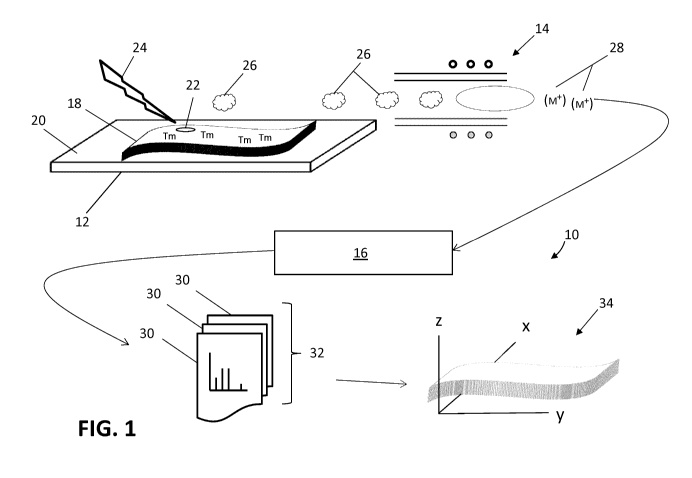
DESCRIPTION OF VARIOUS EMBODIMENTS
[0008] It should be understood that the phrase "a" or "an" used in
conjunction with the present teachings with reference to various
elements encompasses "one or more" or "at least one" unless the
context clearly indicates otherwise. Reference is first made to FIG.
1, which shows a pictorial representation of the sample analysis
system, generally indicated by reference number 10. The sample
analysis system 10 comprises an encoded substrate 12 coupled to an
inductively coupled plasma (ICP) ion source 14 of a mass cytometer 16.
Generally, the ICP ion source 14 can be considered as an integral
component of the mass cytometer 16, however for clarity, the ICP ion
source 14 is represented separately from the mass cytometer 16. The
mass cytometer 16 can comprise a computational system (not shown) for
generating corresponding elemental tag data 30. The encoded substrate
12 provides a surface for supporting a sample 18 of interest while
additionally being configured with a substrate coding 20 structure.
The substrate coding 20 can provide a means for representing or
mapping the spatial arrangement or distribution of a location 22 on
the sample 18 during the analysis, as will be described below. The
sample analysis system 10 further comprises a laser ablation system
(not shown) for supplying at least one laser pulse 24 directed at the
location 22 on the sample 18.
[0009] In use, the at least one laser pulse 24, upon being
directed onto the surface of the sample 18, can remove some of the
sample material in the form of a discrete plume 26. Generally, each
CA 02888304 2015-04-13
WO 2014/063246 PCT/CA2013/050797
laser pulse can generate a discrete plume 26 so that a series of laser
pulses can generate a series of corresponding discrete plumes 26. In
various embodiments, the sample 18 of interest can be labeled with
more than one elemental tag Tn, typically selected from the group
comprising transitional metals as described in co-pending United
States Patent Application No. 12/513,011 published as U52010/0144056,
assigned to the assignees of the present teachings. For convenience,
the "n" notation in Tn can be a variable to signify the different
elemental or metal isotope tag Tn. For example, a tissue sample
containing cells of interest can be labeled with more than one type of
metal conjugated antibody. The metal or elemental tag Tn conjugated
to each type of antibody can be a distinct metal isotope of any one or
a combination of Gd, Nd, Tb, Eu, Gd, Dy, Ho, Sm, Er, Yb, to name only
a few. Consequently, the material removed from the location 22 of the
sample 18 for each discrete plume 26 can contain the more than one
elemental tag Tn - such as the combination of Nd and Sm for elemental
tag "Tl" and Gd, Tb and Er for elemental tag "T2", for example.
[0010] While maintaining the spatial separation of each successive
plume 26, each plume 26 can be transported and introduced into the ICP
ion source 14 as discrete and independent entities. As each discrete
plume 26 passes into the ICP ion source 14, each elemental tag Tn can
be ionized into corresponding elemental ions quantitatively related to
each elemental tag Tn. Since there can be more than one elemental tag
Tn in the labeled sample 18, the ICP ion source 14 can generate a
distinct group of elemental ions for each elemental tag Tn.
CA 02888304 2015-04-13
WO 2014/063246
PCT/CA2013/050797
6
Consequently, for each discrete plume 26, the ICP ion source 14 can
generate groups of elemental ions 28, represented generally as (le) in
FIG. 1.
Each of the groups of elemental ions 28 can be detected by
the mass cytometer 16 according to the ions' mass to charge ratio
(m/z). In accordance with the present teachings, the mass cytometer
16 can detect each of the elemental ions simultaneously and, with its
advantageous fast transit time, the mass cytometer 16 can
differentiate between groups of elemental ions originating from
successive lasers pulses. The elemental tag data 30, shown in FIG. 1
as a succession of single data files, represents the data acquired
from simultaneously detecting the groups of elemental ions 28 for the
succession of each plume 26. Hence, the sample analysis system 10 can
detect and identify each of the more than one elemental tag Tn
simultaneously for each laser pulse 24. While a single laser pulse
can generate a plume containing the more than one elemental tag Tn,
there can be some locations 22 on the sample 18 where a series of
laser pulses 24 can be required to reach a certain sample depth before
encountering the presence of the more than one elemental tag Tn.
Furthermore, there can be instances where there can be an absence of
any elemental tag Tn at a location 22 on the sample 18 and
consequently the series of discrete plumes 26 contain no elemental
tags Tn. In this instance, the absence of any elemental tag Tn can be
interpreted to provide a source of information regarding other
potential characteristics of interest. Accordingly, the applicants
recognize that the information from each discrete plume can
advantageously be used in combination with the substrate coding 20 to
CA 02888304 2015-04-13
WO 2014/063246 PCT/CA2013/050797
7
generate elemental tag profiles throughout the thickness of the sample
18 and to identify its location 22 with respect to the area of the
sample 18, as will be described below.
[0011] To help understand how the encoded substrate 12 can be
structured for identifying and mapping out each location 22 on the
sample 18, reference is now made to FIG. 2. For visual clarity, the
sample 18 and the encoded substrate 12 are separated to show details
of the substrate coding 20. The substrate coding 20 can have an array
arrangement comprising differentiating metal compositions or alloys,
generally denoted as Xn, codified at positions across the encoded
substrate 12. For convenience, the "n" notation in Xn can be a
variable to signify distinct and distinguishable compositions Xn.
Thus, each position on the encoded substrate 12 can be represented and
identified by its specific metal composition Xn. For brevity, the
terms substrate coding 20 and the corresponding metal composition Xn,
arranged for making up the coding, are used interchangeably for the
present teachings. In various embodiments, for example, the substrate
coding 20 can be an aggregate of transitional metal isotopes (as noted
above) assembled in predetermined permutations and concentrations to
achieve an array of unique identifiers. For distinguishability, the
choice of the transitional metal isotopes used for each of the metal
compositions Xn can be selected to be sufficiently distinct and
distinguishable from the elemental tags Tn used for labeling a sample.
Consequently, the position coordinates of each unique identifier on
CA 02888304 2015-04-13
WO 2014/063246 PCT/CA2013/050797
8
the encoded substrate 12 can be recorded for future cross reference
and decoding as required.
[0012] The decoding process for detecting or identifying the
unique identifiers can follow a similar technique as described above
for releasing and detecting the elemental tag Tn from the labeled
sample 18. Congruently, when at least one laser pulse 24 is directed
at the encoded substrate 12, some of the substrate coding 20 can be
removed and can be formed into the plume 26.
The plume 26 comprising
the released composition Xn can be directed to the ICP ion source 14
for ionization. Subsequently, the groups of elemental ions 28
generated by the ICP ion source 14 can be identified by the mass
cytometer 10 as having their origins from the encoded substrate 12 and
accordingly, determine its position by cross referencing the
coordinate information associated with the substrate coding 20.
[0013] While in use, a sample 18 of interest can be supported by
the encoded substrate 12 and the area, or layout, of the sample 18 can
be represented by the underlying substrate coding 20 array. In some
instances, the location 22 can be predetermined or selected by
performing a pre visual analysis (such as florescence,
phosphorescence, reflection, absorption, shape recognition or physical
feature) of the labeled sample 18 to identify locations 22 expressing
certain quality of interest. However, according to the present
teachings, the location 22 of interest can be selected without a pre
analysis of the labeled sample 18. In various embodiments, for
example, the location 22 of interest can be based on a raster pattern,
CA 02888304 2015-04-13
WO 2014/063246 PCT/CA2013/050797
9
a structured sampling technique employing Monte Carlo methods for
instance, or a basic random selection method. During the analysis, as
each laser pulse 24 removes sequential layers of the labeled sample 18
from the location 22 of interest, groups of elemental ions 28
corresponding with the more than one elemental tag Tn can be
simultaneously detected by the mass cytometer 16. Each of the
detected groups of elemental ions 28 can represent the material
removed at each layer of the sample 18. As noted above, some of the
discrete plumes 26 can contain no elemental tag or some of the
discrete plumes 26 can comprise a gradation of elemental tags.
Furthermore, in various embodiments, some of the discrete plumes 26
can comprises overlapping information from each of the more than one
elemental tag Tn. Thus, for each of the simultaneous detection
performed by the mass cytometer 16, the data 30 can contain
qualitative and quantitative information based on the presence and in
some instances the absence, of the one or more elemental tag Tn. Each
of the acquired data 30 can provide a piece of the information about
the cross-section or thickness profile of the labeled sample 18.
[0014] As the analysis progresses and the successive laser pulses
24 penetrates through the thickness of the sample 18, at least one of
the laser pulses 24 can remove or begin to remove some of the
substrate coding 20. Accordingly, when the groups of elemental ions
28 detected by the mass cytometer 16 comprises the elemental ions from
the metal composition Xn, the system 10 can determine that the laser
has completed its ablation through the labeled sample 18. Thus, the
CA 02888304 2015-04-13
WO 2014/063246 PCT/CA2013/050797
elemental tag data 30 resulting from each of the previous laser
ablations can be grouped together as a set 32 of data assigned to
represent the information acquired at the location 22, and that the
set 32 of data corresponds with the specific metal composition Xn on
the encoded substrate 12. The system 10 can then codify the location
22 on the labeled sample 18 to correspond with the position of the
detected substrate coding 20. By cross-referencing the detected
groups of elemental ions 28 with the elemental tags Tn and correlating
with the substrate coding 20, each of the elemental tags Tn and their
location 22 on the sample 18 can be identified as a function of the
substrate coding 20. Consequently, the set 32 of acquired elemental
tag data 30 can be used to generate a distribution profile 34
corresponding with the thickness of the labeled sample 18 at its
identified location 22. This process can be repeated, as necessary,
for each subsequent location 22 on the labeled sample 18.
Accordingly, and with the aid of an appropriate algorithm, the
distribution profile 34 can be visualized to represent a 3-dimensional
image of the elemental tag profile of the labeled sample 18.
[0015] While the present teachings are described in conjunction
with various embodiments, it is not intended that the present
teachings be limited to such embodiments. On the contrary, the
present teachings encompass various alternatives, modifications, and
equivalents, as will be appreciated by those of skill in the art. For
example, the present applicants recognize that the codified metal
compositions Xn can be located on the surface of the encoded substrate
CA 02888304 2015-04-13
WO 2014/063246 PCT/CA2013/050797
11
12, embedded in a sub-layer of the encoded substrate 12 or integrated
in the thickness of the encoded substrate 12 as can be generally
fabricated by such methods as molecular beam epitaxy or
microfabrication using photolithography or similar techniques.
Recesses 36 or etched grooves 38 (such as 100 pm deep wells) on the
encoded substrate 12 can be used to provide receiving areas for each
of the distinct metal compositions Xn, as shown in FIG. 3 and FIG. 4
respectively. The material for the construction of the encoded
substrate 12 can be selected from any one or a combination of
stainless steel, glass, quartz, ceramic, polytetrafluoroethylene
(PTFE) and polyetheretherketone (PEEK) to name a few. While each
metal composition Xn can be generally described as discrete substances
detached or isolated from each other, the present applicants have
contemplated that a trace or track of codified metal composition Xn in
the form of a continuous deposit or coating can be used to provide
unique identifiers. In various embodiments, for example, a continuous
deposit can be applied to the encoded substrate 12 in such a manner as
to provide a varying concentration gradient of more than one
transition metal. The decoding process can be based on detecting the
ratio of the metal concentration at a given location of the deposit.
Accordingly, the analysis system 10 can be programed with the deposit
pattern and the corresponding metal concentration ratios for each
encoded substrate 12. The encoding and decoding information can
enable the correlation between the labeled sample 18 and the substrate
coding 20 for identifying the location of the more than one elemental
CA 02888304 2015-04-13
WO 2014/063246 PCT/CA2013/050797
12
tag Tn with respect to the area of the labeled sample 18 as described
above.
[0016] In various embodiments, the metal codified composition Xn
can be further characterized as having luminescence properties. For
example, the encoded substrate 12 can be made from a transparent
material, such as glass, and the metal codified composition Xn can be
a metal or non-metal fluorescent material (such as, for example,
europium complexes or fluorophores respectively) codified on the
surface, embedded in a sub-layer or integrated in the thickness of the
encoded substrate 12. In use, as the laser pulses 24 penetrates
through the thickness of the sample 18, at least one of the laser
pulses 24 can illuminate the codified fluorescent material at the
location 22 of the sample 18 and produce a distinguishable
fluorescence emission spectra. With an appropriate optical detector
positioned beneath the encoded substrate 12, for example, the detected
emission spectra can be used as the detected substrate coding 20 for
correlation as described above.
[0017] Alternatively, according to FIG. 5, the substrate coding 20
can be based on particles 40, such as beads, or other forms of
carriers to which unique metal identifiers can be incorporated. In
various embodiments, for example, the particles 40 can reside in the
recesses 36 according to FIG. 3. The metal composition Xn can be
attached on to the surface or imbedded within the carrier. The
carriers can be arranged on the encoded substrate in an array pattern
of a predetermined orientation, such as a grid formation, so that the
CA 02888304 2015-04-13
WO 2014/063246 PCT/CA2013/050797
13
carrier's position codifies the encoded substrate. In use, the
energy from the at least one laser pulse can removed the metal
composition Xn, along with or without the material of the carrier,
into the formation of the discrete plume 26 as previously discussed.
[0018] In various embodiments the metal composition Xn can
comprise a reference element (such as, for example, the element Rh or
Ir or a combination thereof) for which the analysis system 10 can
detect and use as a standard for system calibration. Alternatively,
the reference element can be introduced to the sample in the form of a
reference label. The label can be non-specifically attached to the
sample 18 thus providing a reference standard throughout the sample.
[0019] The applicants of the present teachings recognizes that in
order for each acquired elemental tag data 30 to correspond with each
layer of the labeled sample 18, the spatial separation of each
successive plume 26, and the corresponding ions, during their travel
along the path between the encoded substrate into the ICP ion source
14 and between the ion source 14 and the ion detector (not shown) of
the mass cytometer 16 is maintained. For example, a solid state laser
typically used for laser ablation, such as a femtosecond pulsed laser
can be configured to operate with a pulse rate between 10 and 100 Hz.
At this frequency, a plume 26 can be generated every 10 to 100 msec.
Considering the lower limit, it can be required to minimize the delay
time within the system 10 to a level of the order of 10 msec in order
to maintain plume separation. In accordance with various embodiments
of the present teachings, the mass cytometer 16 can be characterized
CA 02888304 2015-04-13
WO 2014/063246 PCT/CA2013/050797
14
as a "flow-through" analytical device comprising a linear ion path
with electrostatic lenses and an ion detector capable of parallel
elemental ion detection. In this configuration, a delay time in the
order of 10 msec can be achieved so that the groups of elemental ions
(le) can undergo acceleration and pass within the mass cytometer 16 for
simultaneous detection. Consequently, the likelihood of the ion
detector to separately detect each of the groups of elemental ions 28
can be realized.
[0020] To maintain a corresponding spatial distinctiveness
upstream of the mass cytometer 16, the configuration of the path
between the laser ablation location, at the encoded substrate 12, and
the entrance to the plasma can be chosen to maximize the plume 26
separation while minimizing flow turbulence. At the lower limit, a
delay time of the order of 10 msec for maintaining the separation of
each plume 26 before ionization can be achieved with the path having a
minimum distance of plume travel and a corresponding means of
accelerating the same. Generally, the ICP ion source 14 utilizes an
injector tube 42, as indicated in FIG. 6, and a flow of carrier gas
(not shown) can be applied appropriately to direct each discrete plume
26 into the plasma 44. Accordingly, the injector tube 42 can be
configured to provide a laminar or near laminar flow geometry, having
a Reynolds number below 2000 for instance, for receiving the plume 26
and for the carrier gas to flow with the plume 26 such that any
turbulence can be minimized. Thus, in various embodiments, the
combined delay time corresponding to the total path between the
CA 02888304 2015-04-13
WO 2014/063246 PCT/CA2013/050797
encoded substrate 20 and the ion source 14 and between the ion source
14 and the ion detector of the mass cytometer 16 can be between 20
msec and 200 msec.
[0021] Furthermore, in various embodiments, the encoded substrate
12 can be positioned relative to the ICP ion source 14 such that the
travel time for each plume 26 can be minimized. For example, the ICP
ion source 14 can be structured to encompass the encoded substrate 12
for providing a closely coupled laser-ablation-ICP ion source. The
laser-ablation-ICP ion source can be configured with an integrated
enclosure having an optical entrance for the laser pulses 24, a
carrier gas for capturing and transporting the plume and the ICP ion
source for generating the groups of elemental ions 28. The carrier
gas flow (typically argon gas at 0.1 to 1 liter per minute for
example) can be configured to sweep off each discrete plume 26 at the
ablation location 22 and pass each plume 26 directly into the plasma
44.
[0022] While efforts have been described to create the conditions
for maintaining the spatial separation of each plume 26 and the
corresponding groups of elemental ions 28 throughout the sample
analysis, the applicants of the present teachings recognizes that some
spatial spreading or overlapping can be present. Accordingly, the
applicants have contemplated combining the acquired elemental data 30
from two or more pulses 26 together to represent information for a
"hybrid" layer of the labeled sample 18. The hybrid method can
potentially produce a distribution profile 34 without significantly
CA 02888304 2015-04-13
WO 2014/063246 PCT/CA2013/050797
16
reducing its resolution. Alternatively, different forms of noise
analysis algorithms, such as FFT, can be applied to the resulting set
32 of acquired elemental data 30 to achieve the necessary resolution
for generating the desired distribution profile 34. The different
forms of algorithms as mentioned above can be operated within the
analysis system 10 or can be applied post data acquisition as is
generally known.
[0023] While in various embodiments the term "sample" is generally
in reference to thinly sectioned biological tissue samples, the
present teachings can be equally applied to samples of greater
thickness than generally practiced. In various embodiments, for
example, in addition to sample sections of up to 100 micrometer thick
produced by a typical sectioning instrument, tissue samples in the
order of millimeters can be analyzed according to the present
teachings. Under some circumstances, un-sectioned tissue sample
blocks having bulk properties of interest can be accommodated for use
with the present teaching.