Note: Descriptions are shown in the official language in which they were submitted.
CA 02888308 2015-04-13
WO 2014/063247 PCT/CA2013/050798
1
CELL ANALYSIS BY MASS CYTOMETRY
FIELD
[ 0 0 0 1] This invention relates to apparatus and methods for cell
analysis by mass cytometry.
INTRODUCTION
[0002] One area of cell biology research involves the
interrogation of cellular samples by the identification of biological
properties indicative of a cell function, cellular processes or a
response due to certain reactions. Some of these properties can be
observed with traditional cell-based imaging techniques such as
microscopy for visualizing the appearance of structural features of
the cell or by visualizing markers in immunocytochemistry and
immunohistochemistry utilizing luminescent or radioactivity detection.
[0003] Alternatively, a technique for single cell analysis using
mass cytometry can be applied to cells labeled with metal conjugated
antibodies and metallointercalators and introduced individually into
an Inductively Coupled Plasma (ICP) ion source, where the cells are
vaporized, atomized and ionized for simultaneous elemental analysis.
As a consequence of the large number of distinguishable element tags
and the simultaneous detection and quantification by the mass
cytometer, the cellular properties determined by this multiplexed
technique can be used to extend the dimension of cellular analysis
above the capabilities of the traditional cell-based imaging or
visualization techniques.
CA 02888308 2015-04-13
WO 2014/063247 PCT/CA2013/050798
2
[0 0 04] However, the cell-based imaging/visualizing techniques and
the mass cytometry techniques require separate and dedicated sets of
cell samples for their analysis. Thus, combining the results of the
various independent cellular analysis techniques based on discrete
samples to increase the dimension of cellular information can be
subjected to inherent uncertainties.
SUMMARY
[0005] In view of the foregoing and in accordance with the present
teachings, the applicants recognize that a multi dimension analysis of
a group of cells can be performed with a combination of techniques by
taking advantage of the fact that each technology perform their
interrogation based on different processes. A group of cells can be
initially prepared according to the conditions required by each
technique so that each process can be accumulatively applied to the
same group of cells, according to each property of interest, without
substantial interference from the conditions imposed by each process.
The sequence for the interrogation processes can be selected in the
order which tends to preserve the conditions for each subsequent
technique, resulting with the interrogation performed by the mass
cytometry detector for the final investigation. A laser ablation mass
cytometry process can be configured to target only candidate cells
that have been previously identified as having properties of interest.
A direct correlation between the results from the mass cytometry
CA 02888308 2015-04-13
WO 2014/063247 PCT/CA2013/050798
3
analysis for each candidate cell and the corresponding properties of
interest can be established for the same group of cells.
[0006] Another aspect of the teaching is a method for cellular
analysis by mass cytometry. The method includes providing a group of
cells labeled with more than one distinct elemental tag and selecting
a candidate cell in the group of cells by identifying the location of
the candidate cell having a property of interest. The location of the
candidate cell according to its position within the group of cells is
recorded such that when at least one laser pulse is directed onto the
candidate cell at the recorded location a discrete plume for each of
the at least one laser pulse is generated. Each of the discrete plume
comprises the more than one distinct elemental tag. The method
further comprises introducing each of the discrete plume into an
inductively coupled plasma and generating groups of elemental ions
corresponding with each of the more than one distinct elemental tag
which can be simultaneously detected by mass cytometry for each
discrete plume. The detected elemental ions are correlated with the
property of interest.
[0007] Yet another aspect of the teaching is an elemental tagged
cell analysis system. The system has at least one interrogator
configured to identify the location of a candidate cell and a data
source formatted to record the location of the candidate cell. A
laser ablation system is interfaced with the data source in which the
laser ablation system is configured to direct at least one laser pulse
at the location of the candidate cell. The system further comprises a
J 4
4
mass cytometer coupled to the laser ablation system in which the
mass cytometer is configured to detect the elemental tag associated
with the candidate cell.
[0007a] There is provided a method for cellular analysis by mass
cytometry comprising: providing a group of cells labeled with more
than one distinct elemental tag (Tn); selecting a candidate cell in
the group of cells by identifying a location of the candidate cell,
the candidate cell having a property of interest; recording the
location of the candidate cell according to its position within the
group of cells; directing at least one laser pulse onto the
candidate cell at the recorded location and generating a discrete
plume for each of the at least one laser pulse, each of the discrete
plume comprises the more than one distinct elemental tag (Tn);
introducing each of the discrete plume into an inductively coupled
plasma and generating groups of elemental ions corresponding with
each of the more than one distinct elemental tag (Tn); detecting
each of the groups of elemental ions simultaneously for each of the
discrete plume by mass cytometry; and correlating the detected
elemental ions with the property of interest.
[0007b] There is also provided a system for elemental tagged cell
analysis comprising: at least one interrogator configured to
identify a location of a candidate cell; a data source having a
format to record the location of the candidate cell; a laser
ablation system interfaced with the data source, the laser ablation
system configured to direct at least one laser pulse at the location
of the candidate cell; and a mass cytometer coupled to the laser
CA 2888308 2020-01-31
i s
4a
ablation system, the mass cytometer configured to detect an .
elemental tag associated with the candidate cell.
DRAWINGS
[0008] The skilled person in the art will understand that the
drawings, described below, are for illustration purposes only. The
drawings are not intended to limit the scope of the applicant's
teachings in any way. In the accompany drawings, in which like
reference numerals indicate like parts:
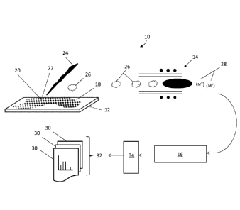
FIG. 1 is a pictorial representation of the system and
process according to one embodiment of the present teaching;
FIG. 1A is a schematic diagram of the system illustrated in FIG. 1;
Fig. 2 is an exemplary matrix containing the multi dimension
cellular information according to the present teaching;
FIG. 3 is close up view of the base according to an embodiment of
FIG. 1;and
FIG. 4 is a schematic view of an embodiment of the ICP ion source
according to the present teaching.
DESCRIPTION OF VARIOUS EMBODIMENTS
[0009] It should be understood that the phrase "a" or "an" used in
conjunction with the present teachings with reference to various
elements encompasses "one or more" or "at least one" unless the
context clearly indicates otherwise. Reference is first made to FIG.
CA 2888308 2020-01-31
CA 02888308 2015-04-13
WO 2014/063247 PCT/CA2013/050798
1, which shows a pictorial representation of the cellular analysis
system, generally indicated by reference number 10. The cellular
analysis system 10 comprises a base 12 coupled to an inductively
coupled plasma (ICP) ion source 14 of a mass cytometer 16. Generally,
the ICP ion source 14 can be considered as an integral component of
the mass cytometer 16, however for clarity, the ICP ion source 14 is
represented separately from the mass cytometer 16. The base 12, a
glass microscope slide for instance, provides a surface that can be
configured to hold a group of cells 18, from which candidate cells 20
can be identified, or have been identified by an interrogator 19, as
having properties of interest and subsequently for analysis by the
mass cytometer 16. The cellular analysis system 10 further comprises
a laser ablation system 23 for supplying at least one laser pulse 24
directed at the candidate cell 20 at its location 22 within the group
of cells 18. The mass cytometer 16 can comprise a control system 34
for controlling the laser ablation system and for generating
corresponding elemental tag data 30. A schematic of this system is
illustrated in FIG. 1A.
[0010] Generally, in mass cytometry, for simultaneous multi
parameter analysis within a single cell, a group of cells 18 can be
labeled with more than one distinct elemental tag Tn. The distinct
elemental tag Tn can be typically selected from the group comprising
transitional metals as described in co-pending United States Patent
Application No. 12/513,011, published as US2010/0144056, assigned to
the assignees of the present teachings. For convenience, the "n"
notation in Tn can be a variable to signify the different transitional
CA 02888308 2015-04-13
WO 2014/063247 PCT/CA2013/050798
6
elementals or metal isotopes. In various embodiments, for example, a
group of cells 18 can be labeled with more than one affinity reagents,
such as in the case of the different types of elemental conjugated
antibodies where each type of antibody being tagged with one or more
distinct elemental tag Tn. The distinct metal or elemental tag Tn
conjugated to each type of antibody can be a metal isotope of any one
or a combination of Gd, Nd, Tb, Eu, Gd, Dy, Ho, Sm, Er, Yb, to name
only a few. Each type of elemental conjugated antibodies can be
uniquely distinguishable by its distinct elemental tag Tn. As
generally known, the cells in the group of cells 20 which express an
affinity to the metal conjugated antibody can remain labeled with the
more than one elemental tag Tn. Thus, upon elemental analysis by the
mass cytometer 16, the elemental signature of the cell is represented
by the distinct element tags Tn associated with the antibodies.
[0011] As noted above, the applicants of the present teachings
recognizes that the interrogation process for detecting different
properties of interest can be combined in a multi dimension cellular
analysis with the same group of cells 18 according to the provision
that a) at the outset, the group of cells exhibit or can be prepared
with the condition(s) to exhibit the property of interest
corresponding with each interrogation process; b) the sequence of
analysis can be selected to maintain the integrity of each property of
interest for subsequent interrogation process; and c) the results from
each analysis can be cross correlated. Since the labeled cells
containing the elemental tag(s) is vaporizes during ionization by the
IC2 ion source 14, the mass cytometry detection process can be the
CA 02888308 2015-04-13
WO 2014/063247 PCT/CA2013/050798
7
basis of the final interrogator of the multi dimension cellular
analysis. Accordingly, the labeled group of cells 18 can undergo
initial interrogation processes to identify the candidate cells 20
having properties deemed to be of interest, other than those
associated with the more than one distinct elemental tag Tn. In
various embodiments, for example, a property of interest can be a
viability determination to identify candidate cells 20 that can be
either live or dead and, another property of interest can be based on
the physical shape or a physical feature inherent in the live or dead
candidate cell. A further property of interest, dependent or
independent of either of the aforementioned property of interest can,
for example, correspond with other luminescent properties associated
with the condition of immunostaining techniques to identify candidate
cells with certain proteins of interest. Thus, it is anticipated that
each property of interest can be represented by its specific physical
location(s) within the same or different candidate cells 20. Although
the process of the initial interrogation disclosed generally
correspond with optical or fluorescence microscopes, other cell-based
imaging/visualizing type of interrogators 19 such as that employing
electron or confocal microscopes have been considered and that their
use can be applied independently or in combination thereof. Thus for
the candidate cell section process, the initial interrogation can be
performed by at least one interrogator 19 configured to identify
candidate cells with corresponding properties of interest. Once the
candidate cells 20 have been identified, the location of the candidate
cells 20, as can be defined by its coordinates, with respect to their
CA 02888308 2015-04-13
WO 2014/063247
PCT/CA2013/050798
8
position within the group of cells 18 can be recorded against each
property of interest independently or in combination thereof. As
illustrated in FIG. 2, the information in a data source 21 concerning
the candidate cell's location 22 and the corresponding properties of
interest can be coded in a format which can be accessible by the
control system 34 and can be made part of the matrix containing the
multiple dimension of cellular information 36.
[0012]
Subsequently, by having access to the information in the
data source 21, for each recorded location 22, the control system 34
can direct at least one laser pulse 24 onto the corresponding
candidate cell 20 so that some of the cell material in the form of a
discrete plume 26 can be removed. Generally, each laser pulse can
generate a discrete plume 26 so that a series of laser pulses can
generate a series of corresponding discrete plumes 26. Consequently,
the material removed from the candidate cell 20 for each discrete
plume 26 can contain the more than one distinct elemental tag Tn.
Upon mass cytometry analysis, the detection of the more than one
distinct elemental tag Tn from the candidate cell 20 can represent the
presence of the associated affinity reagent and can be correlated with
the property of interest for the candidate cell 20, as exemplified in
FIG. 2.
[0013] While maintaining the spatial separation of each successive
plume 26, each plume 26 can be transported and introduced into the ICP
ion source 14 as discrete and independent entities. As each discrete
plume 26 passes into the ICP ion source 14, each elemental tag Tn can
CA 02888308 2015-04-13
WO 2014/063247
PCT/CA2013/050798
9
be ionized into corresponding elemental ions quantitatively related to
each elemental tag Tn. Since there can be more than one distinct
elemental tag Tn in the candidate cell 20, the ICP ion source 14 can
generate a distinct group of elemental ions for each elemental tag Tn.
Consequently, for each discrete plume 26, the ICP ion source 14 can
generate groups of elemental ions 28, represented generally as (le) in
FIG. 1. Each of
the groups of elemental ions 28 can be detected by
the mass cytometer 16, according to the ions' mass to charge ration
(m/z). In accordance with the present teachings, the mass cytometer
16 can detect each type of elemental ions simultaneously and, with the
advantage of a fast ion transit time for minimizing overlap, the mass
cytometer 16 can differentiate between groups of elemental ions
originating from successive lasers pulses. The elemental tag data 30,
shown in FIG. 1 as a succession of single data files in a total set 32
of data, represents the data acquired from simultaneously detecting
the groups of elemental ions 28 for the succession of each plume 26.
Hence, the cellular analysis system 10 can detect and identify each of
the more than one distinct elemental tag Tn simultaneously for each
laser pulse 24 and generate elemental tag data 30 that can be recorded
against the candidate cell's property of interest.
[0014] While generally, a single laser pulse 24 can completely
ablate a candidate cell 20 and generate a plume containing the more
than one distinct elemental tag Tn, there can be some instance of
candidate cells 20 requiring a series of laser pulses 24 to
penetrating into the cell, or through an adjacent surface, in order to
achieve maximum intensity of the more than one distinct elemental tag
CA 02888308 2015-04-13
WO 2014/063247 PCT/CA2013/050798
Tn. Furthermore, cellular profiling can be achieved with an
appropriate laser configuration having the capability of delivering
energy with temporal and spatial precision. A number of laser pulses
24 can be used to resolve the elemental tag Tn contained within the
candidate cell 20. For instance, during the analysis, as each laser
pulse 24 removes sequential layers of the labeled candidate cell 20,
groups of elemental ions 28 corresponding with the more than one
distinct elemental tag Tn can be simultaneously detected by the mass
cytometer 16. Each of the detected groups of elemental ions 28 can
represent the material removed at each layer of the candidate cell 20.
As noted above, some of the discrete plumes 26 can contain no metal
elements or some of the discrete plumes 26 can comprise a progression
of metal elements of various concentrations. Thus, for each of the
simultaneous measurements performed by the mass cytometer 16, the data
30 in the set 32 can contain qualitative and quantitative information
based on the presence and in some instances the absence, of the one or
more elemental tag Tn. Each of the acquired data 30 can provide a
piece of the information about the cross-section or thickness profile
of the labeled candidate cell 20.
[0015] Furthermore, there can be instances where, for example, the
thickness to the group of cells 18 is greater than an average cell
diameter such as in the case of overlapping layers of cells in the
presence of cell medium or in the case of thick tissue sections or
unsectioned whole tissue specimen, as generally indicated in FIG. 3.
Consequently, the location of one or more of the identified candidate
cell 20 or a part of a candidate cell 20 can be embedded at some
CA 02888308 2015-04-13
WO 2014/063247 PCT/CA2013/050798
11
distance from either the top or bottom surface of the group of cells
18. In various embodiments for example, the embedded candidate cell
or cells 20 can be identified by fluorescence microscopy using a
confocal microscope technique to provide localized cell structural
images. Other techniques for selecting the candidate cells 20 can be
based on identifying their property of interest can comprise of a
phosphorescence, a reflection, an absorption, to name only a few.
Hence, the location or the local details of the embedded candidate
cell or cells 20 can be defined by, for example, a Cartesian
coordinate system in 3-dimensional space, (X,Y,Z), where the value for
the Z axis in FIG. 3 denotes the depth within the thickness of the
group of cells 18. The 3-dimensional coordinates can be recorded (as
exemplified in FIG. 2) and used to represent and identifying the
location of the candidate cell 20 with respect to its position within
the group of cells 18. In this instance, when the location 22 of the
candidate cell 20 undergoes the elemental tag Tn analysis technique in
accordance with the present teaching, the depth coordinate Z can be
regarded as a property of interest and can be used to indicate the
expected number of laser pulses 24, for example, that can typically be
required in order to reach the embedded candidate cell 20. The
succession of discrete plumes 26 generated by the succession of laser
pulses 24 can contain no elemental tags Tn until the at least one
laser pulse 24 reaches the embedded candidate cell 20 at coordinate Z.
Conversely, the actual number of laser pulses deployed to reach the
candidate cell 20 and to generate plumes containing elemental tag Tn
and the detected group of elemental ions 28 can be correlated with the
CA 02888308 2015-04-13
WO 2014/063247 PCT/CA2013/050798
12
recorded depth Z as a way of cross calibrating the two mutually
exclusive cell-based techniques.
[0016] While the present teachings are described in conjunction
with various embodiments, it is not intended that the present
teachings be limited to such embodiments. On the contrary, the
present teachings encompass various alternatives, modifications, and
equivalents, as will be appreciated by those of skill in the art. For
example, the applicants of the present teachings recognize that
concurrently with the elemental tag Tn conjugated label, the group of
cells 18 can be initially prepared with conditions to exhibit the
property of interest detectable by the imaging / visualization process
for identifying the candidate cells 20 as generally known in the art.
As such, a group of cells 18 can be prepared with fluorophores labeled
proteins and/or endogenous expression of fluorescent reporter proteins
in addition to the elemental tag Tn conjugated affinity reagents
labeling. Additionally, the elemental tag Tn conjugated antibody can
be prepared with luminescent characteristic so as to provide dual
properties detectable by more than one mutually exclusive cell-based
analytical techniques. In various embodiments, for example, a 0D34
protein can be tagged with 148Nd elemental isotopes in addition to a
fluorescein for labeling in a biological sample suspected of having a
certain function. With fluorescence microscopy, the biological sample
can be initially examined and, candidate cells expressing 0D34 can be
identified and isolated on the glass microscope slide. The location
of the isolated CD34 expressed candidate cells can be targeted for
elemental detection by mass cytometry according to the present
CA 02888308 2015-04-13
WO 2014/063247 PCT/CA2013/050798
13
teachings. In this example, the detected group of elemental ions 28
can be correlated with the fluorescein detection as a way of providing
quantitation or for confirmatory purposes.
[0017] In various embodiments, the information recorded for the
candidate cell 20, including but not limited to its location 22, can
be in various data source formats and can be accessed by the control
system 34 using various protocols. For example, a visual
representation of the group of cells 18, such as a fluorescence
emission image, can be used to identify and represent each candidate
cell 20. In this instance, the interface between the data source 21
and the control system 34 can be an optical detection system with
appropriate visual recognition software. The software can be
configured for determining the location of the candidate cell 20 from
the emission image and subsequently for directing the at least one
laser pulse 24 at the location of the candidate cell 20.
Alternatively, the information recorded can be represented and
retrieved by various optical machine-readable interfaces such as that
embodied with barcode (1D or 2D) readers/scanners or through other
interfaces that employ radio frequency identification (RFID) or
variations and combinations thereof. Furthermore, the location of the
candidate cell 20 along with its property of interest can generally be
recorded as an alphanumeric data record accessible by the control
system 34 or the data can be manually entered into the operating
controls of the laser ablation system or the mass cytometry system
directly. Irrespective of the interfacing format from which the
candidate cell's information is transferred, the configuration of the
CA 02888308 20104-13
WO 2014/063247 PCT/CA2013/050798
14
laser ablation system can be designed so that recorded information can
provide the location coordinates for directing at least one laser
pulse 24 at the location 22 of the candidate cell 20.
[0018] In various embodiments, while the base 12 has been
described as a glass microscope slide that is generally consistent
with the material requirements for microscopy applications, the
applicants have contemplated the base 12 to be made of other material
such as one of or a combination of stainless steel, quartz, ceramic,
polytetrafluoroethylene (PTFE) and polyetheretherketone (PEEK) to name
a few. Alternatively, referring to FIG. 3, the base 12 can be a
support or a structure that can be separated from a microscope slide
38 holding the group of cells 18. For instance, in optical
microscopy, a group of cells 18 labeled with more than one distinct
elemental tag Tn can be mounted on a microscope slide 38 and
illuminated from below (for typical transilluminated light microscopy)
or illuminated from above through an objective lens (for typical
fluorescence microscopy). An image captured by the microscope optics
can be used for identifying a candidate cell 20 according to a
property of interest, such as a physical shaped or the presence of a
fluorescent probe(s) for example. The location of the identified
candidate cell 20 along with its property of interest can be recorded
to represent information relevant to the cellular analysis of the
biological sample. Generally the identification process can be
repeated as required so that more than one candidate cell 20 can be
located in the group of cells 18. Once the candidate cell 20 or
candidate cells 20 have been identified, the microscope slide 38 and
CA 02888308 2015-04-13
W02014/063247 PCT/CA2013/050798
its contents can be transferred to the base 12 for supporting thereof.
The information regarding the location of each of the candidate cell
20, expressed by Cartesian coordinates represented in either 2-
dimensional or 3-dimensional space within the group of cells 18 for
example, can be utilized to direct the at least one laser pulse 24
onto the candidate cell 20 for generating the discrete plume 26 for
each of the at least one laser pulse 24.
[0019] The applicants of the present teachings recognizes that in
order for each elemental tag data 30 to correspond with each of the at
least one laser pulse 24, the spatial separation of each successive
plume 26, and the corresponding ions, during their travel along the
path between the base into the ICP ion source 14 and between the ion
source 14 and the ion detector of the mass cytometer 16 is maintained.
For example, a solid state laser typically used for laser ablation,
such as a femtosecond pulsed laser can be configured to operate with a
pulse rate between 10 and 100 Hz. At this frequency range, a plume 26
can be generated every 10 to 100 msec. Considering the lower limit,
the system 10 would be required to have a maximum delay time of the
order of 10 msec. In accordance with various embodiments of the
present teachings, the mass cytometer 16 can be characterized as a
"flow-through" analytical device comprising a linear ion path with
electrostatic lenses and an ion detector capable of parallel elemental
ion detection. In this configuration, a delay time in the order of 10
msec can be achieved so that the groups of elemental ions (IC) can
undergo acceleration and pass within the mass cytometer 16 for
simultaneous detection. Consequently, the likelihood of the ion
CA 02888308 2015-04-13
WO 2014/063247 PCT/CA2013/050798
16
detector to separately detect each of the groups of elemental ions 28
can be realized.
[0020] To maintain a corresponding spatial distinctiveness
upstream of the mass cytometer 16, the configuration of the path
between the laser ablation location, at the base 12, and the entrance
to the plasma can be chosen to maximize the plume 26 separation while
minimizing flow turbulence. At the lower limit, a delay time of the
order of 10 msec for maintaining the separation of each plume 26
before ionization can be achieved with a path having a minimum
distance of plume travel and a corresponding means of accelerating the
same. Generally, the ICP ion source 14 utilizes an injector tube 42,
as indicated in FIG. 4, and a flow of carrier gas (not shown) can be
applied appropriately to direct each discrete plume 26 into the plasma
44. Accordingly, the injector tube 42 can be configured to provide a
laminar or near laminar flow geometry, having a Reynolds number below
2000 for instance, for receiving the plume 26 and for the carrier gas
to flow with the plume 26 such that any turbulence can be minimized.
Thus, in various embodiments, the combined delay time corresponding to
the total path between the base 12 and the ion source 14 and between
the ion source 14 and the ion detector of the mass cytometer 16 can be
between 20 msec and 200 msec.
[0021] Furthermore, in various embodiments, the base 12 can be
positioned relative to the ICP ion source 14 such that the travel time
for each plume 26 can be minimized. For example, the ICP ion source
14 can be structured to encompass the base 12 for providing a closely
CA 02888308 2015-04-13
WO 2014/063247
PCT/CA2013/050798
17
coupled laser-ablation-ICP ion source. The laser-ablation-ICP ion
source can be configured with an integrated enclosure having an
optical entrance for the laser pulses 24, a carrier gas for capturing
and transporting the plume and the ICP ion source for generating the
groups of elemental ions 28. The carrier gas flow can be configured
to sweep off each discrete plume 26 at the ablation location 22 and
pass each plume 26 directly into the plasma 44.
[0022] While in
various embodiments the term "group of cells" is
generally in reference to cells contained in thinly sectioned
biological tissue samples or unsectioned tissue whole specimens, the
present teachings can be equally applied to cells typically found in
cell cultures. In various embodiments, the group of cells 18 can be a
mixture of cell subgroups where each subgroup can originate from
different sources or different biological entities. In this instance,
the cell's origins can be characterized by one of the properties of
interest. For example, consider samples taken from more than one
biological entity where each sample corresponds with a cell subgroup
that can be distinctly labeled with more than one distinct elemental
tag Tn. Each of the cell subgroups can be combined into the same
group of cells 18 so that the cells from each sample can collectively
express its distinctly labeled elemental conjugated antibody within
the same group of cells 20. The combined group of cells 18 can be
interrogated and candidate cells 20 can be identified and selected
based on some property of interest without knowledge of the candidate
cell's origin. Upon elemental analysis according to the present
teachings, the elemental tags Tn can be detected and the corresponding
CA 02888308 2015-04-13
WO 2014/063247
PCT/CA2013/050798
18
candidate cells 20 can be identified as being related to the specific
biological entity. Accordingly, the relation between the property of
interest and the detected elemental ions 24 can be correlated to
determine the property of interest indicative of that cell subgroup
and potentially the cell's origin.
Generally, the advantage of the
present teachings can be applied to groups of cells comprising non-
disrupted cells where non-disrupted candidate cells can be identified
and further analyzed for its elemental tags by mass cytometry.
[0023] While efforts have been described to create the conditions
for maintaining the spatial separation of each plume 26 and the
corresponding groups of elemental ions 28 throughout the cellular
analysis, the applicants of the present teachings recognizes that some
spatial spreading or overlapping can be present. Accordingly, the
applicants have contemplated an averaging approach applied to two or
more of the acquired elemental data 30 without substantial loss to the
information generated from each laser pulse 24. Alternatively, an
integrated approach of combining each of the acquired elemental data
30 into the set 32 can be sufficient to represent the mass cytometry
information portion of the multi dimension cellular analysis. In
various embodiments, a data analysis algorithm, such as FFT, can be
used for de-convoluting the integrated data set 32. Different forms
of algorithms can be operated within the analysis system 10 or can be
applied post data acquisition as is generally known.
[0024] Furthermore, the control system 34 can be configured to
serve multifunctional purposes in accordance with the present
CA 02888308 2015-04-13
WO 2014/063247 PCT/CA2013/050798
19
teachings. Although the control system 34 has been described in
connection with controlling the laser ablation system and for
generating corresponding elemental tag data 30, the control system 34,
typically functioning under logic control, can provide various levels
of automation or to perform a sequence of feedback controlled actions
to enable the analysis system 10 to be automated. In various
embodiments, for example, the control system 34 can be a processor
driven system coupled with a automated sample handling system to
perform the interrogation and identification, the information
recording and accessing, the directing of the laser pulses, the mass
cytometry detection process and the data correlation autonomously or
with user intervention.